Chapter One: Epithelium
Introduction
There are four basic types of tissue in the body: 1) epithelial, 2) connective, 3) nervous, and 4) muscular. All of the free surfaces of the body whether external (skin) or internal (tubes and cavities) are lined by an epithelium which serves as a protective barrier between tissues and spaces. These epithelial cells are held together by specialized intercellular junctions. Some cells which do not appear to be lining a surface (namely the secretory parenchyma of glands) are also epithelial by definition (little intercellular space, avascular, junctional complexes, basal lamina).
Epithelium is one of the four primary tissues of the body. It is predominantly a cellular tissue with little intervening space between adjacent cells. The key to the nature of an epithelial layer is the shape of cells that comprise it, particularly the shape of cells upon its free or exposed surface. The principal activities of these tissues in their living condition are related to the thickness of the epithelial layers and the shapes of the cells that comprise them.
Epithelial tissue is avascular and dependent upon its underlying connective tissue for its metabolic requirements. All epithelia have in common these features: 1) avascular; 2) little intercellular material; 3) surface specializations; 4) junctional complexes; 5) basal lamina. The epithelial cells rest on a condensation of extracellular matrix elaborated both by the epithelial cell as well as underlying connective tissue. This condensation is called the basement membranes (old light microscopic term) or basal lamina). It consists of two parts: 1) a layer of collagen (type IV) and amorphous ground substance secreted by the epithelial cells, and 2) reticular fibers which extend into the ground substance from the underlying connective tissue. An electron lucent space separates the basal lamina from the plasma membrane of the epithelial cells.
Epithelial cells have a variety of functions and locations in the body. Their location and function will dictate their shape and the number of layers that will be found. These factors will also determine the type of intercellular junctions that will be present between the epithelial cells. Epithelium can function in: absorption (as in the gastrointestinal tract), secretion (as in a sweat gland), transport and excretion (as in urine formation in the kidney), sensation (taste buds or olfactory cells) as well as for protection.
Epithelia (plural of epithelium) are usually classified according to the shape of the cells in the outermost layer and the number or arrangement of their layers.
- Classification by cell shape (surface)
- squamous - flat
- cuboidal - shaped like a cube
- columnar - tall narrow cell
- Classification by number of layers
- simple - 1 layer
- stratified - multiple layers
There are two primary exceptions to the classification scheme listed above. These are: 1) pseudostratified (falsely stratified) columnar (defined below) which is found in the respiratory system, and 2) transitional epithelium in the urinary system. Since your urinary bladder can be distended, the epithelium changes from a stratified cuboidal appearance to a stratified squamous, thus this transition between types leads to the name transitional epithelium.
 Top
Top
Types of Epithelium
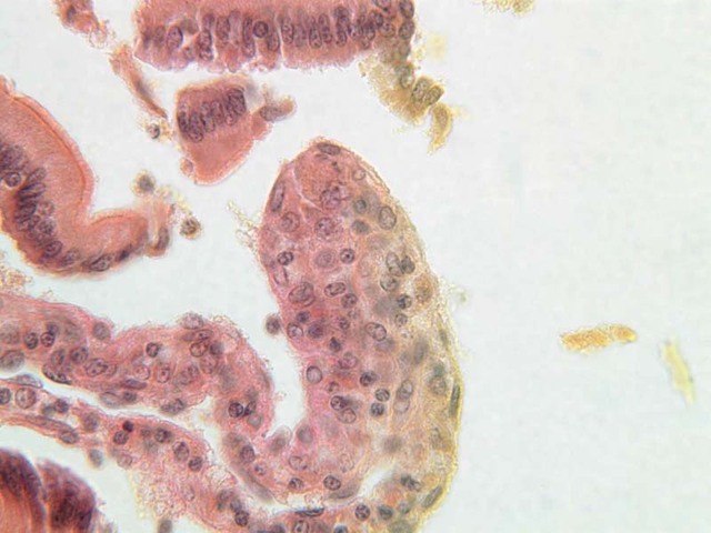
Simple Squamous Epithelium
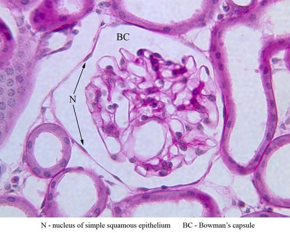
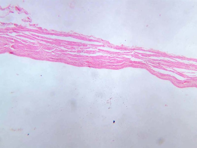
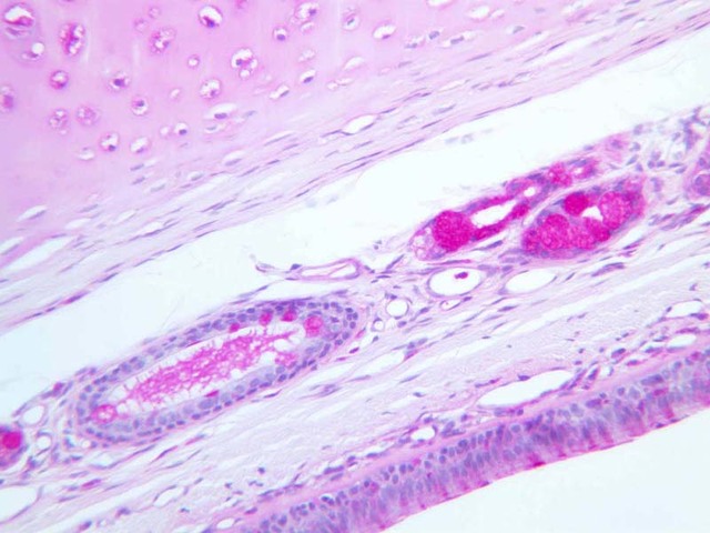
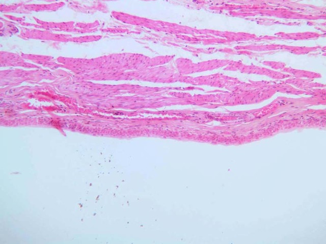
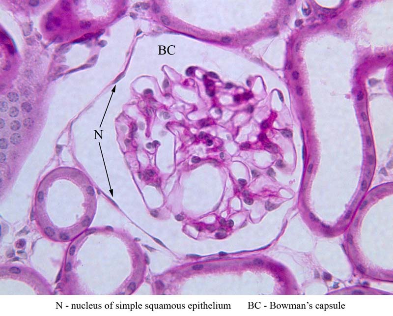
Simple Squamous Epithelium is composed of flattened polygonal cells. Simple squamous epithelial cells line the heart and the blood and lymph vessels (where they are called endothelium) as well as the outside of many visceral organs where they are called mesothelium (from embryological mesoderm). They are also found in body organs, e.g., lining the pulmonary alveoli (air sacs) or the renal glomerular capsule (Bowman's
capsule).
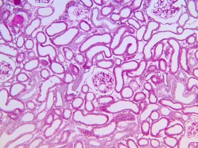
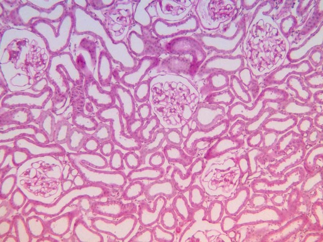
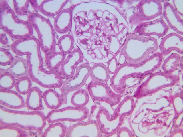
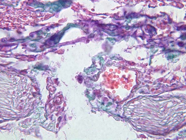
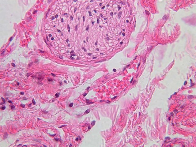
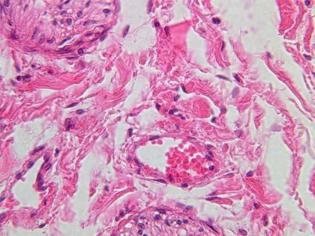
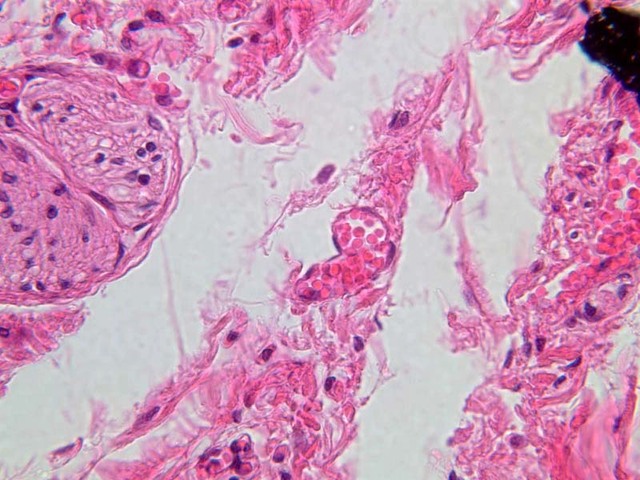
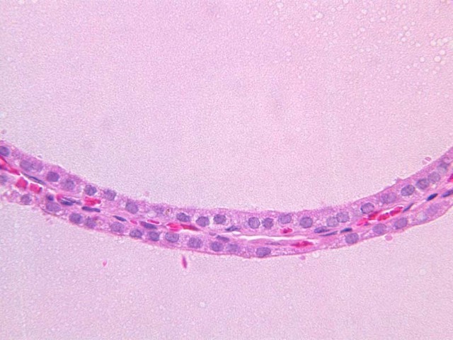
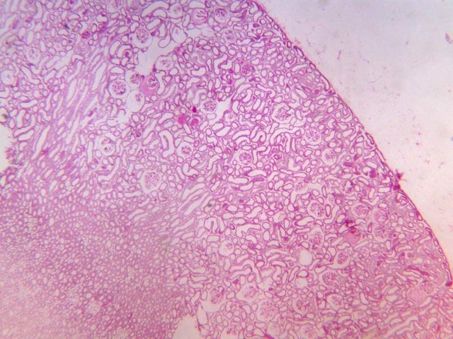
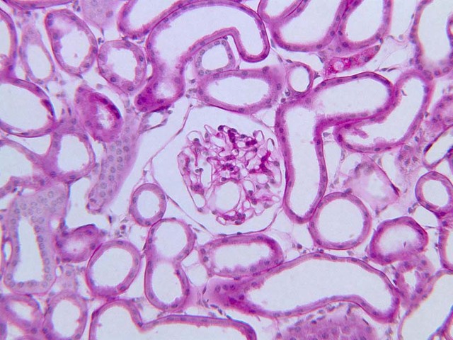
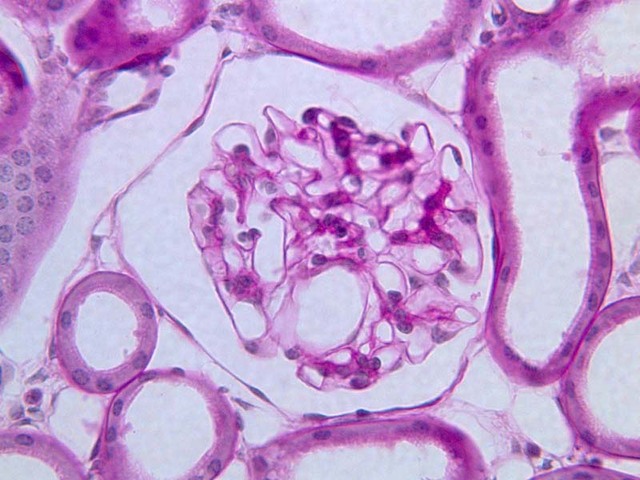
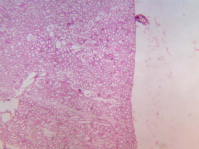
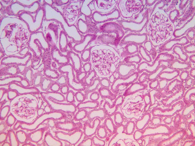
This type of one-layered epithelium serves as a filter in many places in the body. It is composed of very thin sheets of flattened, polyhedral (squamous) cells and thus is called simple squamous epithelium. Look for cells arranged in a single layer surrounding a glomerulus in the kidney. Within the cortex (the outer, or peripheral zone of the kidney) identify capillary tufts called glomeruli located within a circular structure (slide B-68, kidney, H&E [
10x,
20x,
40x-labeled]). The lining of the circular structure (called Bowman's capsule) is simple squamous epithelium. You may only appreciate the flattened nuclei of these cells. The epithelium, cut perpendicular to its plane of spread, is visible as a chain of nuclei connected by very thin lines of cytoplasm. This attenuated sheet of cells with their semipermeable membranes offers a minimum barrier to the passage of many substances, but completely halts others: hence its efficacy as a living filter.
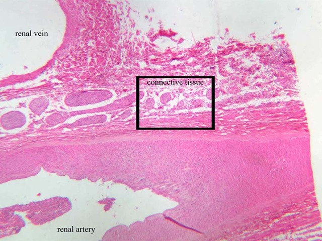
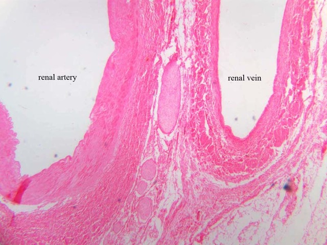
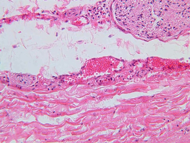
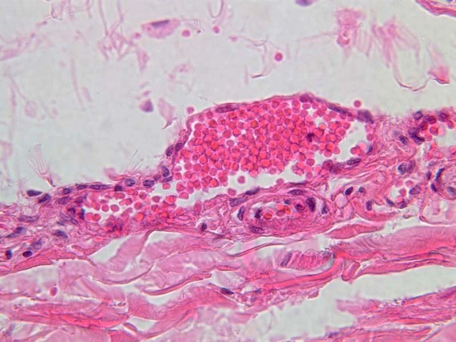
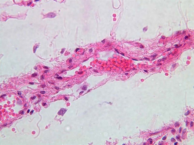
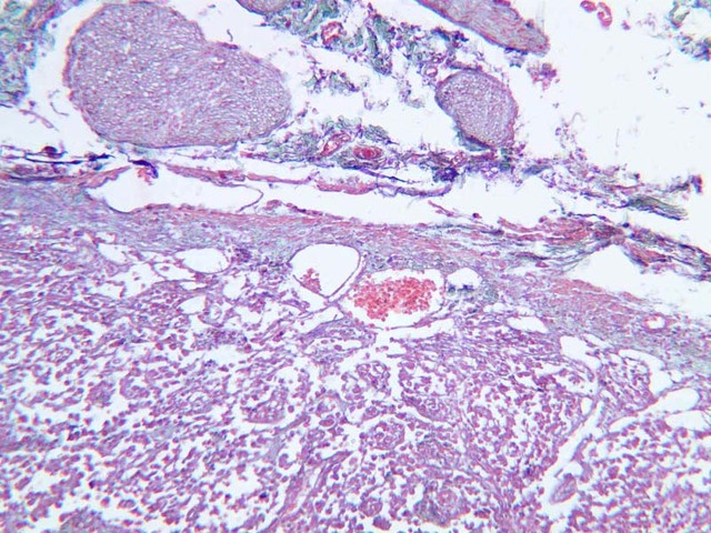
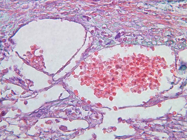
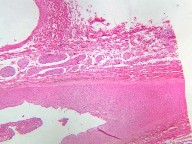
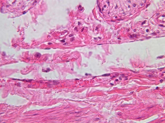
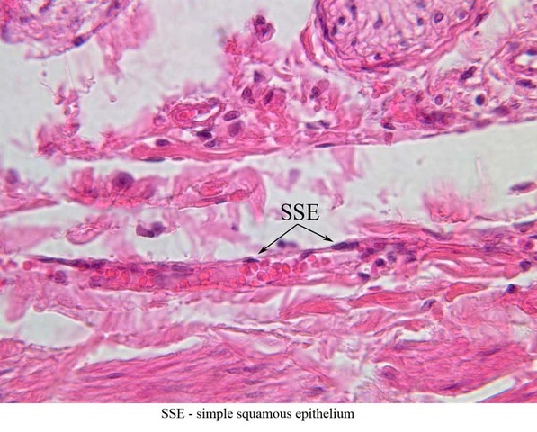
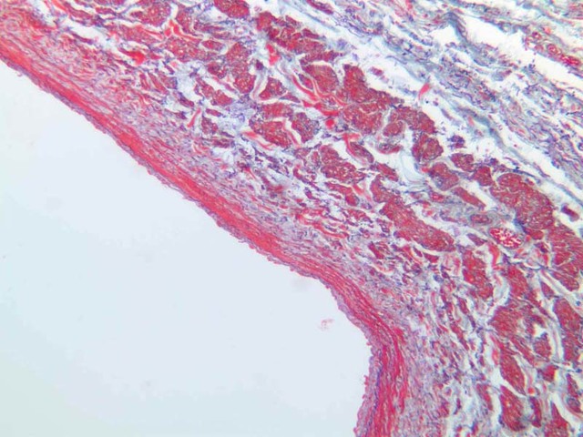
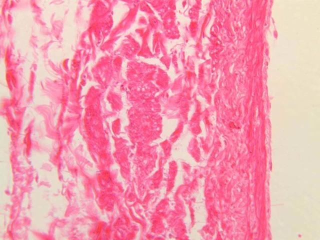
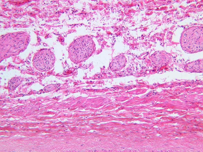
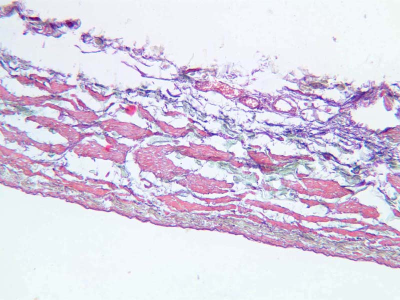
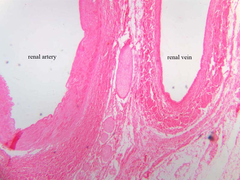
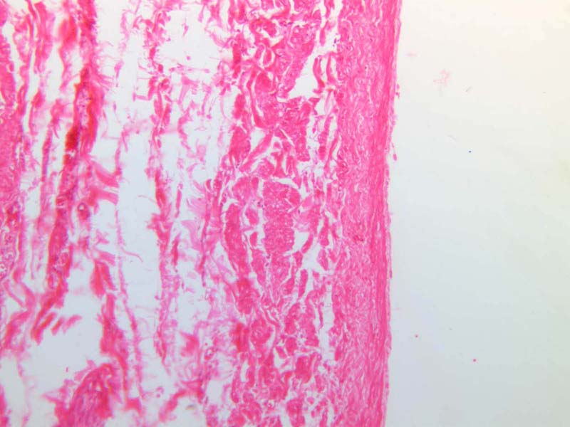
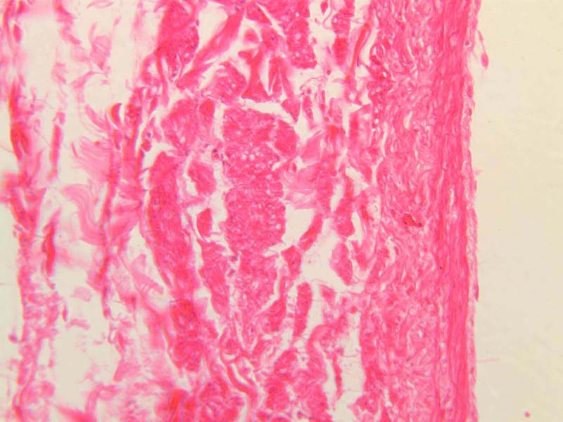
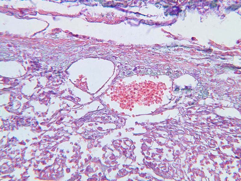
Look for a similar connected chain of nuclei lining the inside of a large vein (see slide A-28, renal artery and vein, H&E [
2.5x-labeled,
10x,
20x,
40x-labeled]; AF [
10x,
20x,
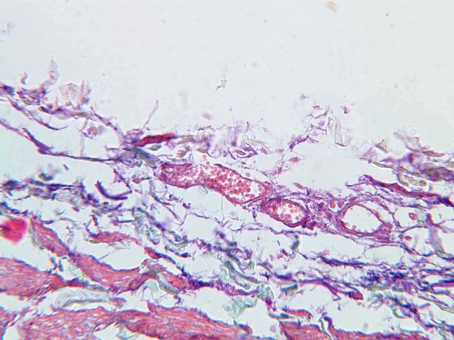
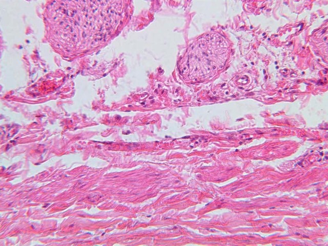
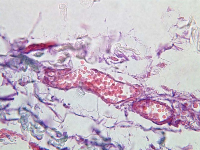
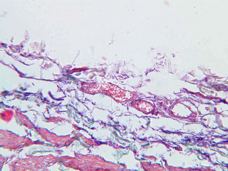
40x]), or in capillaries (slide A-28, connective tissue, H&E [
2.5x-labeled,
10x,
20x,
40x-labeled] [
20x,
40x,
40x,
40x]; AF [
10x,
20x,
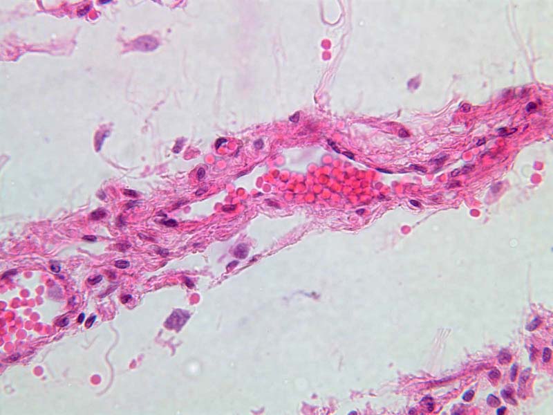
40x]). The inner lining of the blood vessels, called the endothelium is a simple squamous epithelium. The lining may appear interrupted in the section, in part because the cytoplasm between nuclei is drawn out so thinly. In life, of course, the membrane is unbroken and lines the vessel completely. The simple squamous epithelium that lines the abdominal cavity has the special name of mesothelium.
Simple Squamous Image Gallery
Simple Squamous Table of Identifications
| Row |
Structure |
Abbreviation |
Optimal Stain |
Representative Section |
Note |
| 1 |
Renal Artery |
(none) |
H&E |
 A28, Renal Artery and Vein, 2.5x A28, Renal Artery and Vein, 2.5x |
|
| 2 |
Renal Vein |
(none) |
H&E |
A28, Renal Artery and Vein, 2.5x |
|
| 3 |
Connective Tissue |
(none) |
H&E |
A28, Renal Artery and Vein, 2.5x |
|
| 4 |
Bowman's Capsule |
BC |
H&E |
 B68, Kidney, 40x B68, Kidney, 40x |
|
| 5 |
Nucleus of Simple Squamous Epithelium |
N |
H&E |
B68, Kidney, 40x |
|
| 6 |
Simple Squamous Epithelium |
SSE |
H&E |
 A28, Renal Artery and Vein, 40x A28, Renal Artery and Vein, 40x |
|
Top
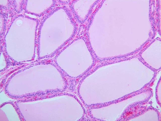
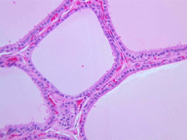
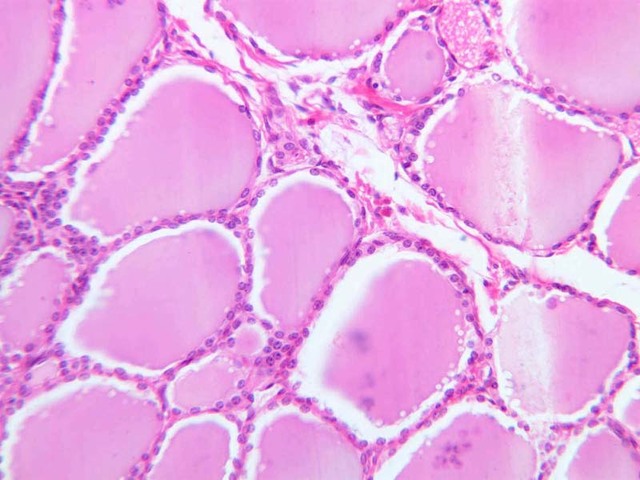
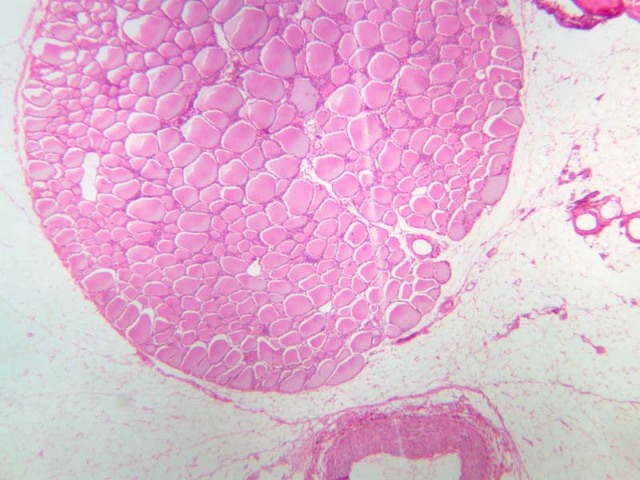
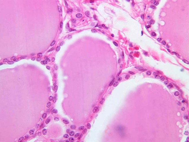
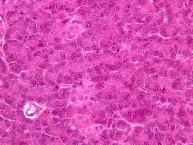
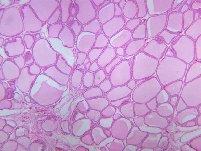
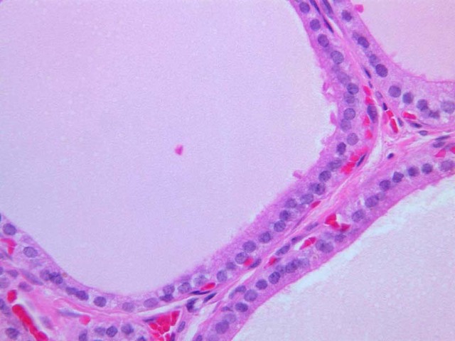
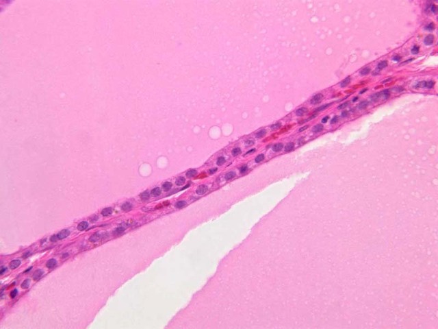
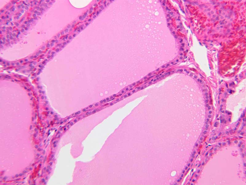
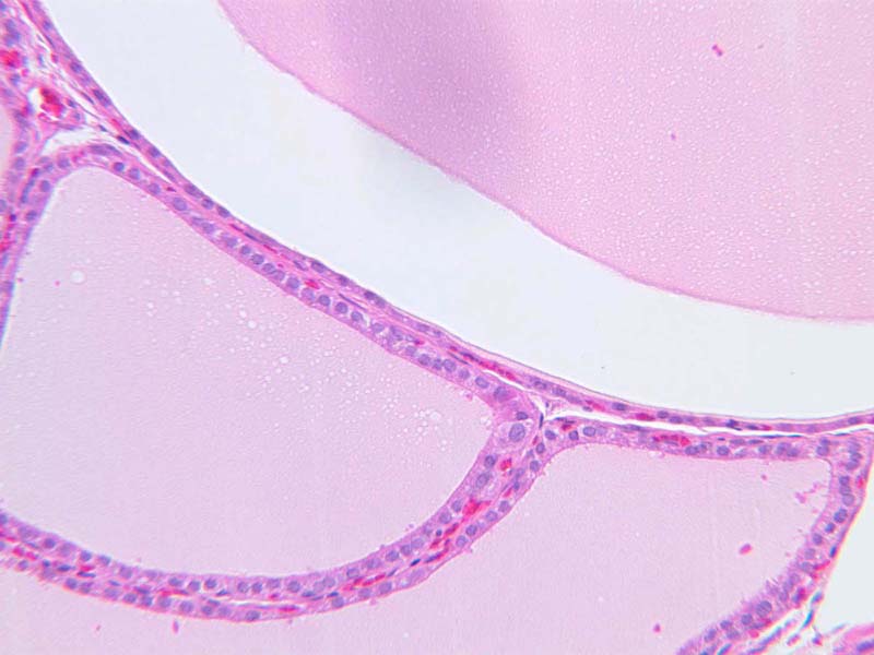
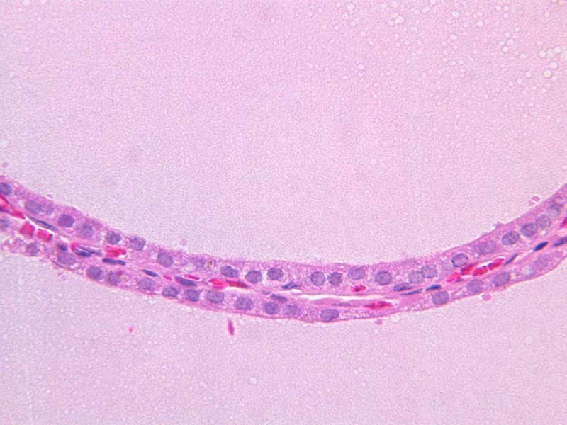
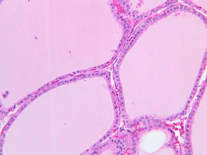
Simple Cuboidal Epithelium
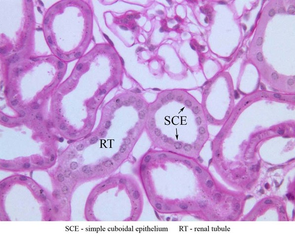
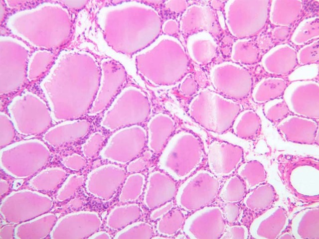
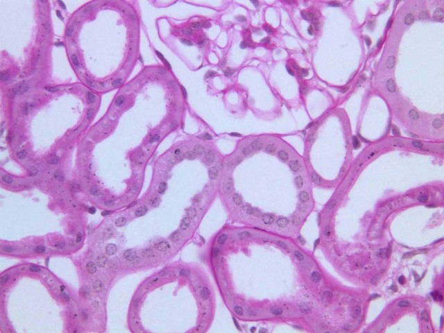
Simple Cuboidal Epithelium lines the ducts of many glands, kidney tubules and the follicles of the thyroid gland (slide B-68, H&E [
2.5x,
10x,
20x,
40x-labeled]). Observe the cuboidal cells lining many of the renal tubules. Here you can find tubular cells in which the height of the cell is approximately the same as the basal diameter of the cell. Slide B-52 (H&E [
2.5x,
10x,
20x,
40x-labeled] [
20x,
40x] [
20x,
40x] [
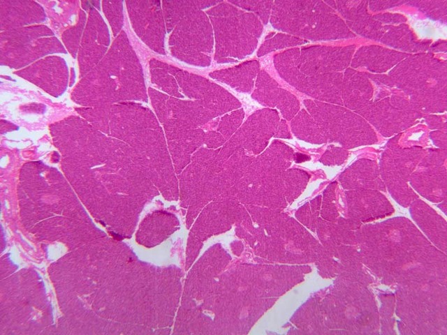
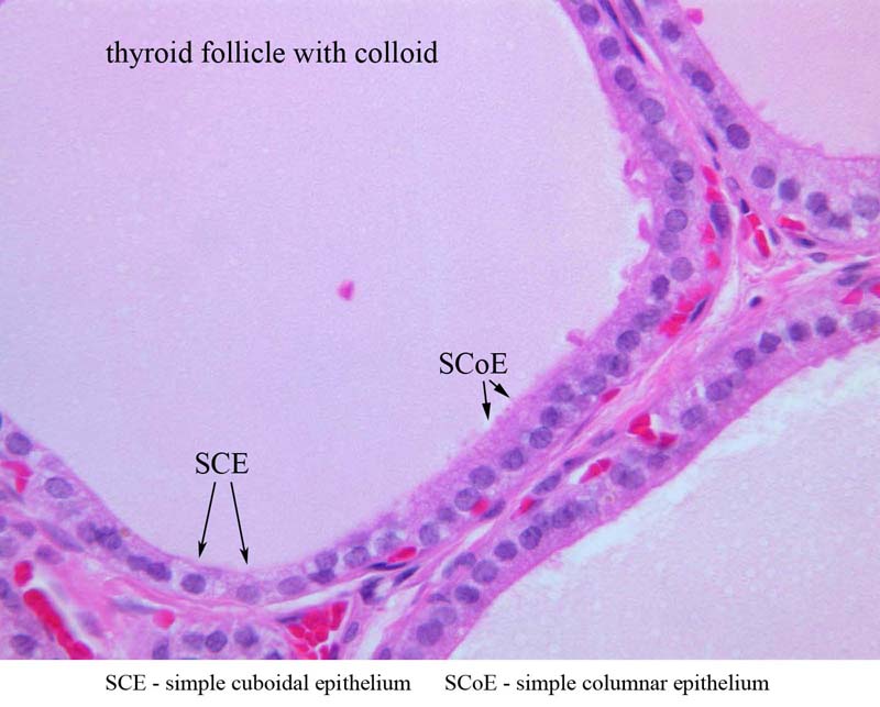
20x,
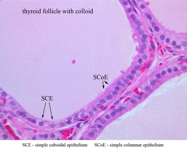
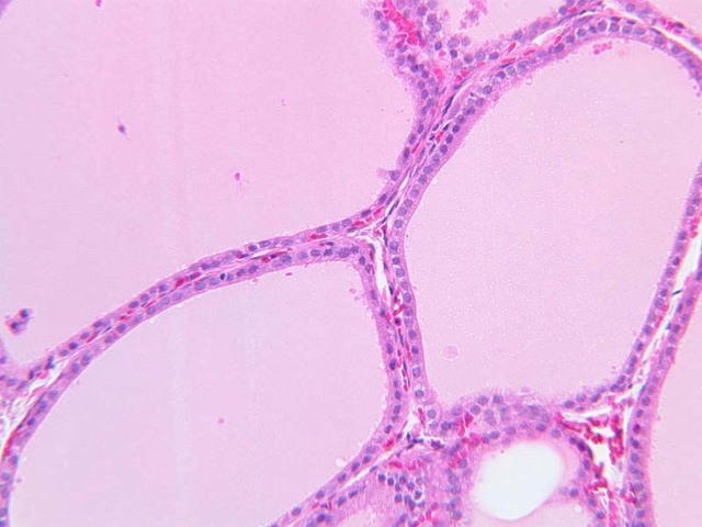
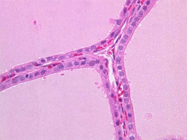
40x]) is a section through the thyroid gland showing many follicles of various sizes. The cells lining the follicles are approximately cuboidal in shape. The pink amorphous material inside the follicle is colloid.
Simple Cuboidal Image Gallery
Simple Cuboidal Table of Identifications
Top
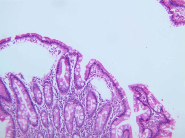
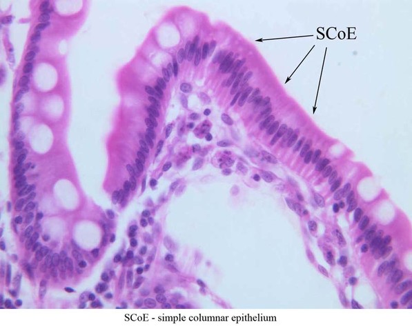
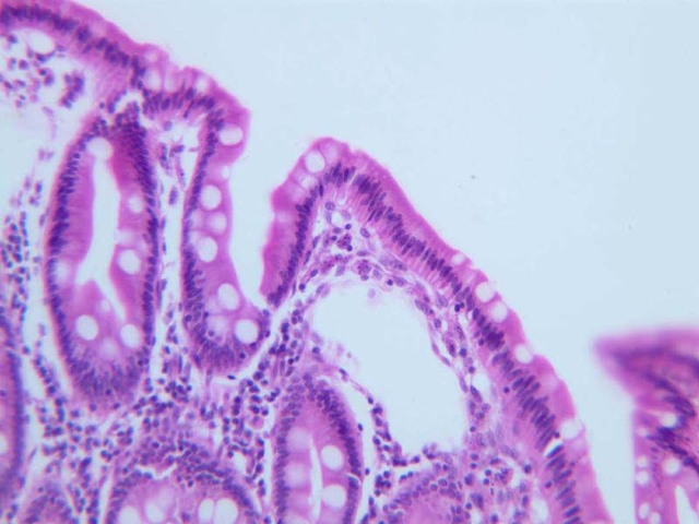
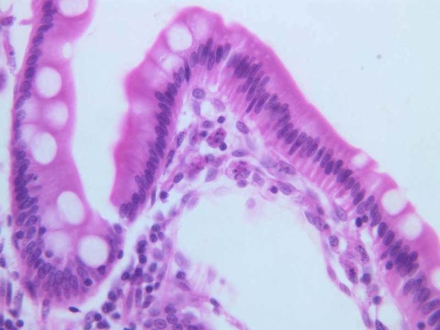
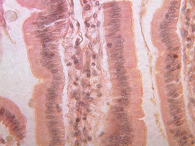
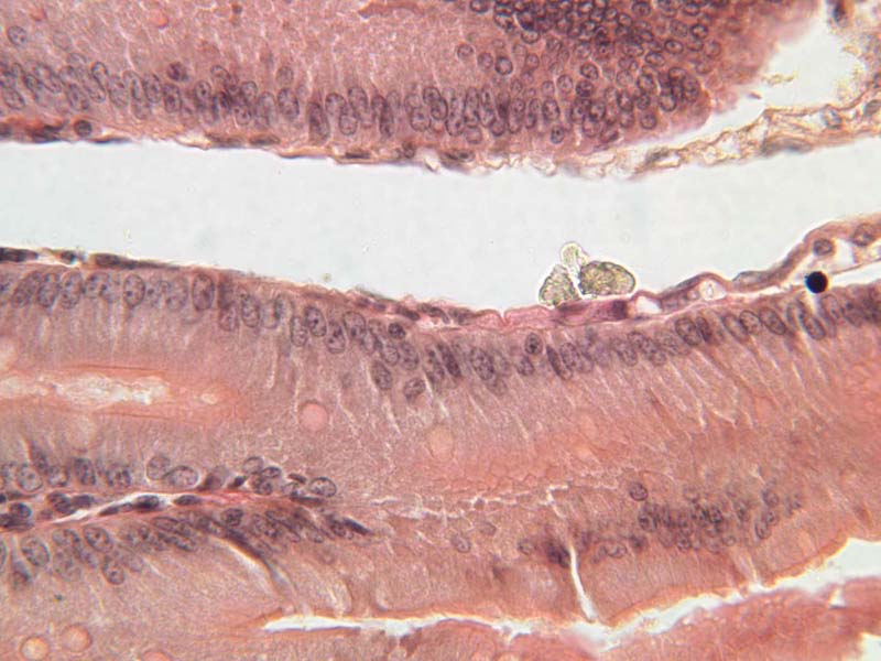
Simple Columnar Epithelium
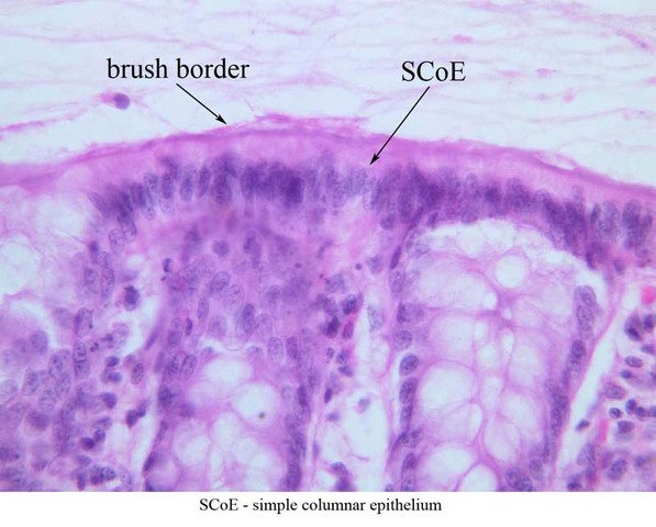
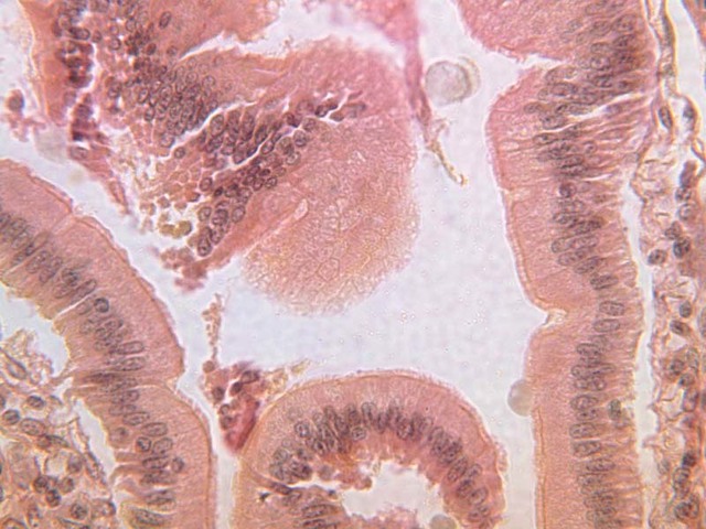
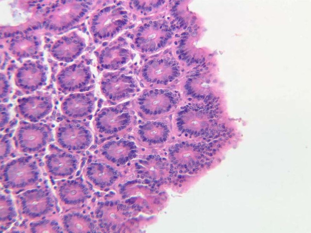
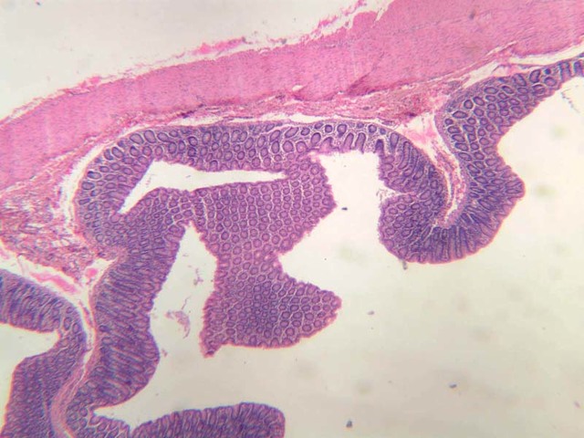
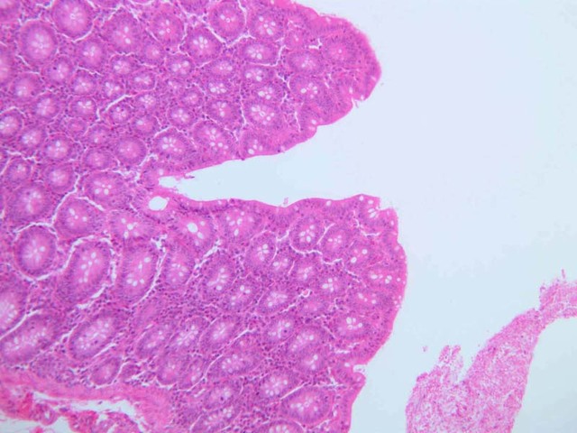
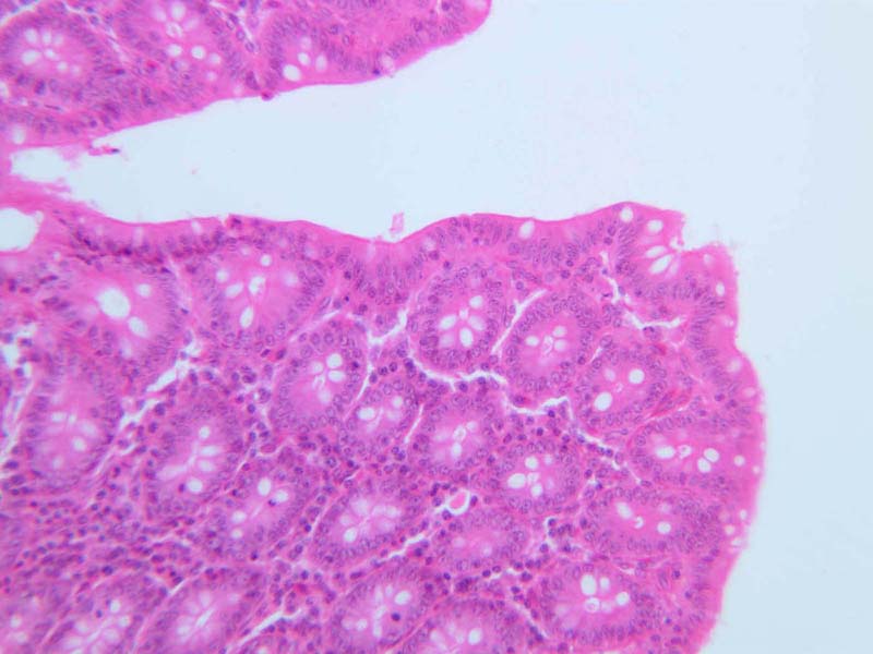
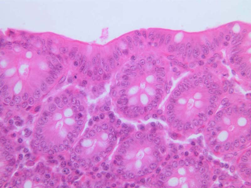
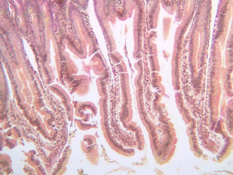
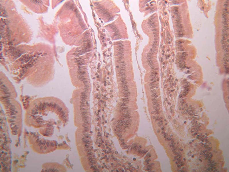
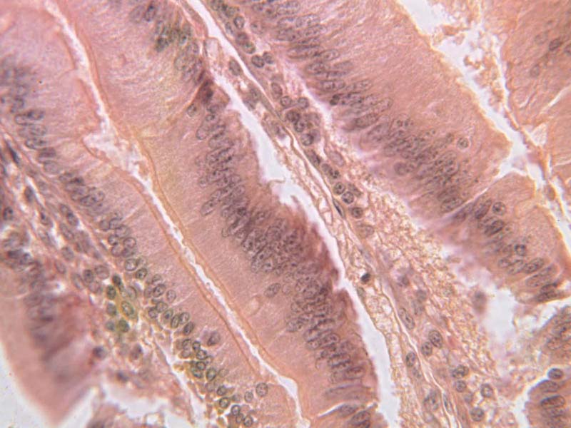
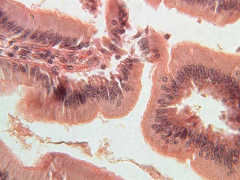
Simple Columnar Epithelium lines much of the human gastrointestinal tract, such as the stomach, intestines, gallbladder, and the major ducts of the digestive glands (biliary, pancreatic, and salivary). These columnar cells are characterized by tall cells with oval nuclei placed usually in the basal region of the cell.
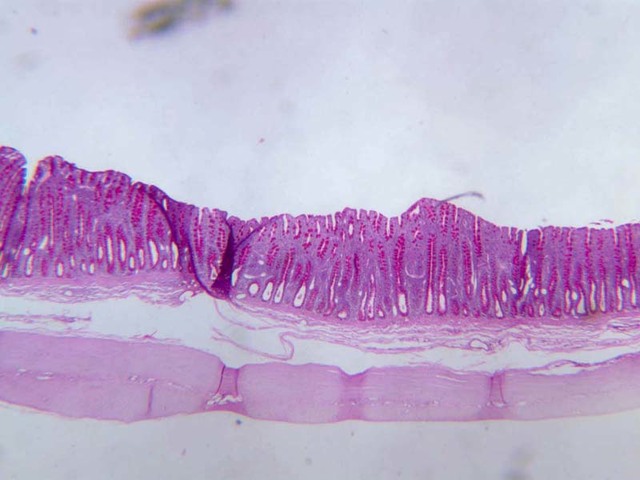
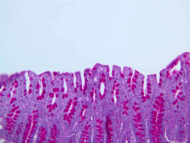
The shape of the columnar cell is more readily observed in places where it occurs in single rather than many layers. A single-layered epithelium is called simple in contrast to a complex (that is, stratified) epithelium. Look at a section from the small intestine (slide B-12, H&E [
2.5x,
10x,
20x,
40x-labeled] [
40x,
40x,
40x]) and study the simple columnar epithelium that lines its inner surface. The epithelial cells are taller than wide and have oval nuclei basally located in the cell.
Cytoplasmic organelles that are particularly active in cell secretion and absorption lie superficial to the nucleus; these organelles, though not usually seen in preparations for the optical microscope, are readily demonstrated in electron micrographs and their location confers a polarity on the epithelial cells. Thus, one end of the cell is active in processes that digest and absorb foodstuffs while the other is separated from these processes and is involved in cell metabolism itself. A simple epithelium with its constituent cells exposed to the contents of an organ makes a much more effective membrane for digestion than a stratified epithelium with dying surface cells. A simple epithelium, however, is particularly vulnerable to mechanical injury which can expose the underlying tissues.
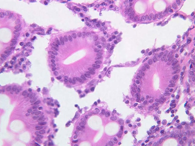
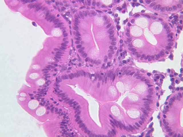
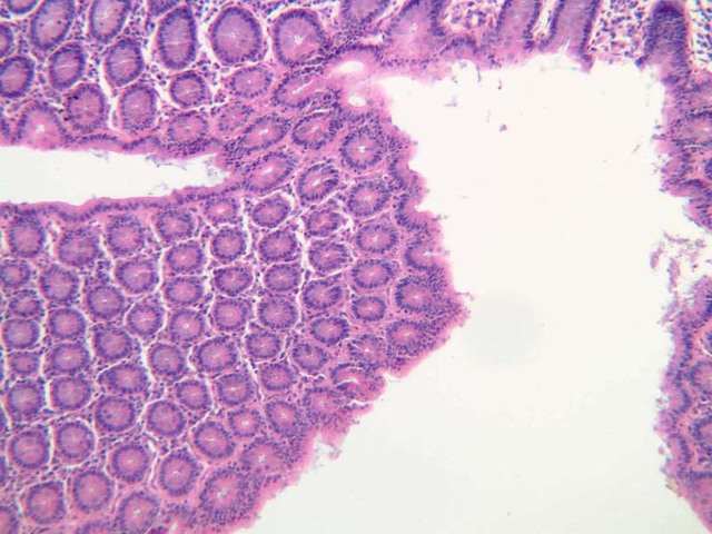
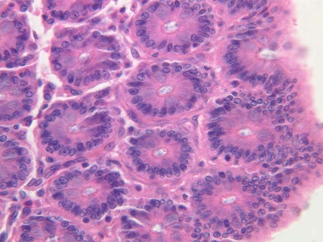
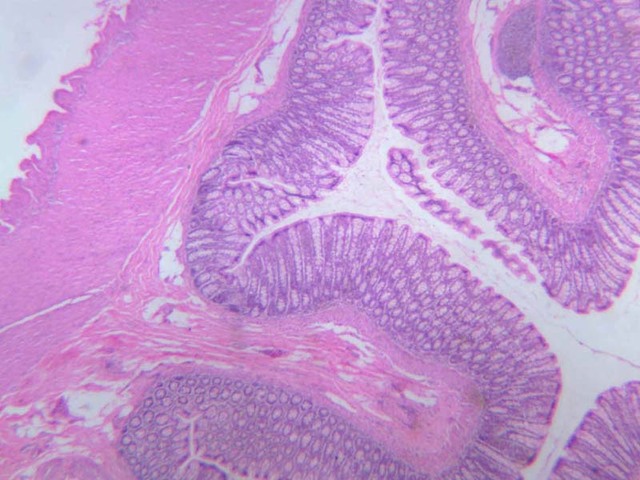
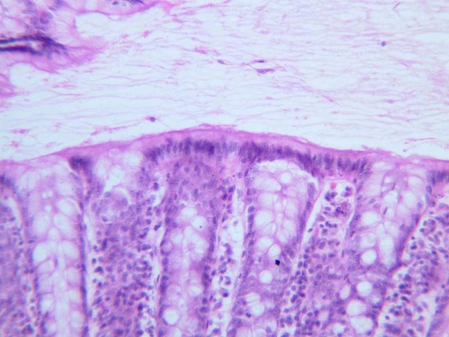
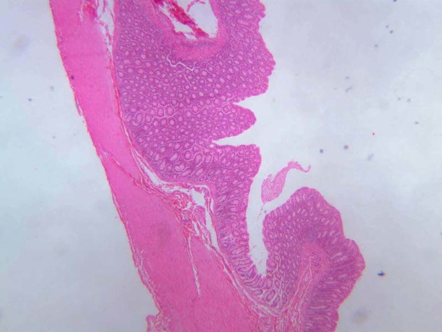
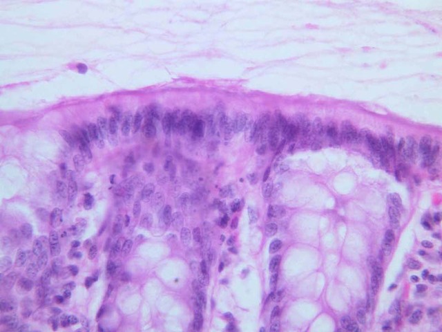
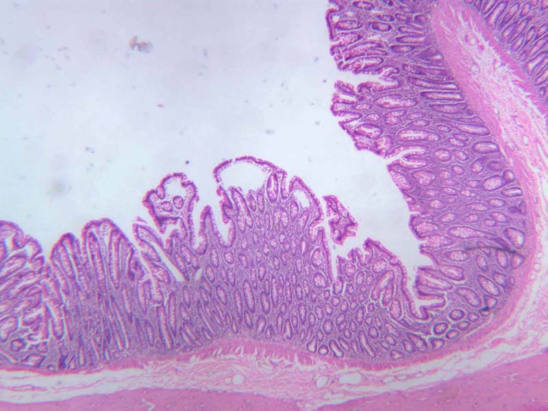
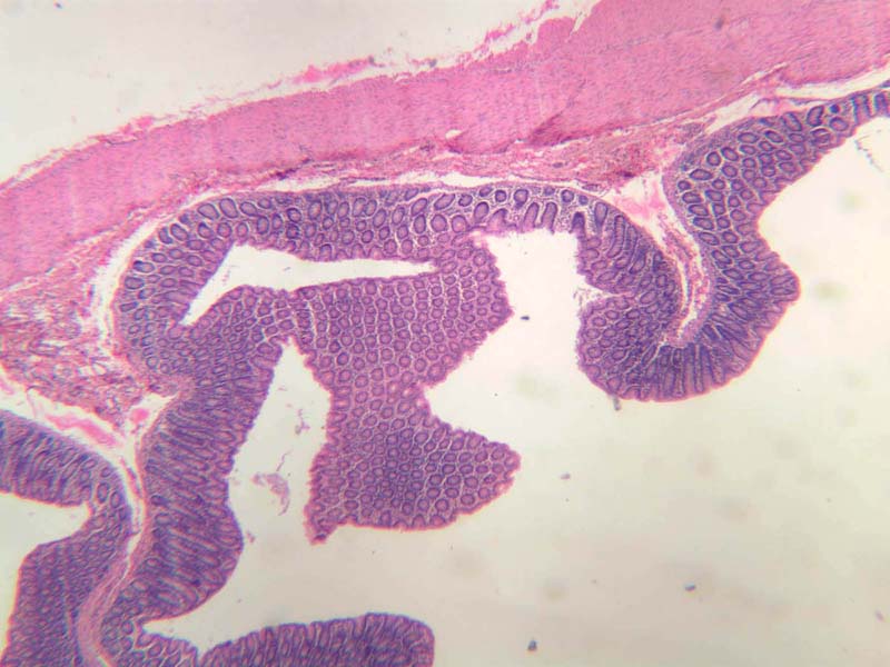
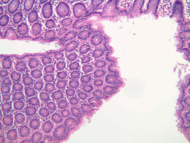
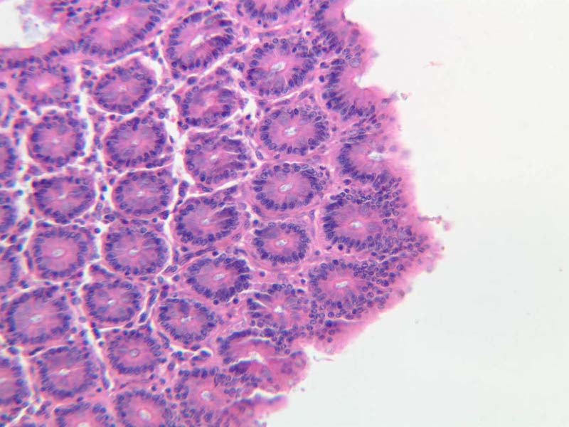
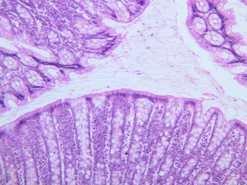
Slide B-24 illustrates simple columnar cells very well (B-24, colon, H&E [
2.5x,
10x,
20x,
40x]). Note also the striate or brush border ([
2.5x,
10x,
20x,
40x-labeled] [
2.5x,
10x,
20x,
40x]) which can be seen (under high-dry) as a thin line along the luminal surface of the cell.
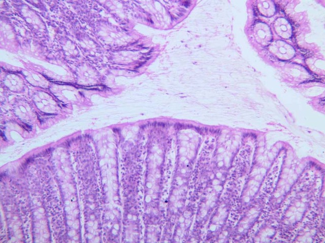
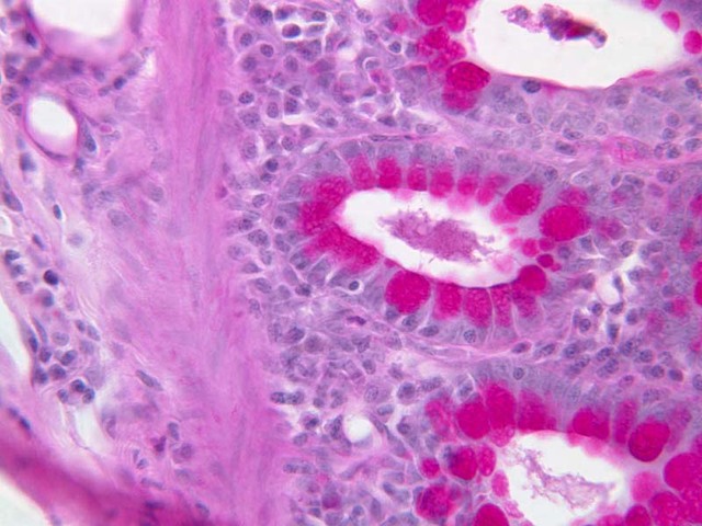
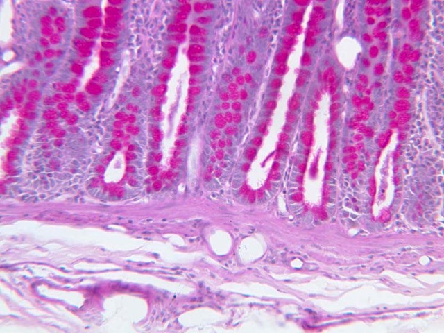
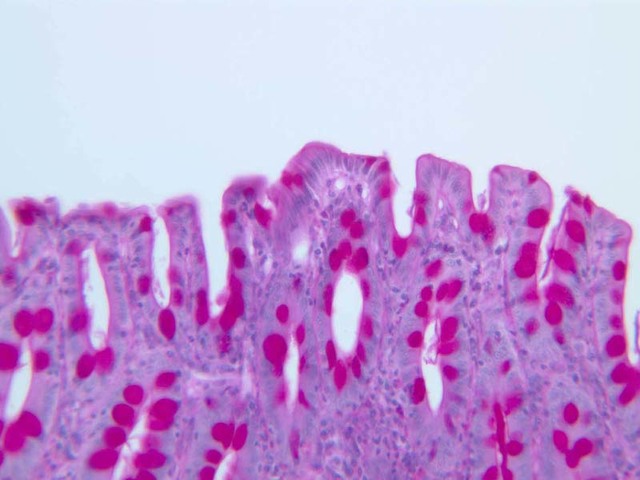
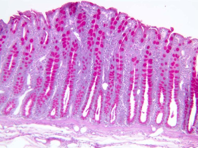
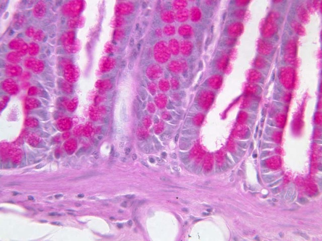
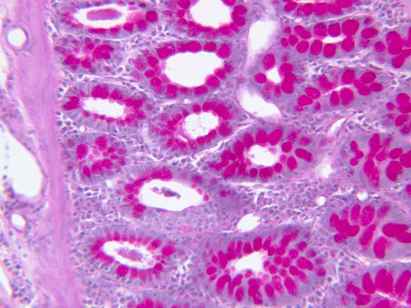
In the lining epithelium of the colon will be found a variant of simple columnar epithelium (slide
B-24 and
B-25). Slide B-25 is stained with the PAS technique ([
2.5x,
10x,
20x,
40x] [
20x,
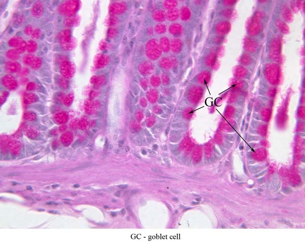
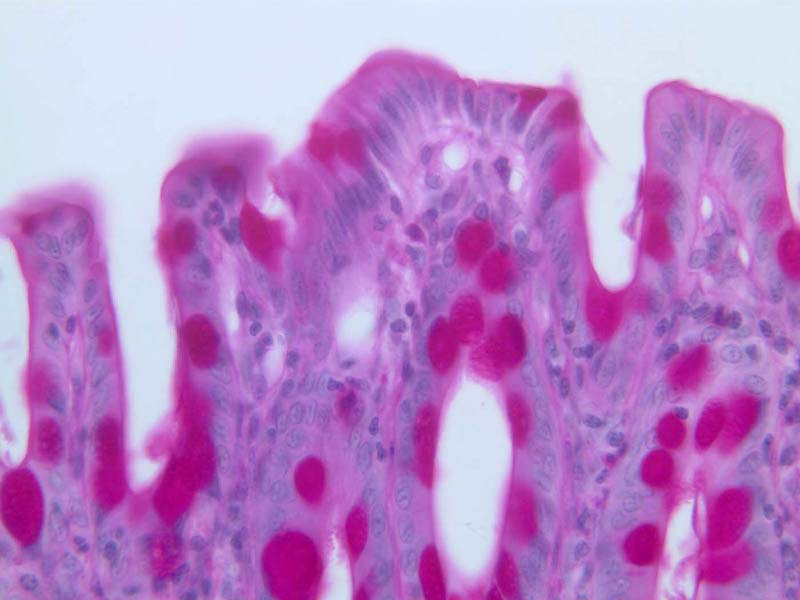
40x]). This procedure is specific for demonstrating carbohydrate residues. Most of the cells that comprise the inner lining of this organ are goblet cells, a unicellular gland that secretes viscid mucus ([
10x,
20x,
40x-labeled]). Goblet cells derive their name from the "goblet" of mucus that occupies the apex of an engorged cell; the nucleus is usually flattened basally in what would be the stem region of a goblet. The relationship of the mucous goblet to other parts of the cell can be more readily seen with the electron microscope. In general, goblet cells occur where a viscous lubricant is useful in protecting a delicate epithelial surface.
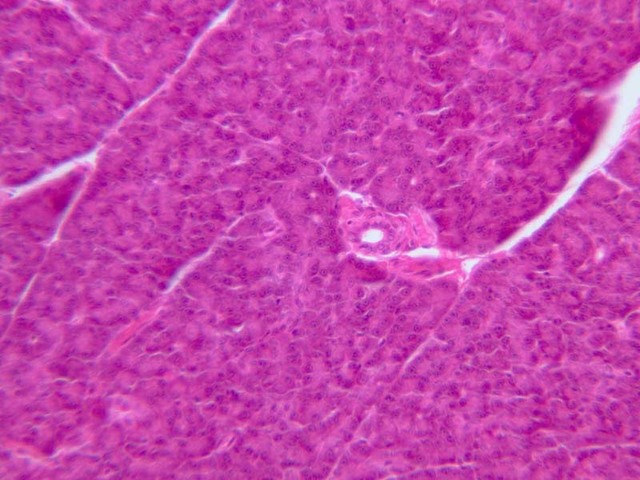
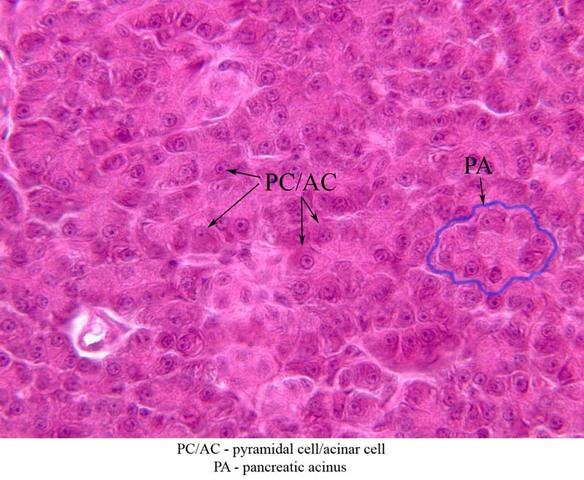
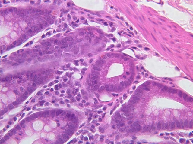
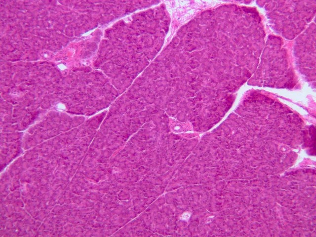
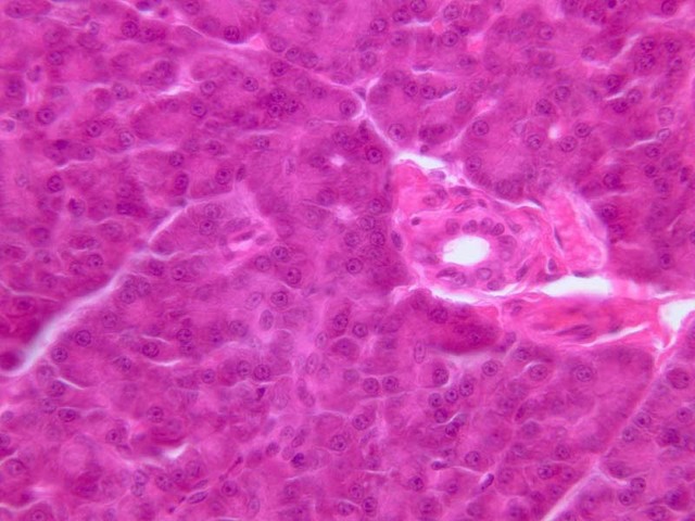
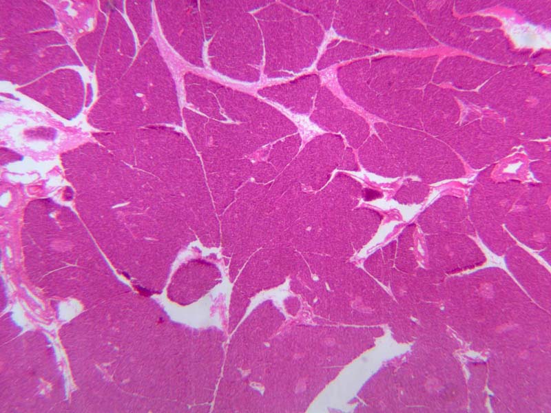
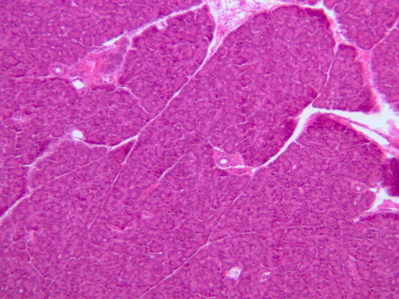
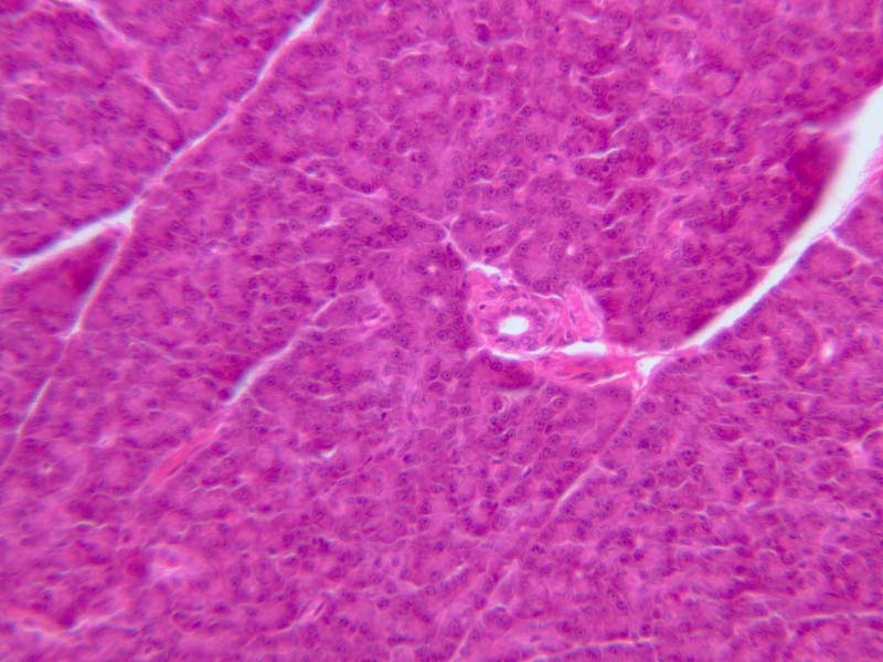
Next examine a section of the pancreas (slide B-36). Find a clump of cells whose profile in section is circular and determine the shape of the individual cells in the clump (called an acinus) ([
2.5x,
10x,
20x,
40x] [
20x,
40x-labeled]). In section, the cells appear to be roughly triangular; the name of the epithelium they form is called pyramidal,however, in reference to their 3-dimensional shape. The clustering together of such pyramidal cells to form an acinus gives it a spherical shape. Pyramidal (also known asglandular) epithelium is another variant of simple columnar epithelium, and as such its cells are similarly polarized. The shape of the glandular cells is an adaptation for secretion into a narrow duct rather than a wide lumen like the intestinal canal. You should note the polarity and arrangement of organelles and trace the pathway by which the secretory material is synthesized and released.
Simple Columnar Image Gallery
Simple Columnar Table of Identifications
| Row |
Structure |
Abbreviation |
Optimal Stain |
Representative Section |
Note |
| 1 |
Simple Columnar Epithelium |
SCoE |
H&E |
 B12, Jejunum, 40x B12, Jejunum, 40x |
|
| 2 |
Brush Border |
(none) |
H&E |
 B24, Colon (Brush Border), 40x B24, Colon (Brush Border), 40x |
|
| 3 |
Goblet Cell |
GC |
PAS |
 B25, Colon (Goblet Cells), 40x B25, Colon (Goblet Cells), 40x |
|
| 4 |
Pyramidal Cell/ Acinar Cell |
PC/AC |
H&E |
 B36, Pancreas (Acinus), 40x B36, Pancreas (Acinus), 40x |
|
| 4 |
Pancreatic Acinus |
PA |
H&E |
B36, Pancreas (Acinus), 40x |
|
Top
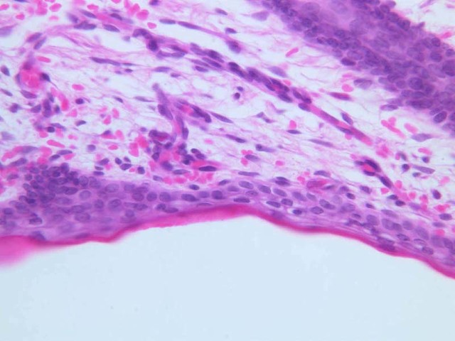
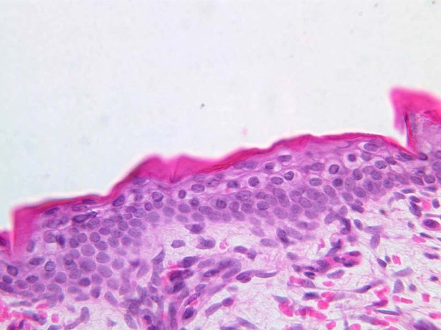
Stratified Epithelium
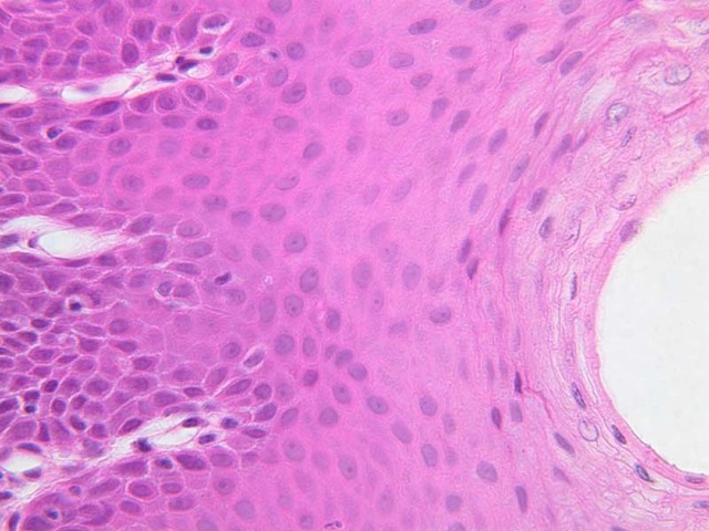
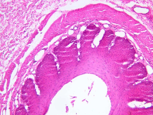
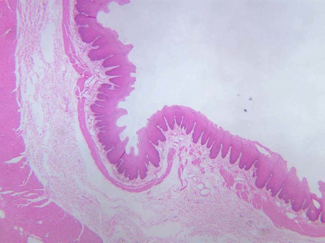
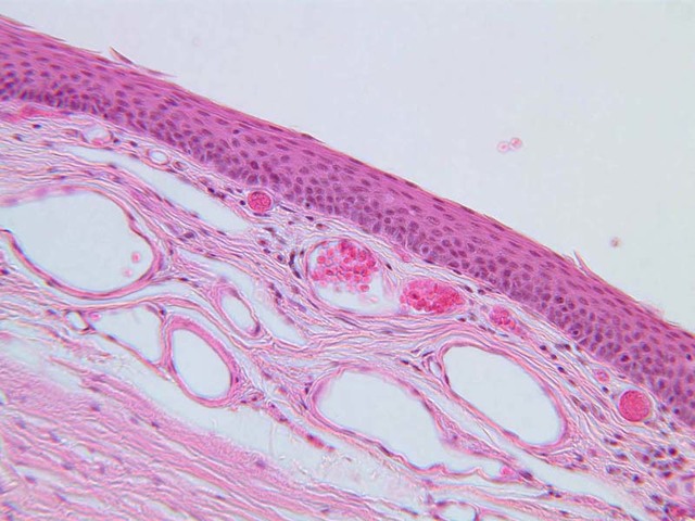
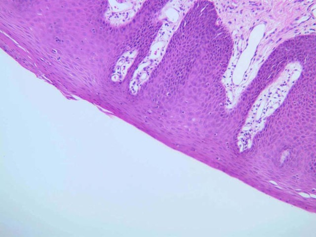
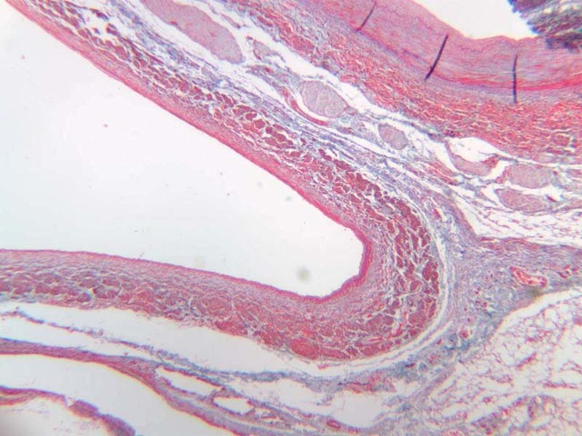
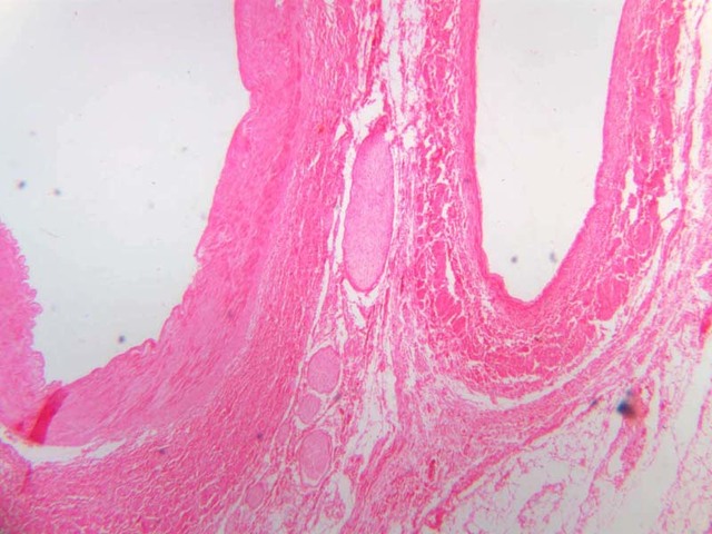
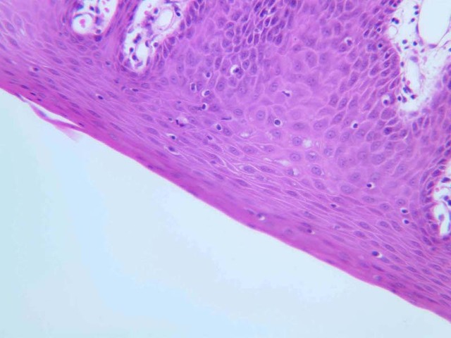
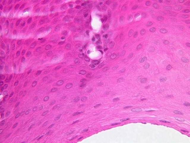
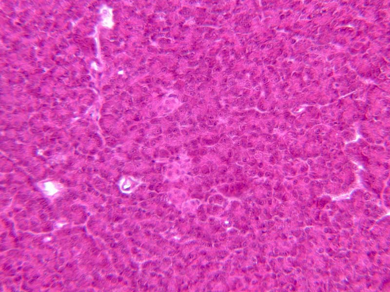
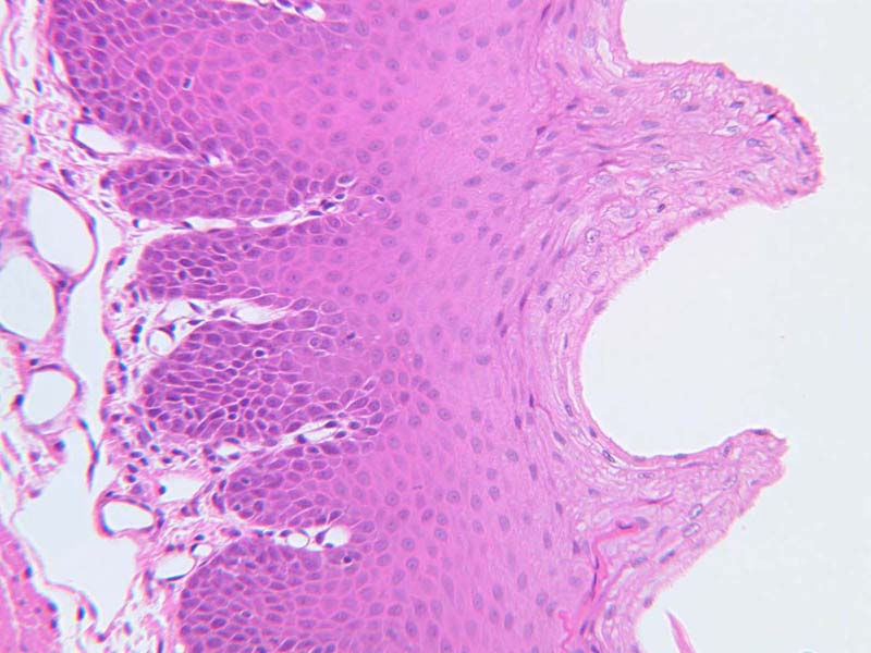
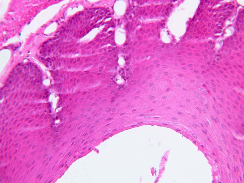
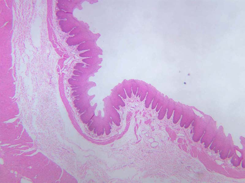
Stratified epithelium. The study of stratified epithelium may be started by looking at a cross-section of the stratified squamous epithelium of the esophagus (slide B-2 [
20x,
40x] [
10x,
20x,
40x] [
2.5x,
10x,
20x,
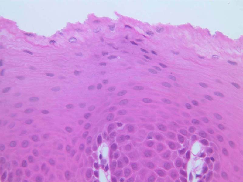
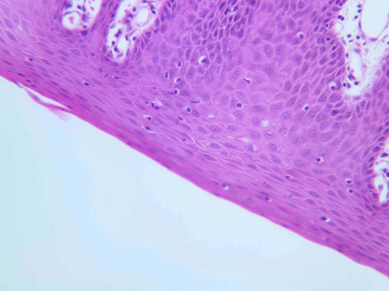
40x]). Make a low power examination of the band of tissue forming its inner lining. The most noticeable feature of this tissue band is its basophilic nuclei. Observe their distribution in numerous layers. Nuclear shapes reflect the shapes of individual cells in the epithelium. By studying these epithelial cells with the high power objective, look for boundaries between cells. If they are not apparent, an imaginary line drawn midway between neighboring nuclei will mark their approximate location.
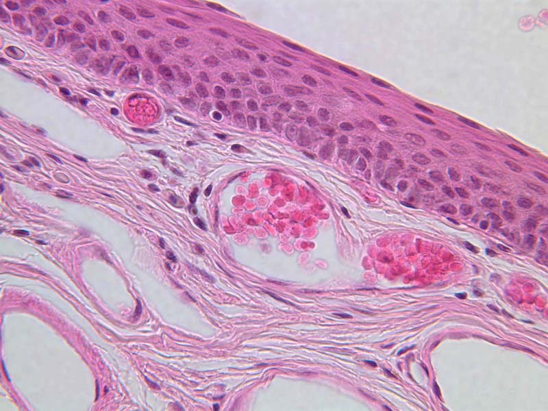
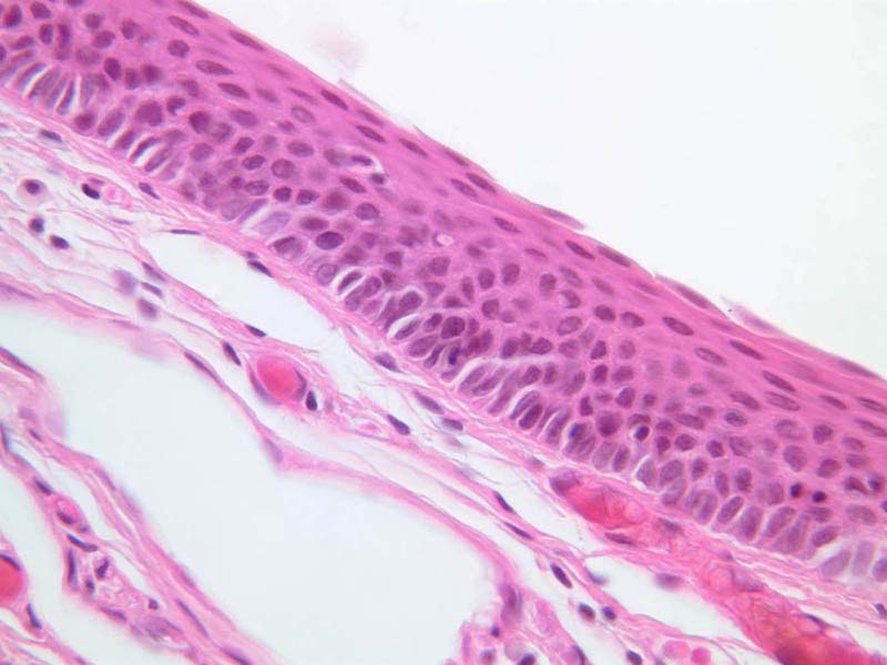
Note that the cells that make up the epithelium have varied shapes, but that at the free surface of the epithelium they are flattened in the plane of the surface and appear elongated. Another good example of this can be seen in the vagina (slide B-98 [
2.5x,
10x,
20x,
40x]), or on the epiglottis (slide A-67 [
20x,
40x,
40x]). In surface view, the cells would appear to be polyhedral and contain round nuclei. These dimensions taken together give such cells a scale-like (squamous) appearance. The correct name for this complete epithelium, then, is stratified squamous, named for its layered structure and the shape of its superficial cells. Note that cells throughout the epithelium have similar stained nuclei. This is a characteristic of soft stratified squamous epithelium.
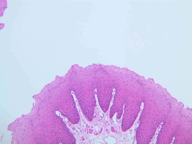
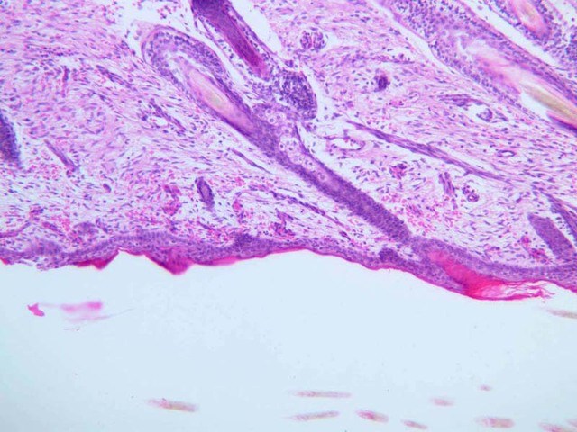
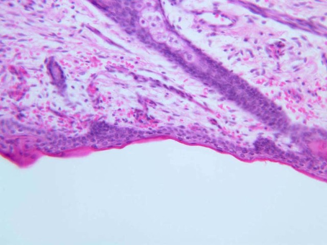
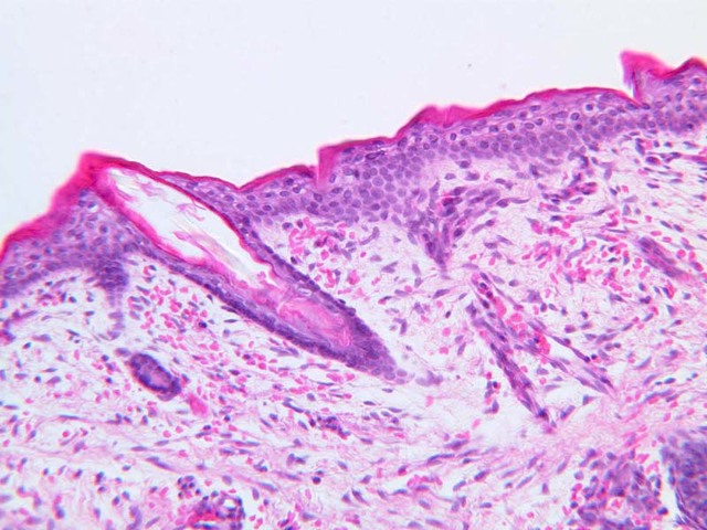
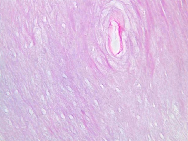
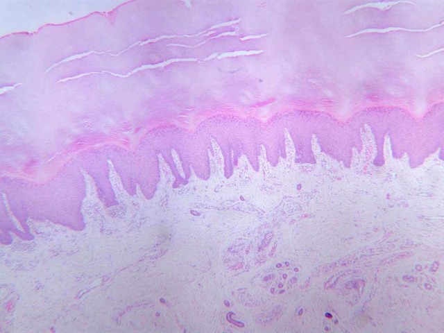
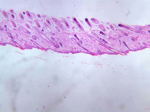
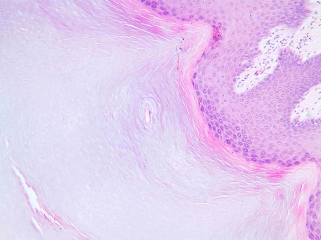
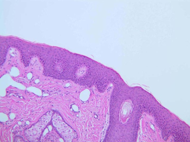
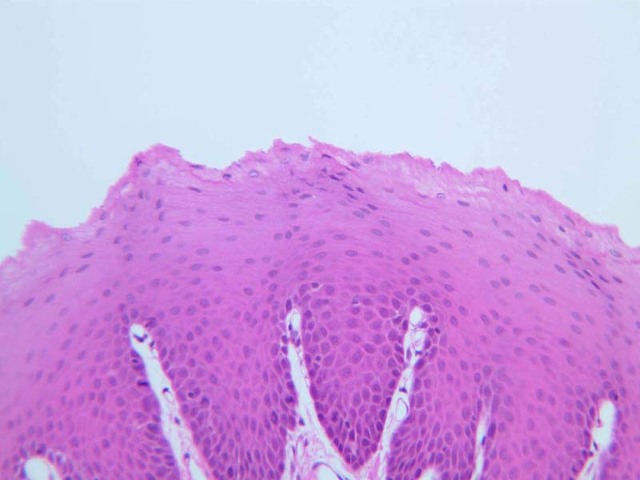
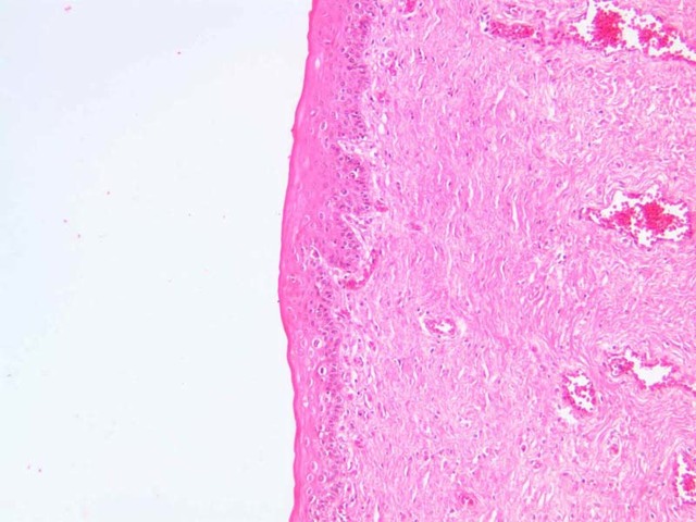
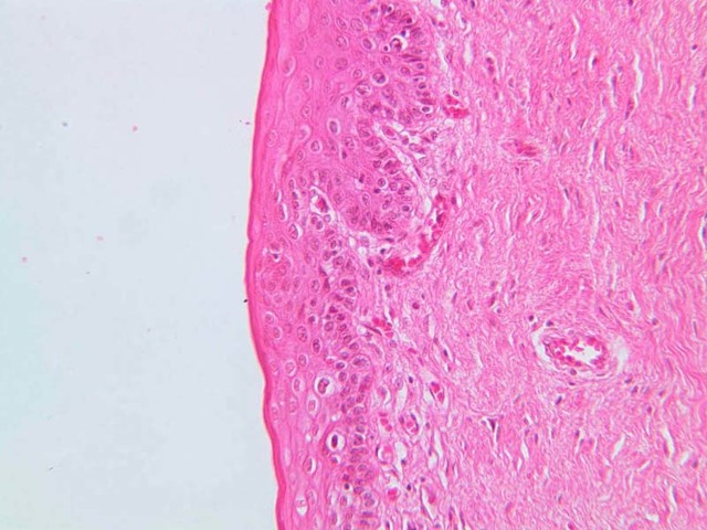
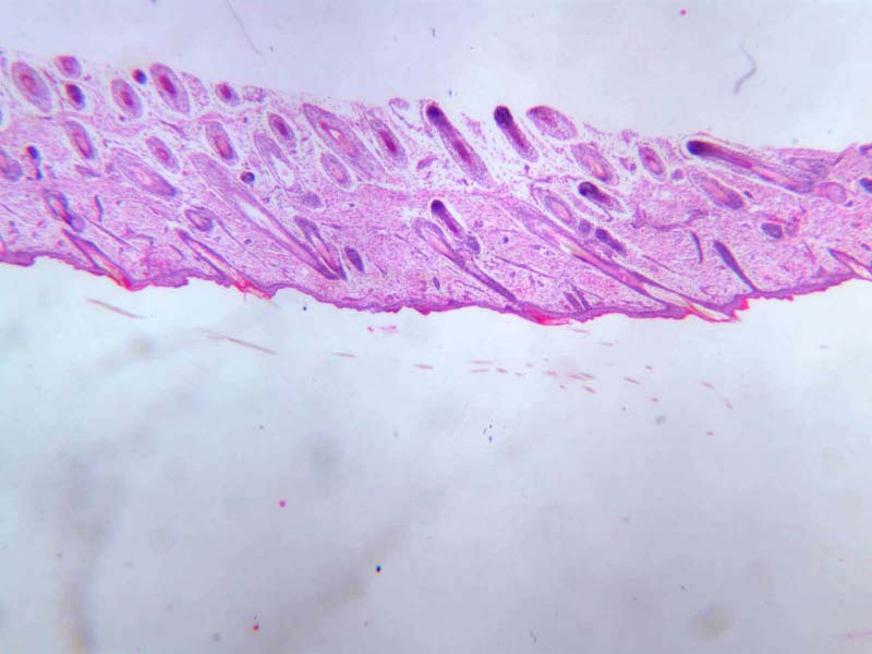
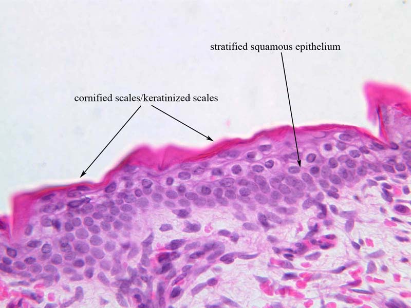
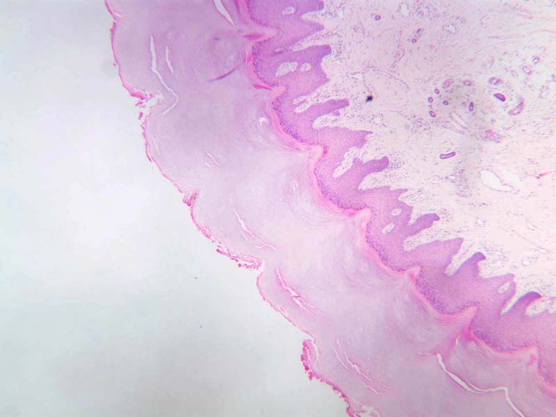
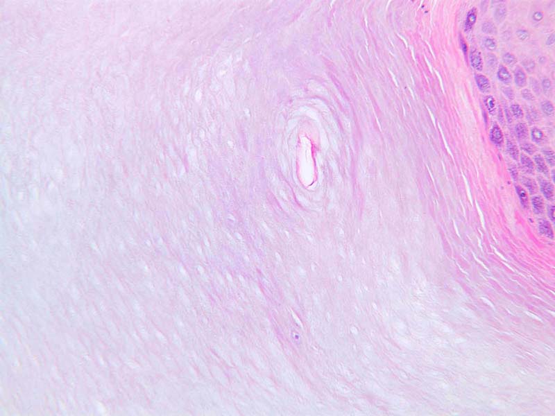
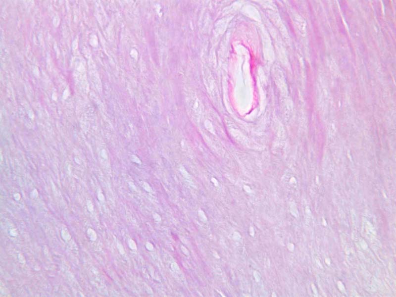
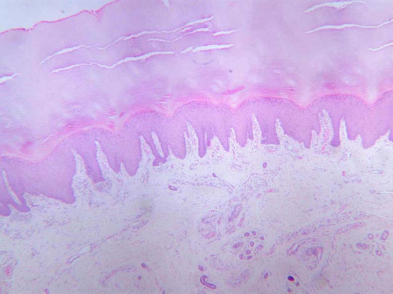
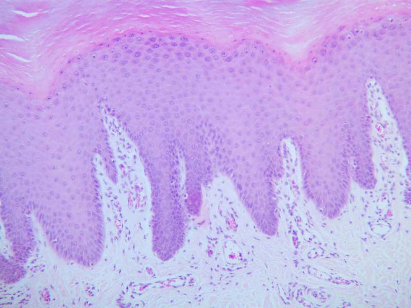
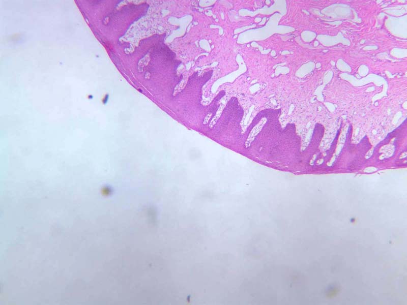
Now examine a slide of skin (slide A-49, scalp [
2.5x,
10x,
20x,
40x] [
20x,
40x-labeled]; A-50, fingertip [
2.5x,
10x,
20x,
40x] [
2.5x,
10x]), again looking for the deeply stained epithelial band at the free surface of the section. Not only the basophilic cells, but also the more superficial, somewhat stringy layers of acidophilic material comprise this epithelium (epidermis). The acidophilic layers are cornified (or keratinized) scales, and form a surface barrier that is relatively tough and impervious to water. A layer composed of cells like this is called cornified or keratinized, stratified squamous epithelium. Note that the thickness of the keratinized layer varies significantly depending on the location of the skin.
Compare the appearance of this stratified squamous epithelium with that which lines the esophagus. The stratification of these epithelia gives some protection to underlying tissues against physical abrasion; it reflects the continuing regeneration of their surface scales in the skin; it prevents desiccation and may provide insulation against heat loss from closely subjacent blood vessels; and in the esophagus, it provides a moist, lubricated layer to aid in food passage. Stratified squamous epithelium can thus exist in a keratinized or non-keratinized form as the local conditions demand. Compare the structure of the stratified squamous epithelium on either side of the lip (slide A-55- internal [
2.5x,
10x,
20x]; A-55-external [
2.5x,
10x,
20x,
40x]).
Stratified Image Gallery
Stratified Table of Identifications
Top
Transitional Epithelium
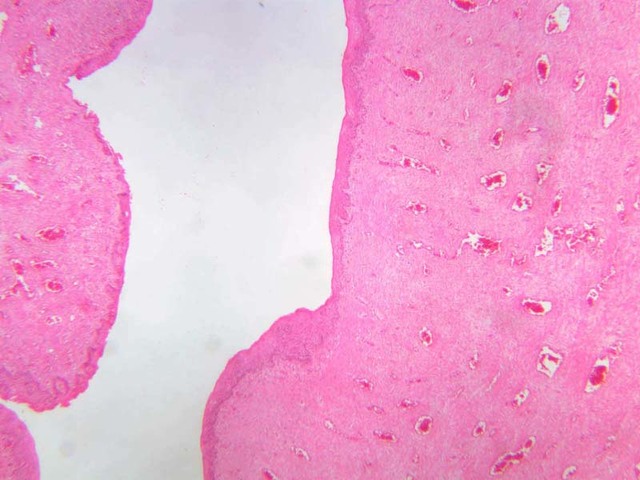
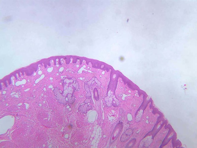
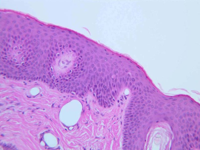
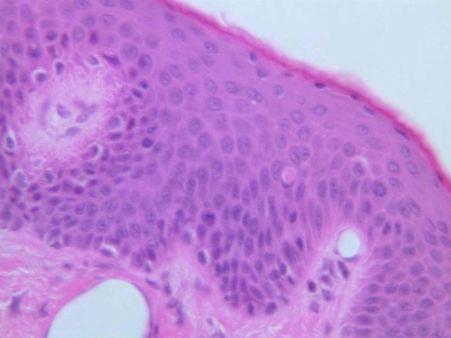
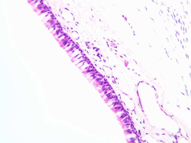
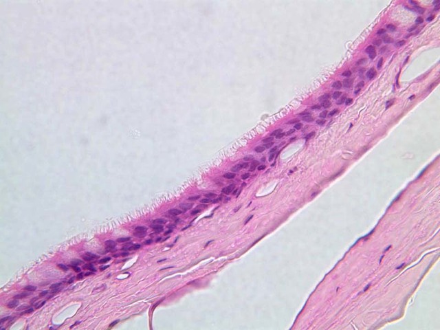
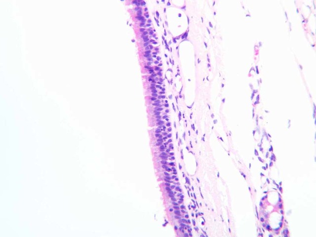
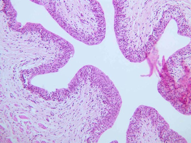
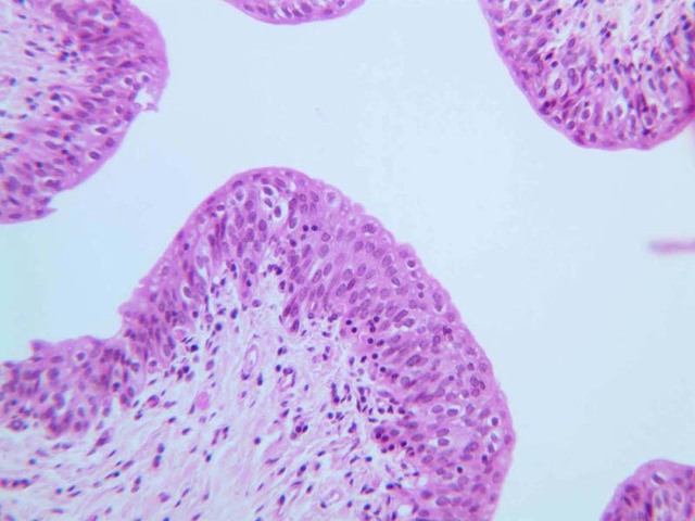
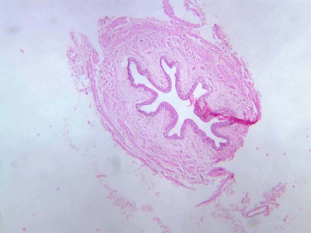
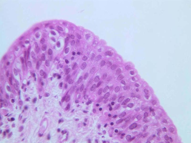
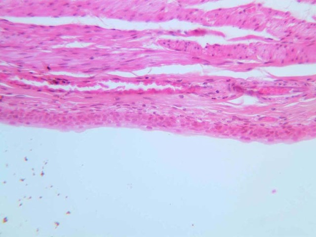
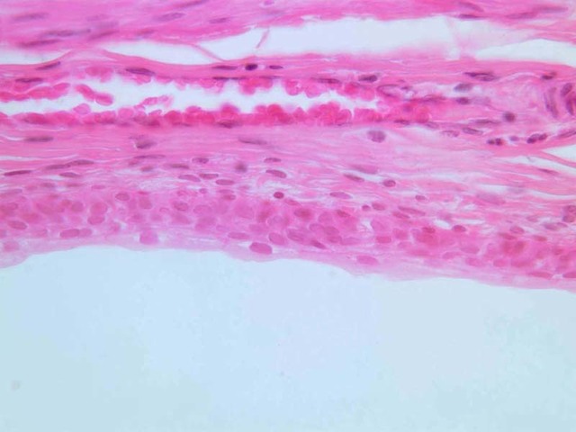
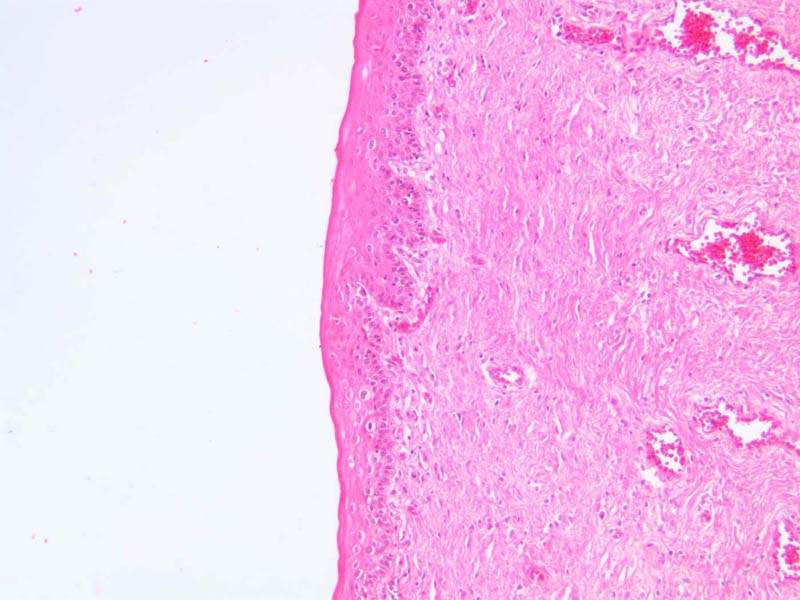
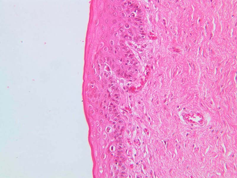
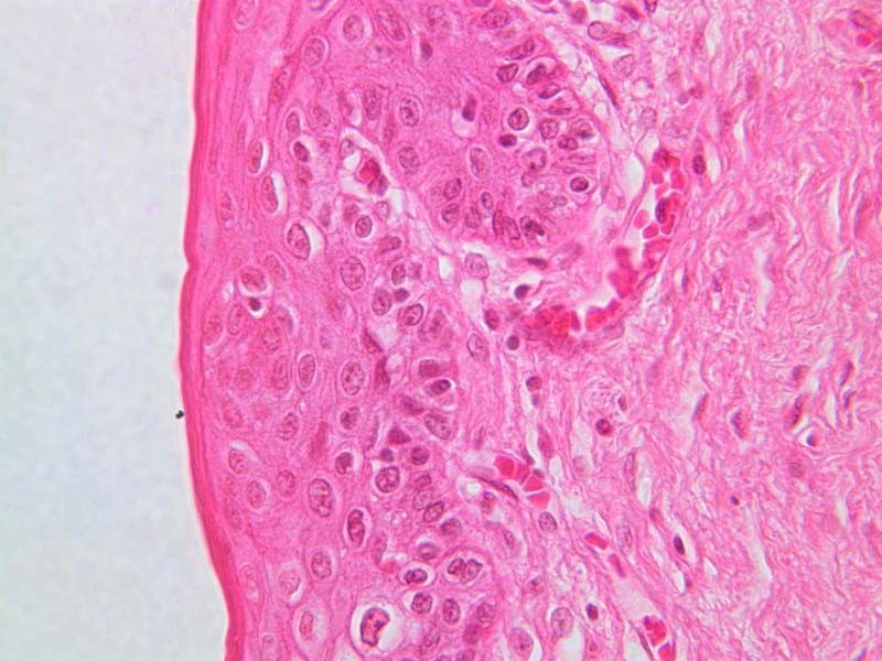
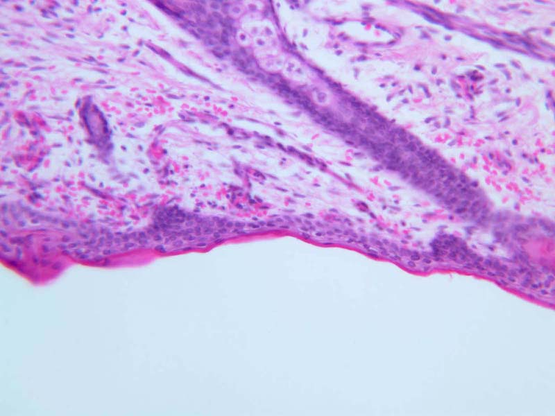
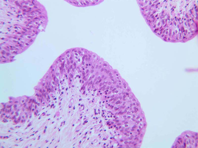
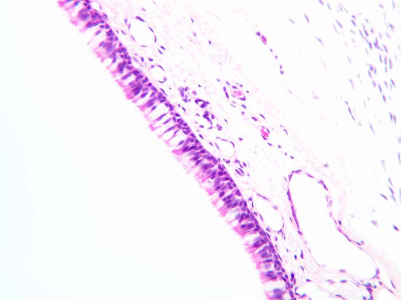
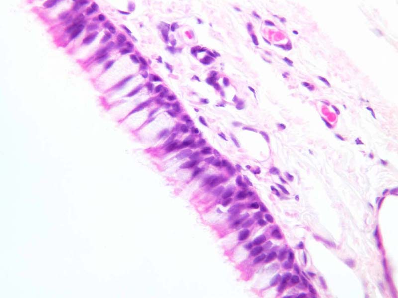
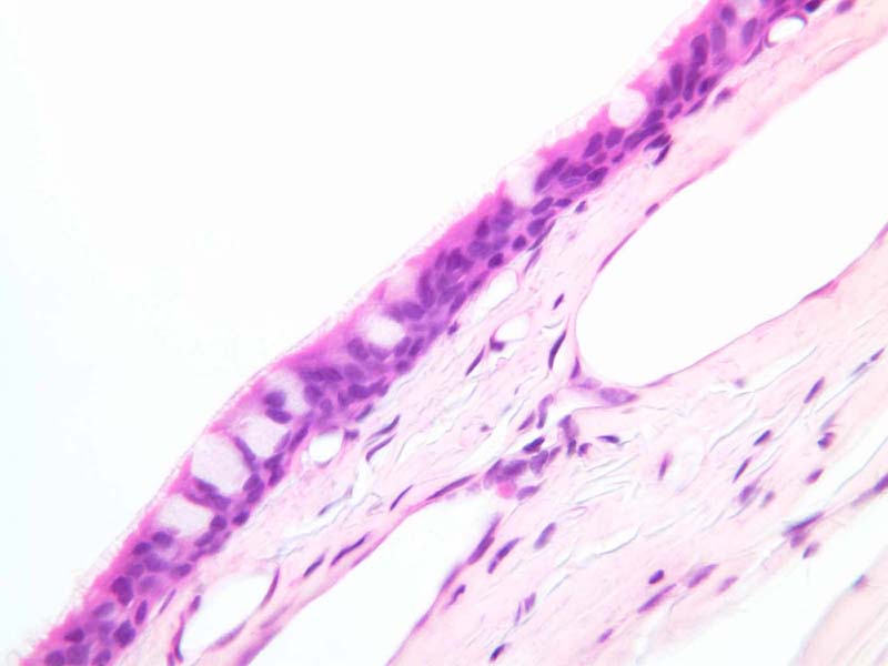
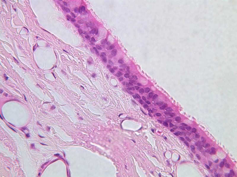
Transitional epithelium. A variation of stratified epithelium occurs in the body such that it resembles stratified squamous epithelium (that is, it has several layers and its surface cells are flattened). This modified epithelium lines organs that undergo changes in volume, and thus the epithelium is stretched into a few layers or is compacted into many layers as the organ expands or contracts. Because of these changes in layering, the epithelium is called transitional.
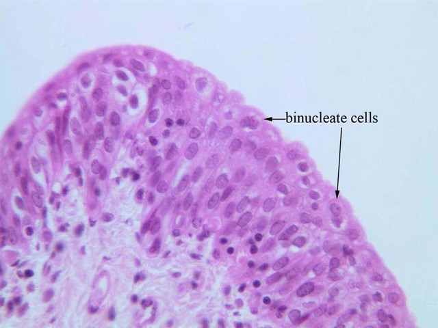
Transitional epithelium is found in certain parts of the urinary system. These organs are subject to distention which is allowed by the epithelium's ability to stretch. Observe the epithelial lining of the inner (luminal) surface of the ureter (slide B-72 [
2.5x,
10x,
20x,
40x-labeled]). Note that this transitional epithelium is comprised of from three to seven layers of cells, with the cells in the outer-most layer being cuboidal and somewhat coned or dome-shaped). It is not uncommon to see binucleate cells in transitional epithelium. The distended bladder may only have two to three layers of flatter cells (slide B-76 [
2.5x,
10x,
20x,
40x]).
Transitional Image Gallery
Transitional Table of Identifications
Top
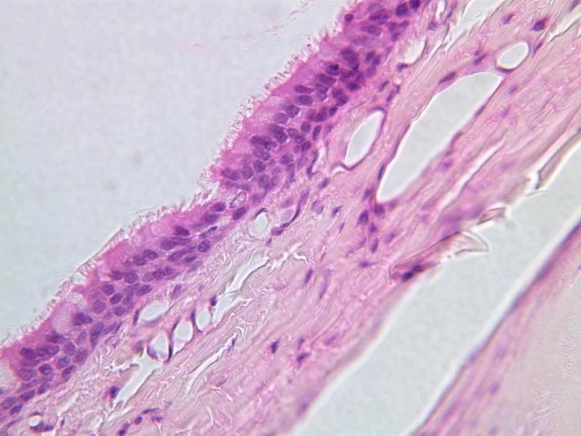
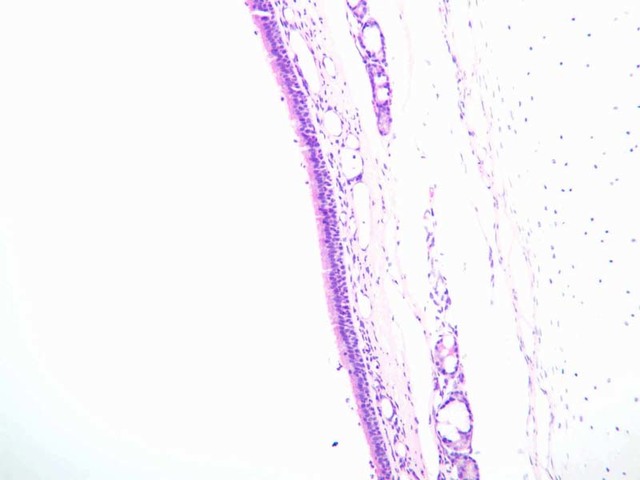
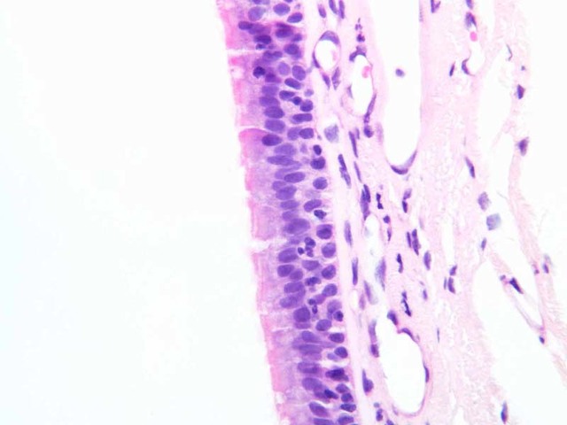
Pseudostratified Epithelium
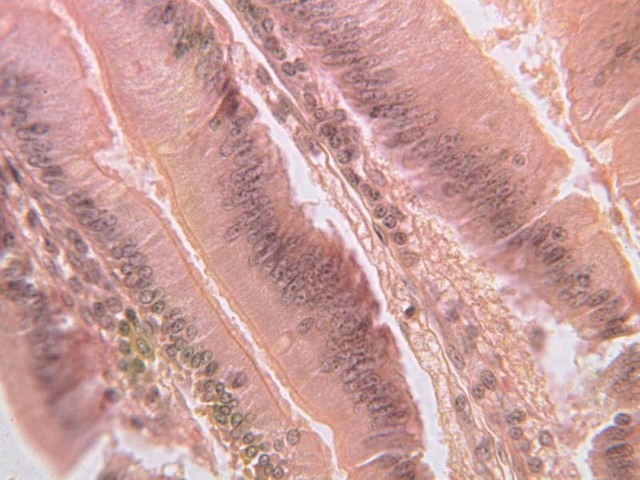
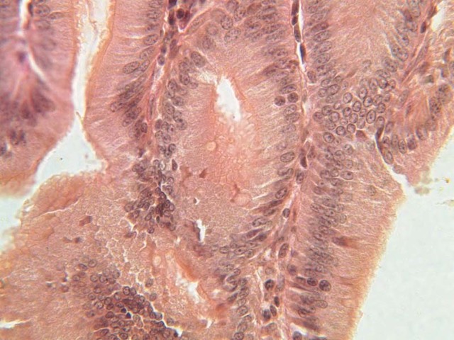
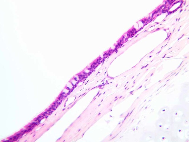
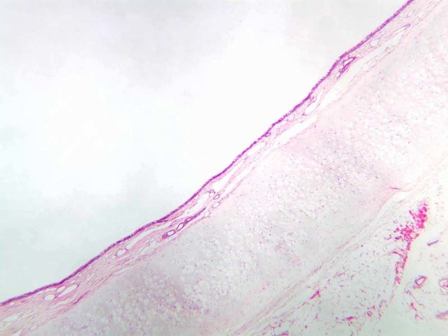
Pseudostratified epithelium. One large category of lining tissue remains. Look at a section of trachea (slide A-75 [
20x,
40x] [
20x,
40x] [
40x,
40x,
40x-labeled]; A-76 [
10x,
20x,
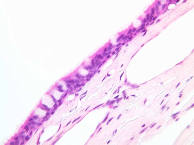
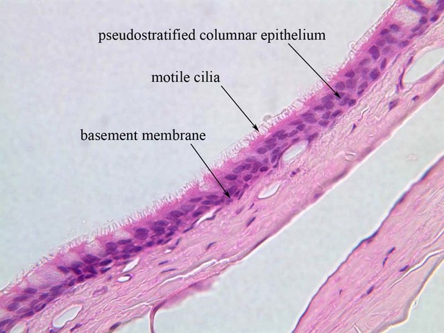
40x]) and observe its inner lining of epithelial cells. The shape of their nuclei suggests columnar cells, and there appears to be more than one layer of nuclei, suggesting that the cells are stratified. However, by maceration, it can be shown that each cell in the epithelium is attached to the underlying basement membrane. Unlike a true simple (single-layered) epithelium, on the other hand, not all of the cells reach the surface (i.e., basal cells). Since it is neither truly a simple nor a stratified membrane, this epithelium is called pseudostratified columnar.
The surface of the tallest cells bear motile cilia, and goblet cells occur interspersed among the tall columnar cells. The structure of the cilia is not readily seen in this section since they are so slender. Electron micrographs show that cilia have a very characteristic pattern of microtubules that appear in cross-sections of cilia as a circumferential ring of nine tubule doublets and a single central pair of tubules. Review the ultrastructural components of cilia. Compare these to the ultrastructure of centrioles/basal bodies.
Pseudostratified columnar epithelium lines the principal respiratory passages. Normally, mucus arising from the goblet cells covers the epithelium like a sticky mat; the mucus tends to trap tiny particulate matter that enters with the inspired air. Ciliary activity sets the mucous mat in motion toward the esophagus, where the "dust trap" is
regularly swallowed. The coughing reflex aids the expulsion of mucus that is not normally moved by the cilia. In the air passages, physical abrasion of the epithelium occurs very rarely, but passing gaseous materials may be harmful to it. The tall columnar cells narcotized or destroyed by such agents may be replaced by growth of the shorter, protected cells.
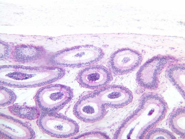
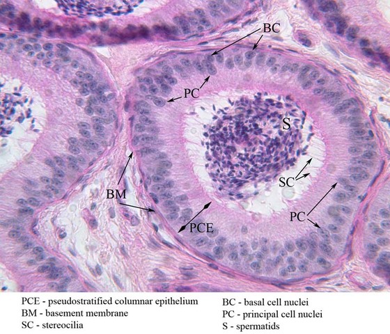
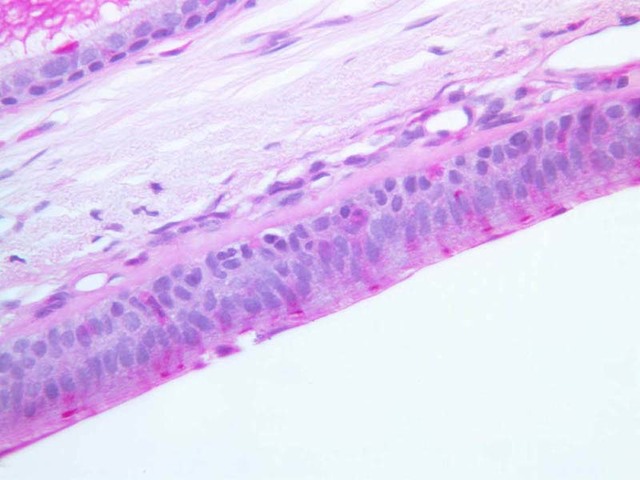
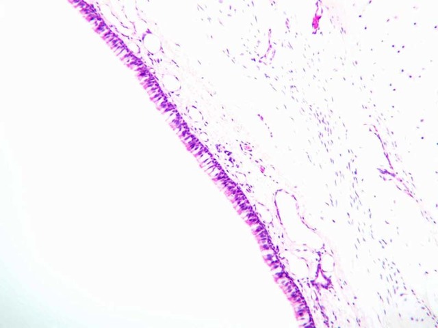
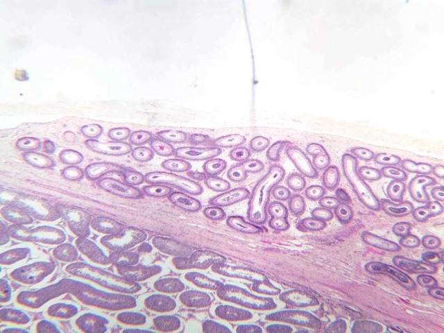
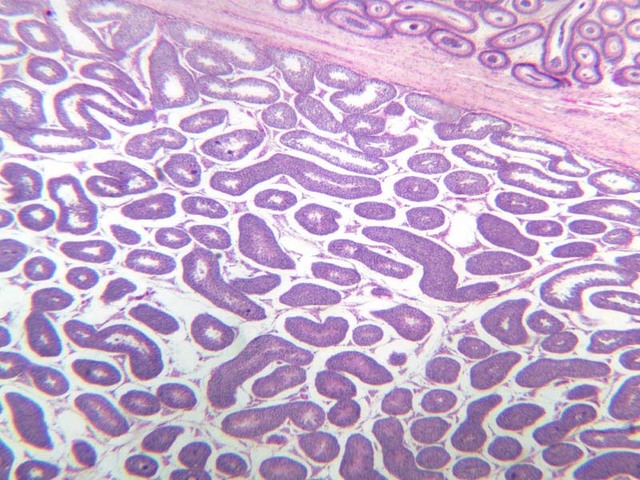
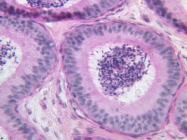
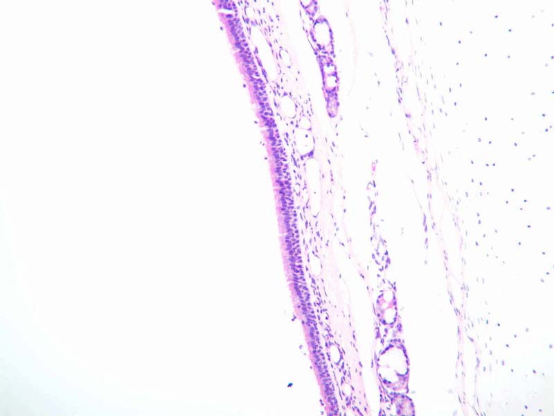
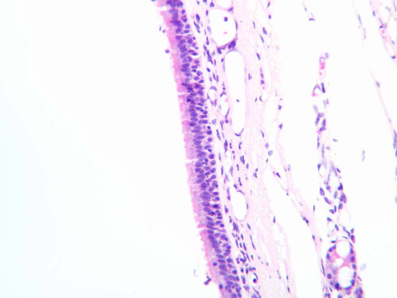
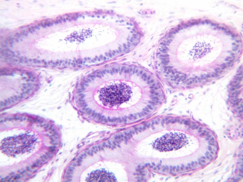
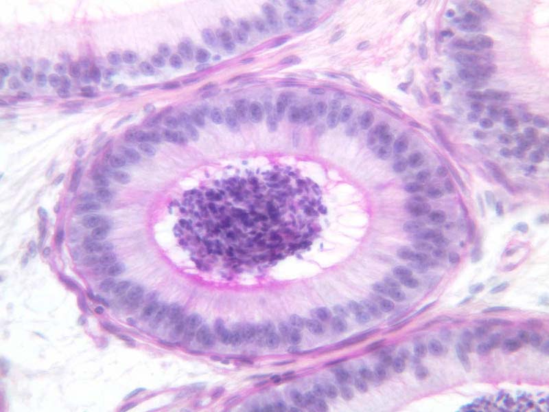
Slide B-81 (Epididymis [
2.5x,
10x,
20x,
40x] [
40x-labeled]) illustrates very tall columnar cells (called principal cells) which have specialized luminal surface appendages called stereocilia). Small basal cells are also present. It is the presence of the nuclei at different heights in adjacent cells that gives this epithelium a "pseudostratified" appearance. All of the cells in this epithelium are in contact with the basement membrane, but not all extend to the surface; those that do are columnar cells, those that don't are basal cells. Are basal bodies present? Are these structures actively motile?
Pseudostratified Image Gallery
Pseudostratified Table of Identifications
| Row |
Structure |
Abbreviation |
Optimal Stain |
Representative Section |
Note |
| 1 |
Pseudostratified Columnar Epithelium |
(none) |
H&E |
 A75, Trachea, 40x A75, Trachea, 40x |
|
| 2 |
Motile Cilia |
(none) |
H&E |
A75, Trachea, 40x |
|
| 3 |
Basement Membrane |
(none) |
H&E |
A75, Trachea, 40x |
|
| 4 |
Stereocilia |
SC |
H&E |
 B81, Epididymis, 40x B81, Epididymis, 40x |
|
| 5 |
Basal Cell Nuclei |
BC |
H&E |
B81, Epididymis, 40x |
|
| 6 |
Principle Cell Nuclei |
PC |
H&E |
B81, Epididymis, 40x |
|
| 7 |
Spermatids |
S |
H&E |
B81, Epididymis, 40x |
|
Top
Glands
Glands are also composed of epithelial cells. Glands that secrete their products onto a surface space or into a duct are exocrine glands, glands that secrete their products into the blood stream are endocrine glands. Exocrine glands can be unicellular like the goblet cell we examined in the trachea and colon, or multicellular. Multicellular exocrine glands are constructed so each cell can excrete its products into a lumen.
Glands can also be classified according to mechanism of secretion:
- Merocrine (eccrine) - cells secrete a secretory product by exocytosis.
- Apocrine - to secrete the product, the apex of the cell pinches off so some cytoplasm is released as well as the secretory product. Mammary glands and some apocrine sweat glands are thought to secrete this way.
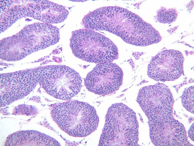
- Holocrine - glands secrete their product by sloughing off of the whole cell. Sebaceous glands of the skin extrude degraded cells to give the oily secretion of the skin. Another holocrine secretion is sperm which are obviously alive when they are secreted. Secretion of whole living cell (like sperm) is also called cytocrine secretion.
Top
Cell Surface Specialization
Specialization of free surfaces is a feature of many types of epithelial cells. The structure of these can only be fully appreciated at the EM level. Some important aspects of each are summarized below.
- Microvilli
- LM - "striated" or "brush" border on absorptive epithelia is PAS positive
- EM - numerous parallel finger-like processes - filaments in core interdigitate with terminal web
- Function - increase surface area for absorption
- Cell coat on surface may function in attachment of molecules to be absorbed
- Microvilli present in smaller numbers on many cell types.
- Cell Coats
- Large variation in thickness, but thought to be present on virtually all animal cells
- Glycoprotein composition
- Integral part of plasma membrane
- Disposition of membrane glycoproteins with carbohydrate groups on outer surface of PM
- Basal Folds
- Basal plasma membrane highly folded in transporting epithelia
- Mitochondria closely associated - energy source
Top
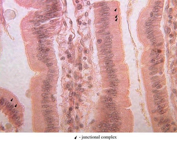
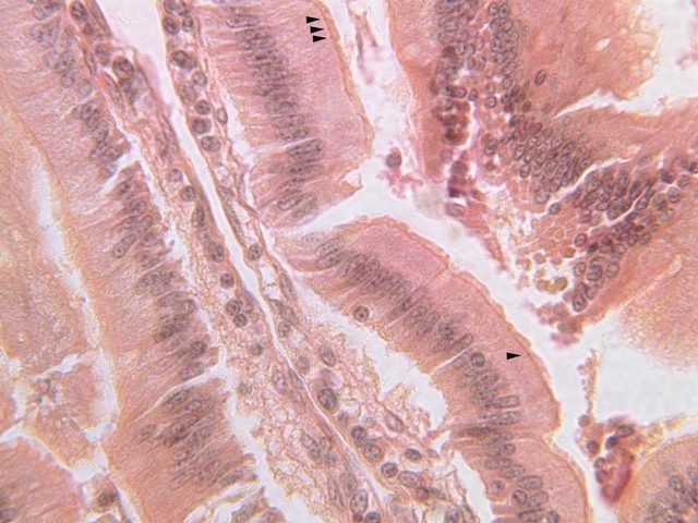
Specialized contacts between cells are a very important aspect of the epithelia. Some of the types of junctions that exist between epithelial cells include: zonula adherens, zonula occludens (tight junction), macula adherens or desmosome, gap junction or nexus.
Unless special stains such as iron hematoxylin are employed, the junctional complexes between cells are difficult to see. In slide B-14 ([
10x,
20x,
40x-labeled] [
40xlabeled,
40x,
40x-labeled,
40x,
40x,
40x]), they are a small dot just below the brush border of the intestinal epithelial cells. In order to see these small structures, carefully adjust the condenser, light, and focus settings of the microscope. In order to fully understand the structure and function of these, it is necessary to utilize the EM. Summarized below are some important aspects of each type. Refer to the
micrographs cited above as you study their comparative structure.
Contact specializations are modifications of cell surface that permit cells to relate functionally in different ways:
- Named by size of area specialized
- macula - spot
- zonula - band (extends around the cell in a belt)
- fascia - large sheet
- Adjective applied describing nature according to distance between apposed cells
- normally epithelial cells are separated by 150-200 \xC5 intercellular space
- adherens - PMs are parallel, lie 200-250 \xC5 apart, and adjacent cytoplasm is specialized.
- occludens or tight - extracellular space obliterated by fusion of outer dense lines of PMs - 5 layered junction.
- gap - 20 \xC5 separation between PMs
- Desmosome (Macula adherens)
- LM - "intercellular bridges"
- EM - no continuity between cells, but a surface specialization.
- PMs of apposed cells are parallel and separated by 250 A.
- Dense plaque in cytoplasm applied to inner aspect of PM's.
- Loops of filaments subjacent to plaque.
- Material in extracellular space contains carbohydrate- "glue" (intraperiod line).
- Function in mechanical attachment of two cells
- Terminal bar (Junctional complex)
- LM - region of increased density between certain epithelial cells (e.g., intestine) next to lumen.
- EM - series of surface specializations
- zonula occludens - next to lumen
- zonula adherens
- desmosomes
- Function
- zonula adherens and desmosomes - mechanical attachment
- zonula occludens - seal off lateral intercellular space from lumen.
- a true tight junction
- ridges in freeze-etch
- important in transport across gut
- Gap Junctions and Electrical Coupling
- In gap junction, two PMs close together but separated by 20 \xC5 space.
- Hexagonal pattern of prisms with 90 \xC5 spacing occupy extracellular space and outer dense leaflet of the 2 PMs.
- Characteristic freeze-etch appearance - hexagonally packed particles with 90 \xC5 spacing (center-to-center).
- Presence of gap junctions correlates with electrical coupling between cells:
- in cells that normally transmit impulses.
- in cells that don't normally transmit - cells of early embryos are extensively coupled, pattern changes with development?
Top
Review of Chapter One
Review of Slides
Review of Identifications
| Row |
Structure |
Abbreviation |
Optimal Stain |
Representative Section |
Note |
| 1 |
Renal Artery |
(none) |
H&E |
A28, Renal Artery and Vein, 2.5x |
|
| 2 |
Renal Vein |
(none) |
H&E |
A28, Renal Artery and Vein, 2.5x |
|
| 3 |
Connective Tissue |
(none) |
H&E |
A28, Renal Artery and Vein, 2.5x |
|
| 4 |
Bowman's Capsule |
BC |
H&E |
B68, Kidney, 40x |
|
| 5 |
Nucleus of Simple Squamous Epithelium |
N |
H&E |
B68, Kidney, 40x |
|
| 6 |
Simple Squamous Epithelium |
SSE |
H&E |
A28, Renal Artery and Vein, 40x |
|
| 7 |
Simple Cuboidal Epithelium |
SCE |
H&E |
 B68, Kidney, 40x B68, Kidney, 40x |
|
| 8 |
Renal Tubule |
RT |
H&E |
B68, Kidney, 40x |
|
| 9 |
Thyroid Follicle |
(none) |
H&E |
 B52, Thyroid, 40x B52, Thyroid, 40x |
|
| 10 |
Simple Columnar Epithelium |
SCoE |
H&E |
B12, Jejunum, 40x |
|
| 11 |
Brush Border |
(none) |
H&E |
B24, Colon (Brush Border), 40x |
|
| 12 |
Goblet Cell |
GC |
PAS |
B25, Colon (Goblet Cells), 40x |
|
| 13 |
Pyramidal Cell/ Acinar Cell |
PC/AC |
H&E |
B36, Pancreas (Acinus), 40x |
|
| 14 |
Pancreatic Acinus |
PA |
H&E |
B36, Pancreas (Acinus), 40x |
|
| 15 |
Stratified Squamous Epithelium |
(none) |
H&E |
 A49, Scalp (Skin), 40x A49, Scalp (Skin), 40x |
|
| 16 |
Cornified scales/ Keratinized scales |
(none) |
H&E |
A49, Scalp (Skin), 40x |
|
| 17 |
Binucleate Cells |
(none) |
H&E |
 B72, Ureter, 40x B72, Ureter, 40x |
|
| 18 |
Pseudostratified Columnar Epithelium |
(none) |
H&E |
A75, Trachea, 40x |
|
| 19 |
Motile Cilia |
(none) |
H&E |
A75, Trachea, 40x |
|
| 20 |
Basement Membrane |
(none) |
H&E |
A75, Trachea, 40x |
|
| 21 |
Stereocilia |
SC |
H&E |
B81, Epididymis, 40x |
|
| 22 |
Basal Cell Nuclei |
BC |
H&E |
B81, Epididymis, 40x |
|
| 23 |
Principle Cell Nuclei |
PC |
H&E |
B81, Epididymis, 40x |
|
| 24 |
Spermatids |
S |
H&E |
B81, Epididymis, 40x |
|
| 25 |
Junctional Complexes |
(arrows) |
Fe |
 B14, Intestine, 40x B14, Intestine, 40x |
|
Top
Top
--
AshleyLPistorio - 27 May 2007


