 |
|
You are here: Medical Histology>Main Web>AtlasContents>MaleReproductiveSystemAtlas15 (20 Jun 2015, LorenEvey)Edit Attach
Chapter Fifteen: Male Reproductive System


- Introduction
- Testis
- Accessory Glands
- Penis
- Chapter Fifteen Review
- Comments
Introduction
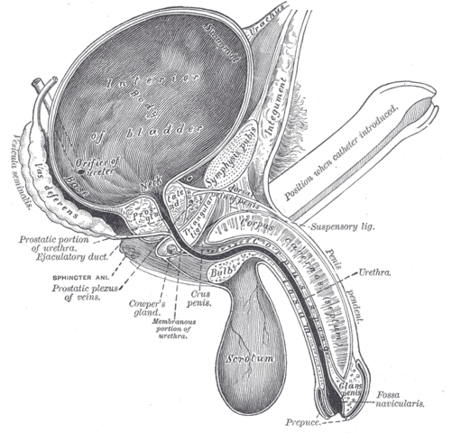
The male reproductive system is responsible for: 1) production, maturation, storage, and transport of the male gamete (spermatogenesis), and 2) synthesis and controlled release of steroid hormones (steroidogenesis). In this lab you will be studying the microscopic anatomy of many components of the male reproductive system, including the testis, the excurrent duct system, a series of accessory exocrine glands that produce much of the seminal volume, and the penis. Prior to learning the microarchitecture of Male Reproductive System, use the table below to review some of the gross anatomy of these tissues:| Structure | Image |
|---|---|
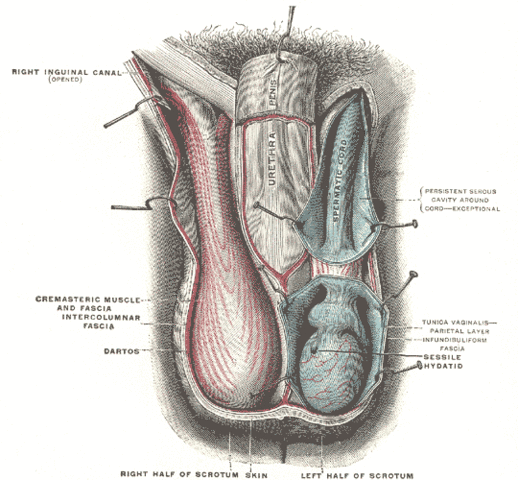
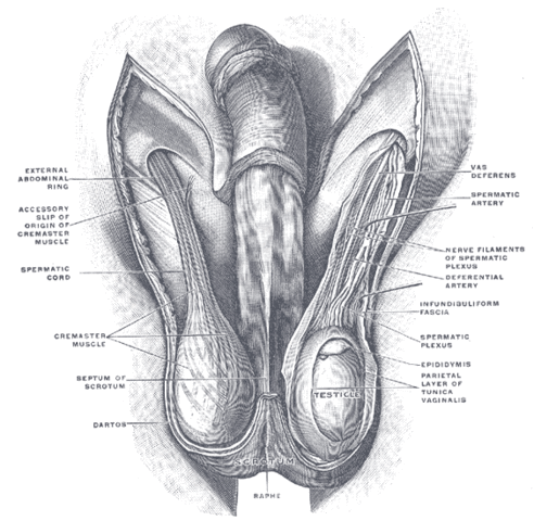
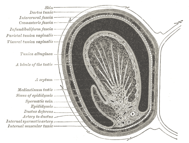
| Fascial Layers Surrounding the Testis | |
| Gross Anatomical Location of the Testis | |
| Cross Section of the Testis | |
| The Ducts of the Male Reproductive System | |
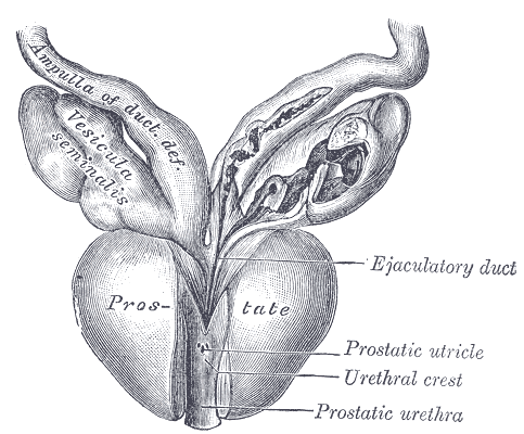
| The Prostate and Seminal Vesicle | |
| Orientation of the Male Urogenital Tract | |
| The Penis | |
| Cross Section of the Penis | |
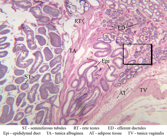
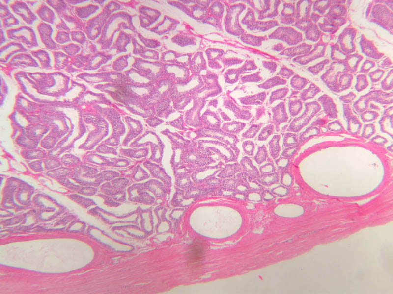
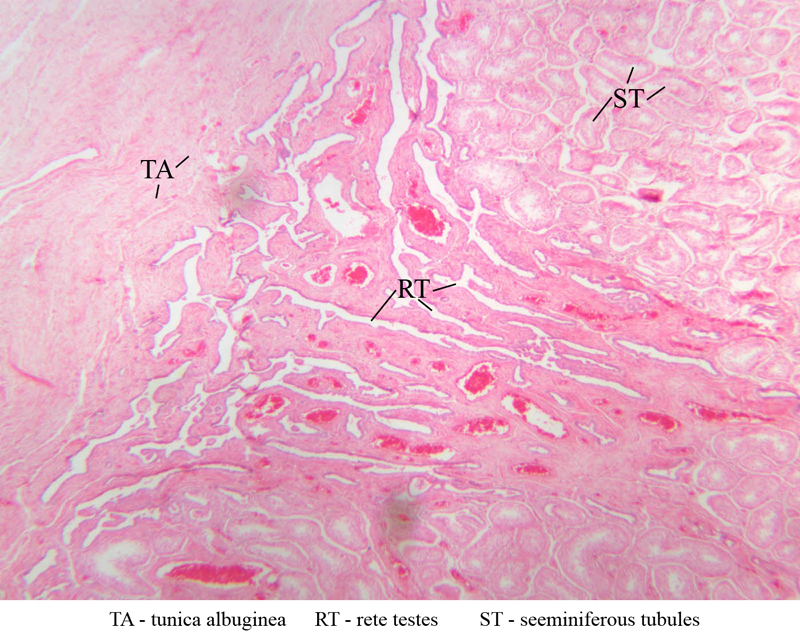
Testis
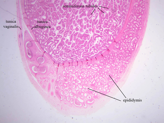
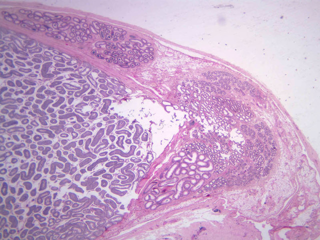
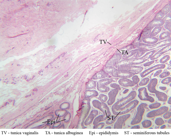
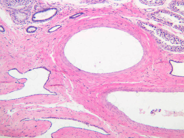
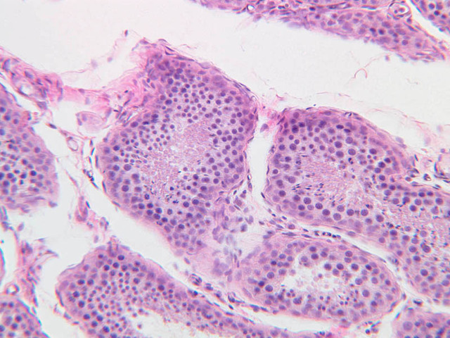
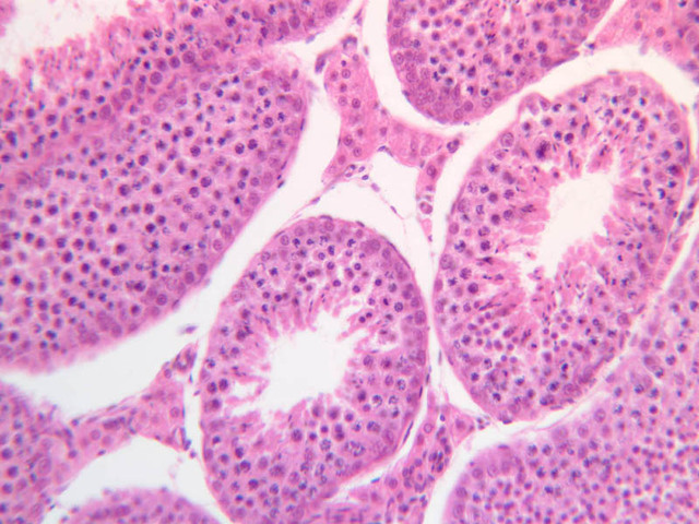
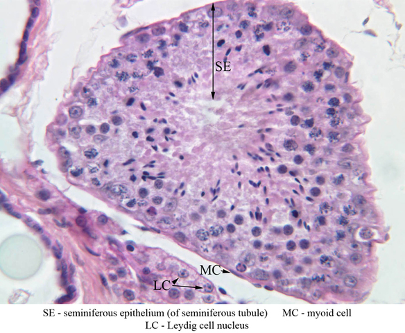
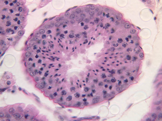
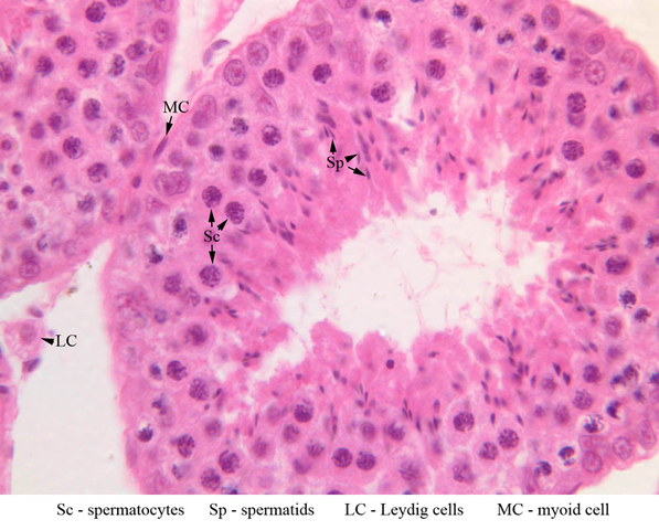
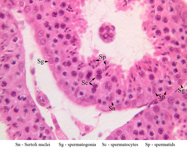
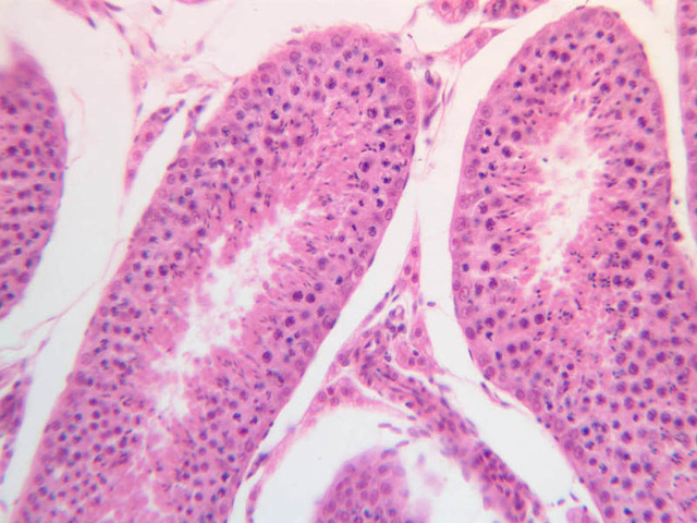
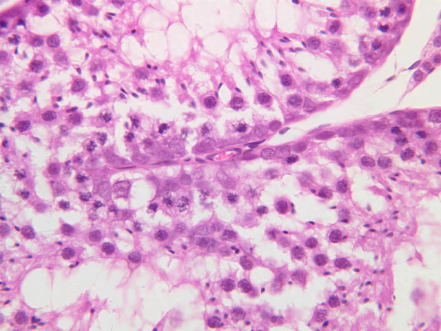
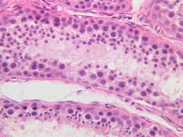
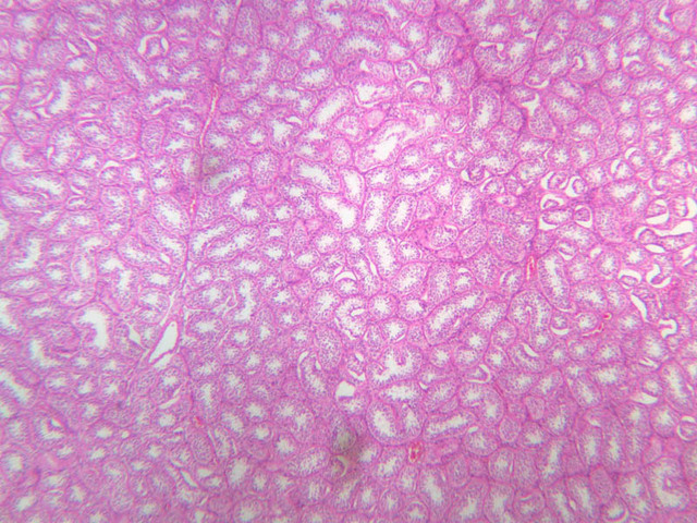
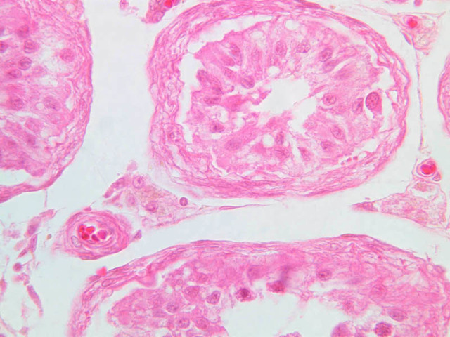
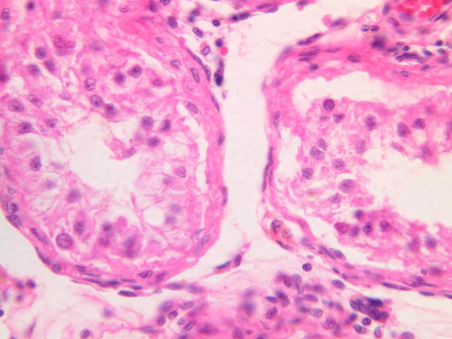
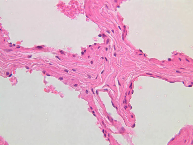
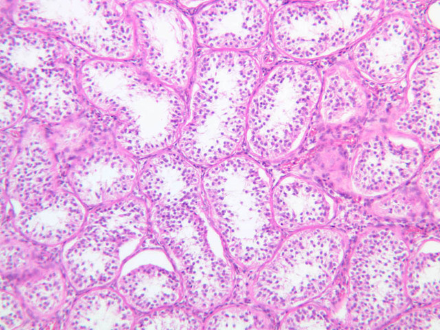
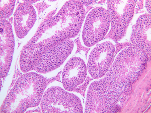
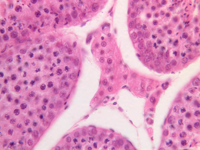
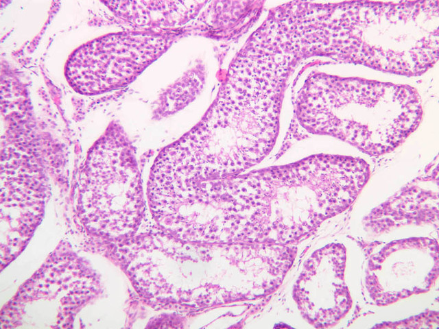
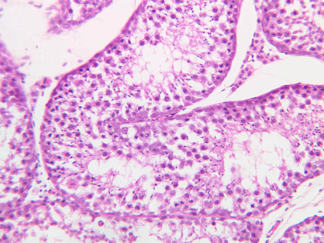
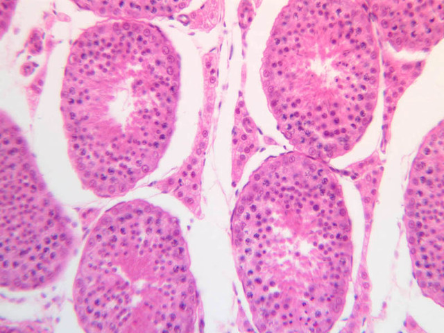
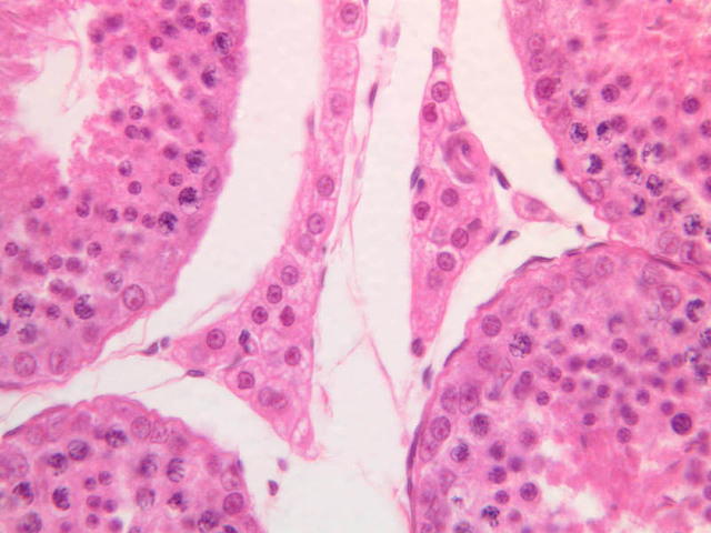
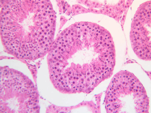
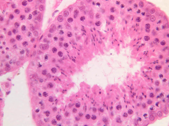
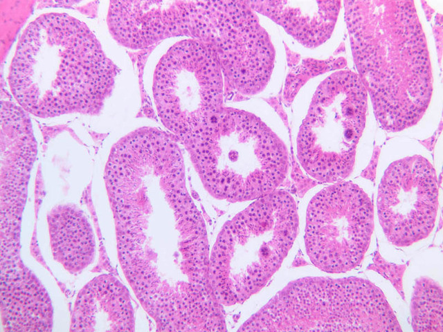
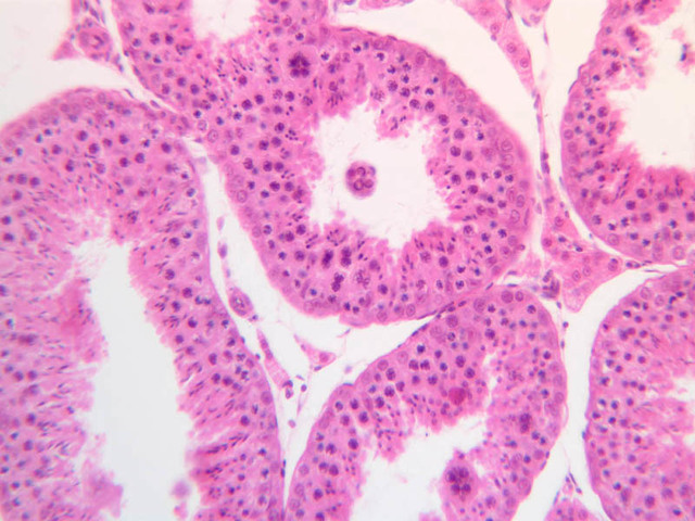
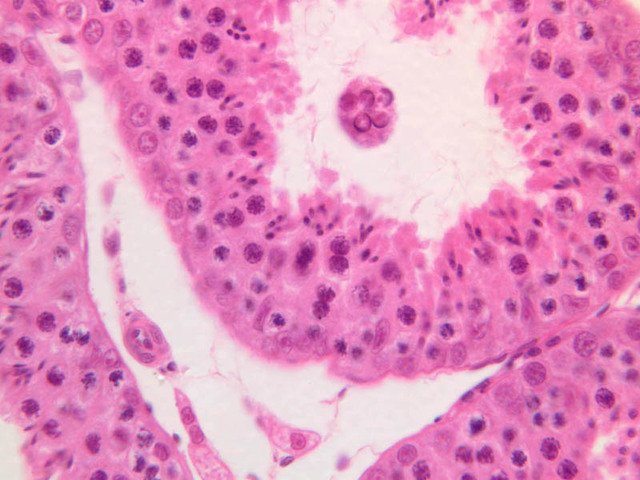
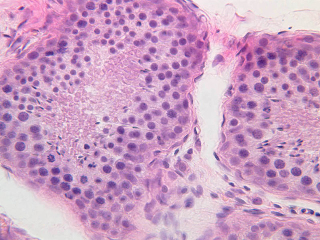
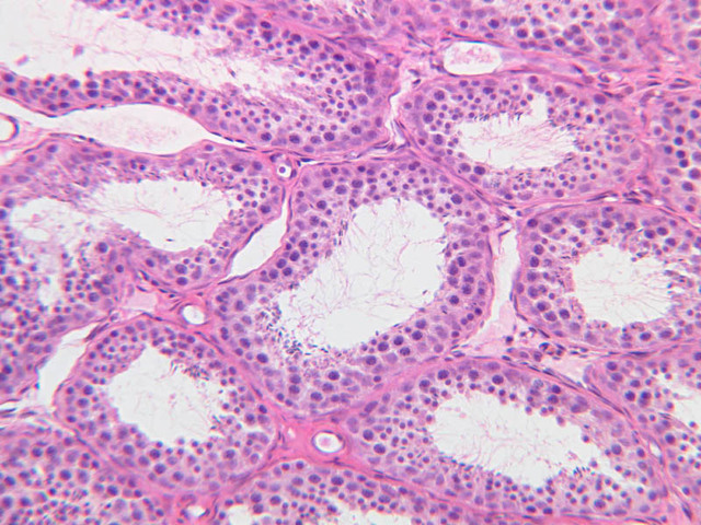
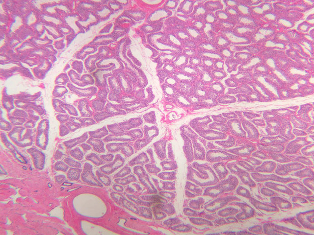
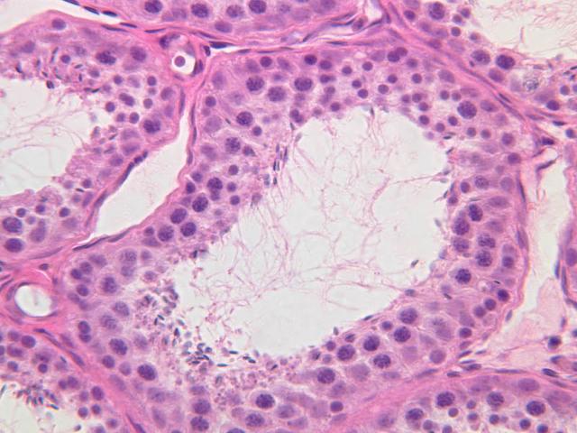
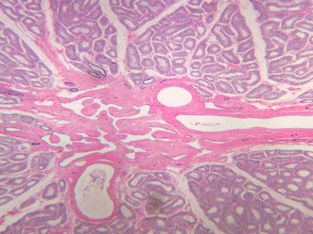
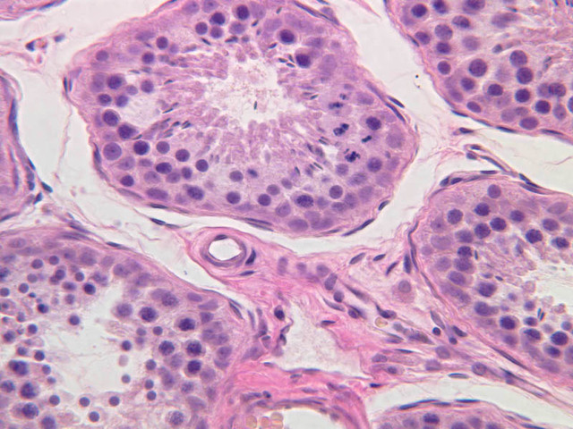
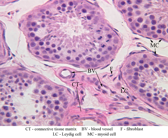
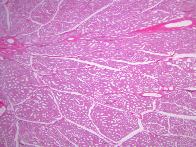
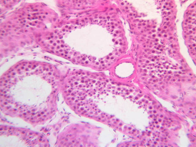
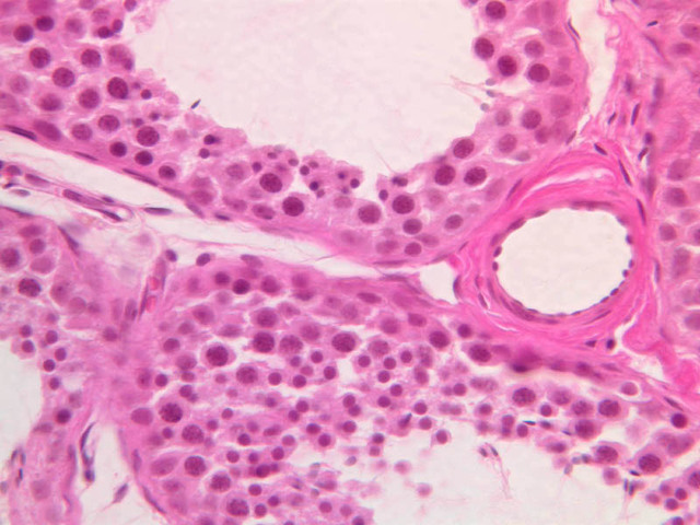
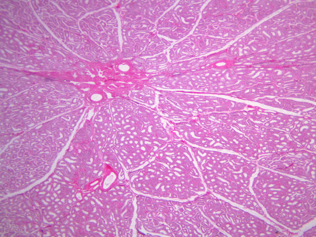
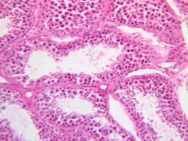
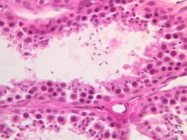
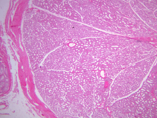
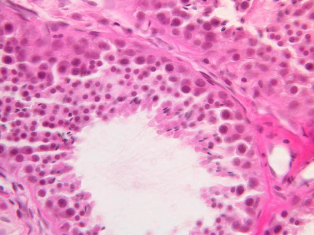
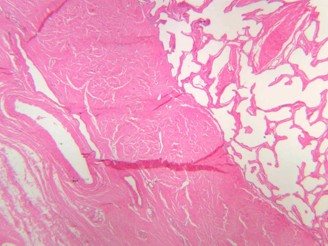
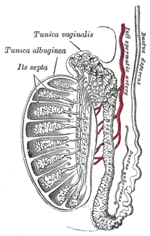
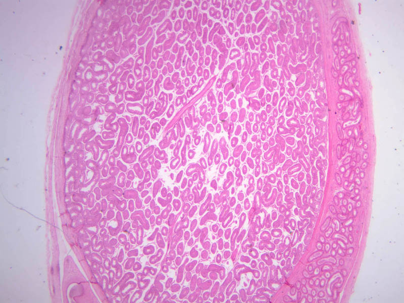
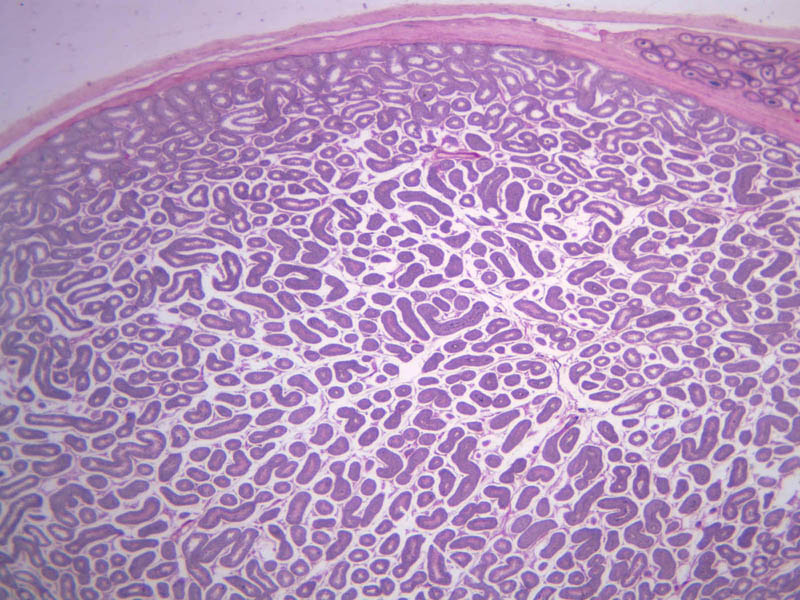
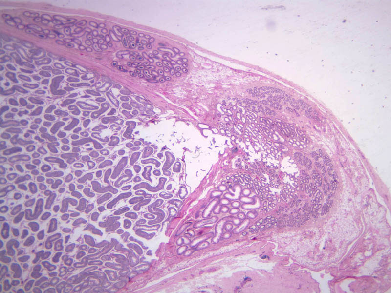
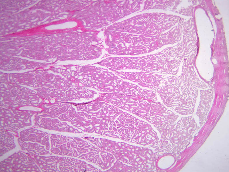
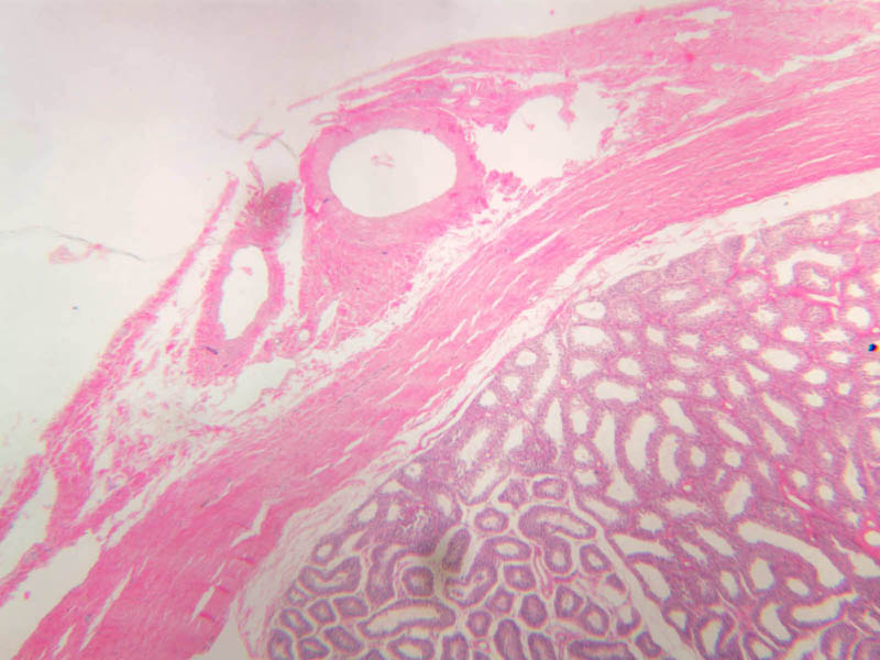
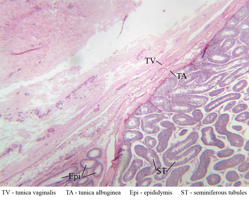
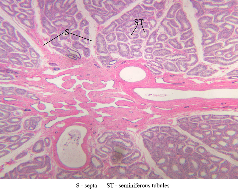
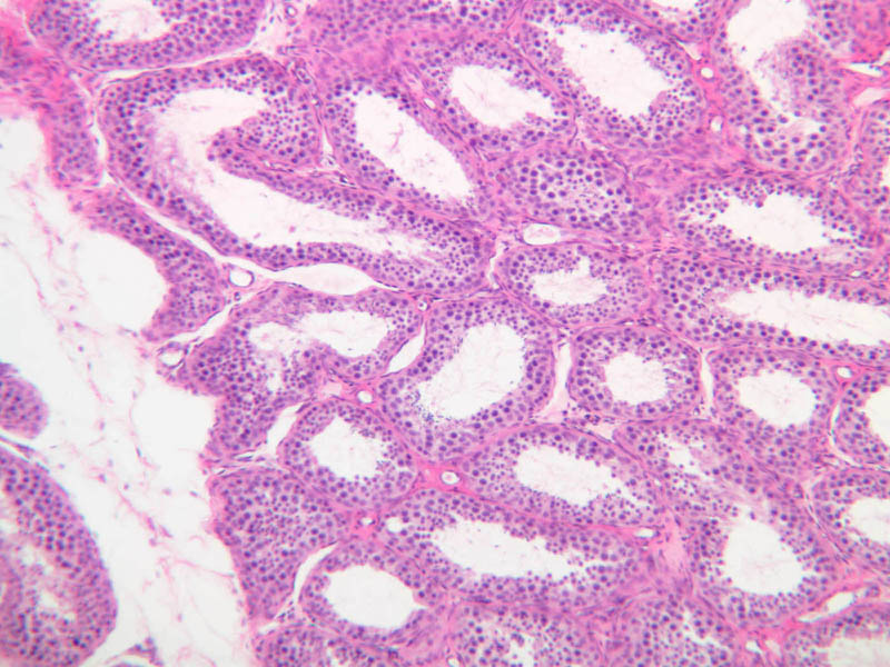
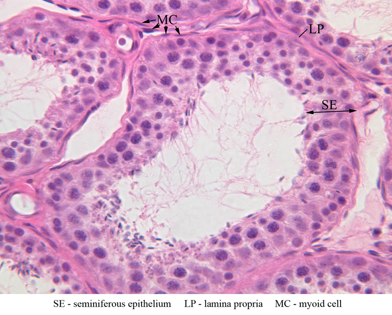
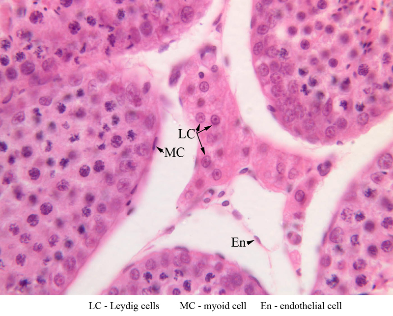
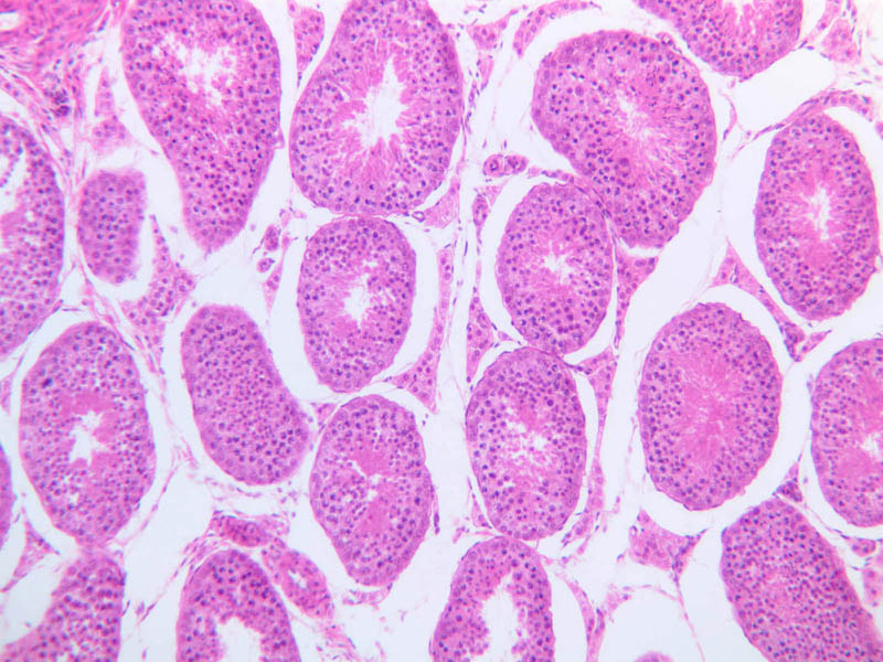
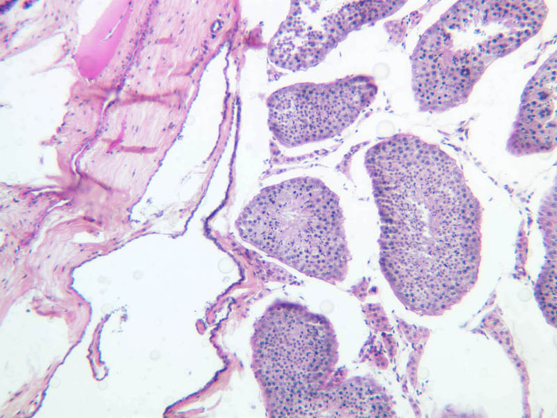
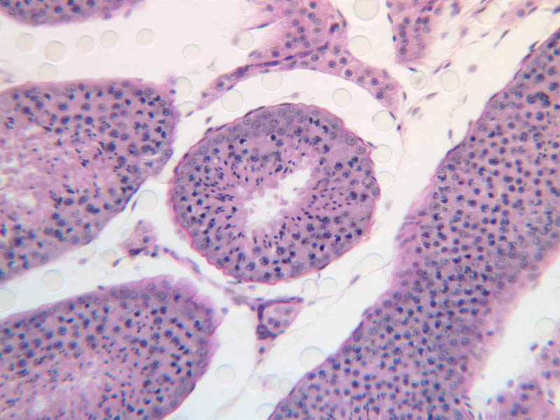
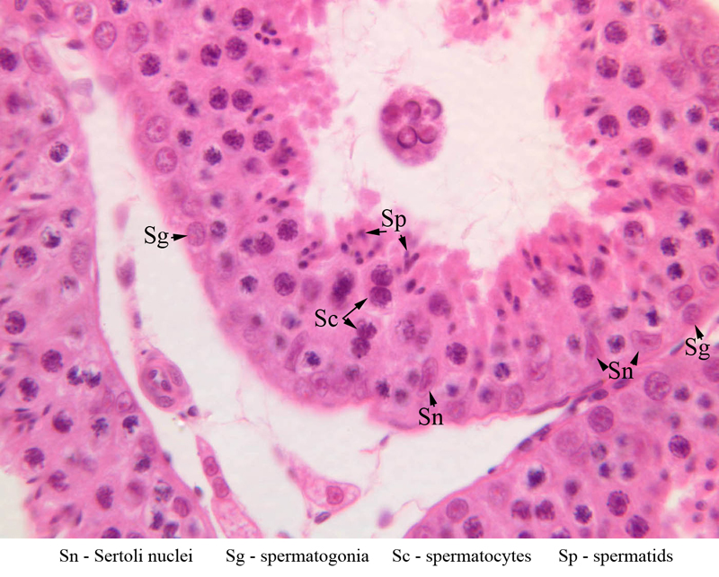
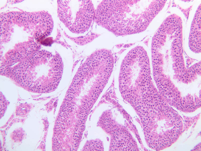
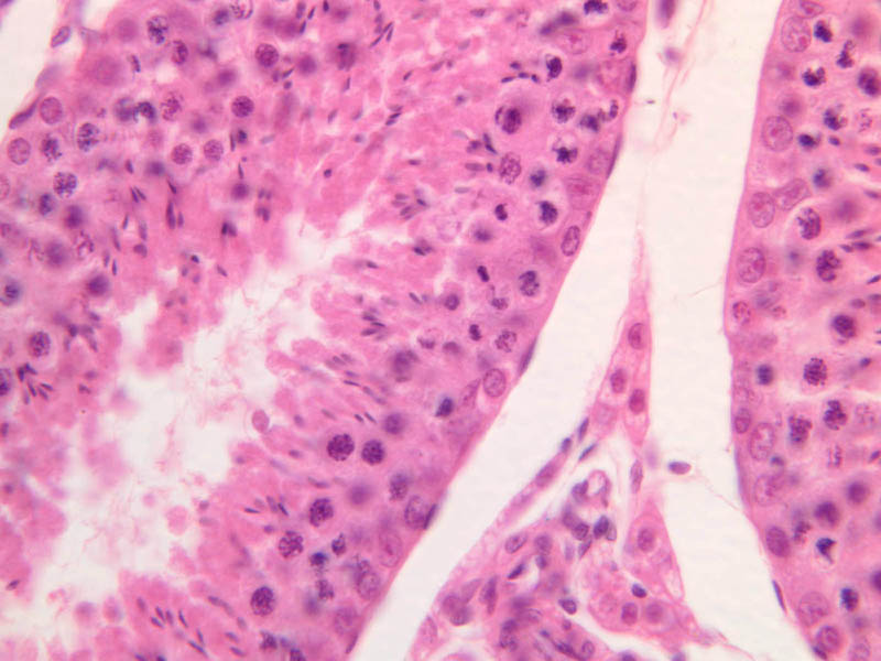
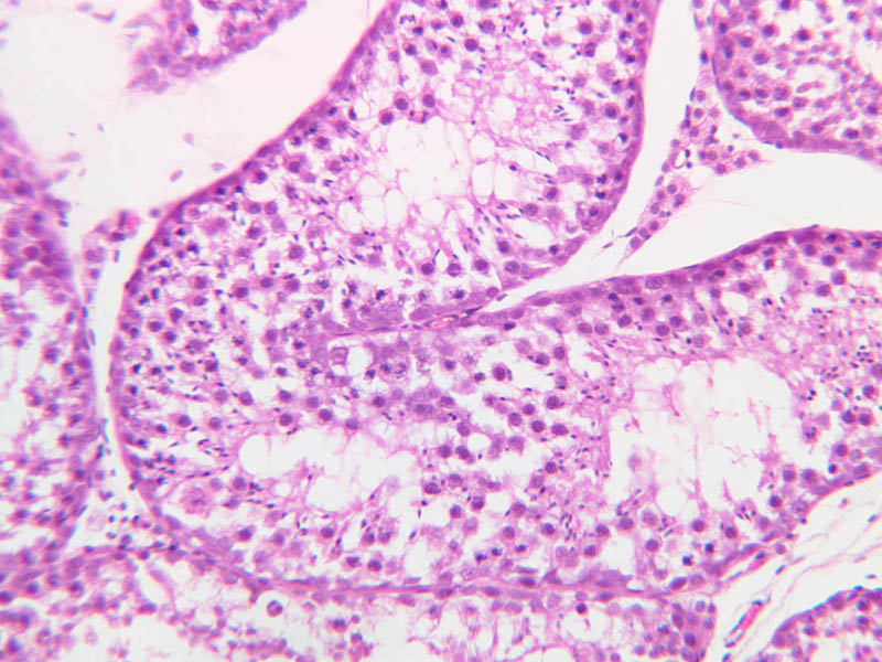
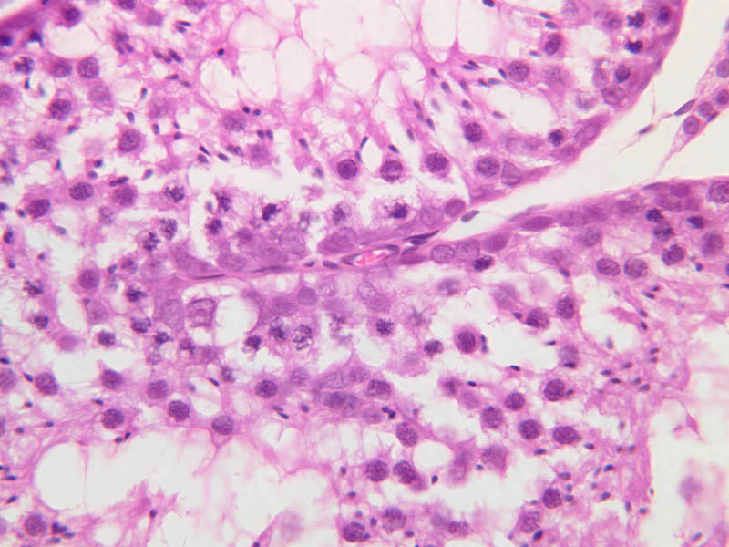
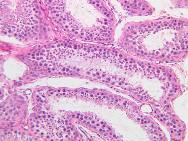
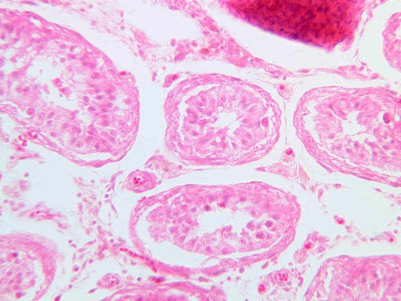
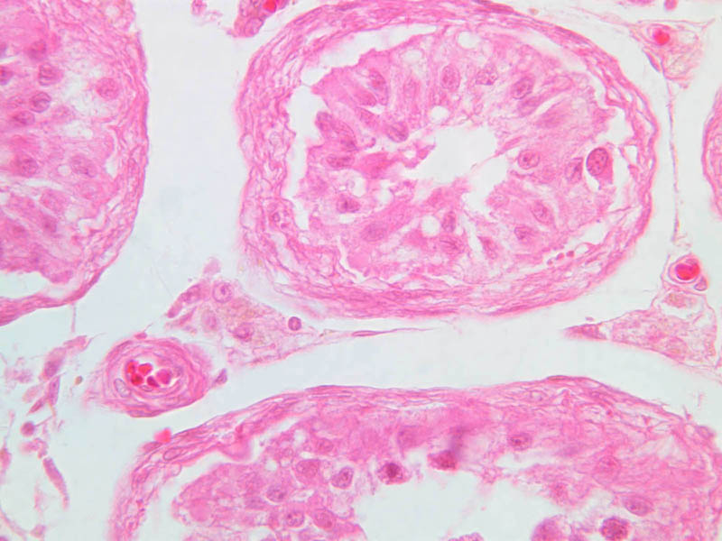
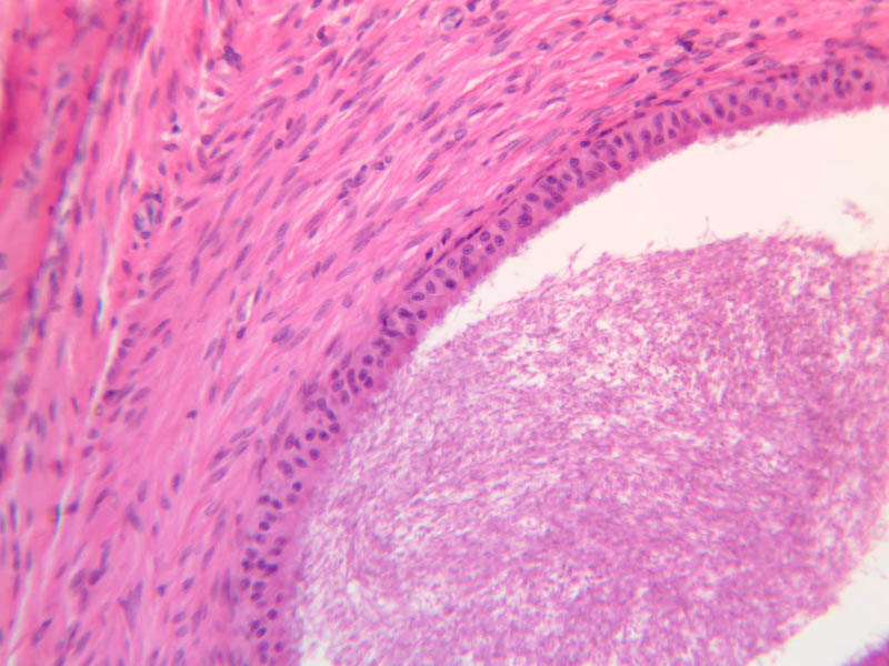
Scan a section of testis from the slide boxes at low magnification, and note the arrangement of the testicular components (slide B-80, H&E [1x-labeled, 1x, 1x]; B-81, PAS [1x, 1x]; B-82, adult monkey testis, H&E [1x, 1x, 1x, 1x, 1x] [2.5x, 10x]). A thick fibroelastic connective tissue capsule, or tunica albuginea, surrounds the testis. Within the tunica and sometimes bulging inward are a number of large blood vessels. The larger, thicker walled vascular profiles are sections through the testicular artery, which executes a highly convoluted course through the tunical plane. Closely applied to the outer surface of the tunica albuginea is the visceral layer of the tunica vaginalis, whose free surface is lined with mesothelium (B-81, PAS [2.5x-labeled]). In some specimens the parietal layer of the tunica vaginalis has been removed, but in others it is possible to find both layers of the tunica vaginalis and the potential serous space between them. How does the tunica vaginalis originate? Connective tissue septa extend inward from the tunica albuginea, subdividing the testis into a number of lobules. Within each lobule are numerous profiles of seminiferous tubules (B-82 [2.5x-labeled, 10x] [20x, 40x]). The interstitial space between the seminiferous tubules is occupied by a loose connective tissue matrix that is permeated by blood and lymphatic vessels and contains clusters of Leydig cells (interstitial cells) (B-82 [40x-labeled]). Both androgens and spermatozoa are produced by the testis. Androgen synthesis is accomplished by Leydig cells. Leydig cells are often arranged in clusters with their distinct nuclei displaying a prominent nucleolus (B-82 [2.5x, 10x, 20x-labeled, 40x-labeled]). These clusters are located in close proximity to small blood vessels or elements of the extensively developed testicular lymphatic system. What might this relationship facilitate? The abundant eosinophilic cytoplasm usually has vacuolated appearance. Observe the interstices at high magnification in slide B-80 (H&E [2.5x, 10x, 20x, 40x-labeled] [10x, 20x, 40x]). Be aware, however, that most of the empty-appearing interstitial spaces in these slides are artifacts of specimen preparation. Sperm production, or spermatogenesis, encompasses two distinct series of events: one affecting mostly the nucleus, the other affecting mostly the cytoplasm. First, there is a series of events, known collectively as spermatocytogenesis, in which rounded diploid stem cells (spermatagonia) give rise to clusters of rounded haploid cells (spermatids). Then there is a series of events, known collectively as spermiogenesis, in which the round spermatid spins a long flagellum, undergoes compaction of its nucleus, transforms its Golgi complex into an acrosomal cap, and sheds nearly all of its cytoplasm to yield a spermatazoa. Bear in mind that if you are to develop a good sense of the dynamics of sperm production, you will have to examine a number of seminiferous tubules. This is because spermatogenesis is typified by complex waves of proliferation and differentiation that sweep both around the circumference and along the length of the seminiferous tubule. Using the high dry objective, search slide B-81 for a transversely sectioned seminiferous tubule (i.e. one that presents a round profile in which a large lumen is surrounded by an epithelial wall of uniform thickness). Observe that the peripheral surface is demarcated by a continuous layer of small flattened cells containing elongated basophilic nuclei. These are contractile myoid cells (similar to the myoepithelial cells of other glands), which aid in propulsion of the luminal contents towards the excurrent duct system (B-81, testis, PAS [2.5x-labeled, 10x, 20x. 40x-labeled]; [10x, 20x, 40x]). The clearly stratified seminiferous epithelium differs in appearance from one tubular profile to another and even from one part of a single tubule to another. The seminiferous epithelium is composed of two populations of cells: proliferating cells of the germinal spermatogenic series and nonproliferating Sertoli cells. Sperm production begins with mitotic division of cells located at the tubule periphery; later events occur progressively closer to the lumen. In typical H&E preparations of seminiferous epithelium it is the distinctive qualities of nuclei that stand out against poorly defined cytoplasm (B-80, testis, H&E [20x, 40x-labeled] [10x, 20x, 40x-labeled] [10x, 20x, 40x] [2.5x, 10x, 20x, 40x]; B-82, testis, H&E [2.5x, 10x, 20x, 40x] [2.5x, 10x, 20x, 40x] [10x, 20x, 40x] [10x, 20x, 40x]). Thus, in your study of seminiferous epithelium, it is nuclear appearance that will enable you to distinguish the following cell types:Top of page
Spermatogonia
Spermatagonia are stem cells that divide mitotically both to maintain the germ cell line (i.e., to produce more spermatagonia) and to yield primary spermatocytes. Although some authors identify three or more different types of spermatagonia, you are not expected to draw such distinctions. You should, however, be able to distinguish between spermatagonia and their neighbors. Spermatagonia are located at the periphery (i.e., in the basal portion) of the seminiferous epithelium. Their nuclei are generally spherical or ovoid, show variable intensity of staining and may or may not have a discernible nucleolus.Primary Spermatocytes
Primary spermatocytes originate from mitotic division of spermatogonia and are usually one or more cell diameters removed from the tubular periphery. Their nuclei are more or less spherical and are conspicuously larger than those of other germ cells; their chromatin is usually condensed into many thread-like structures. Most primary spermatocytes are seen in some stage of the very protracted (21-day) prophase leading up to the first meiotic (reduction) division, in which each primary spermatocyte gives rise to two secondary spermatocytes.Secondary Spermatocytes
Within about 8 hours of its own generation, each secondary spermatocyte completes the second meiotic (equatorial) division to yield two spermatids. Because they are so short-lived, secondary spermatocytes are often difficult to find. When present, they are located toward lumen from primary spermatocytes. They are about 2/3 the diameter of primary spermatocytes and about 1/3 greater in diameter than recently generated, spherical spermatids.Spermatids
Spermatids are located next to lumen of the seminiferous tubule, where they undergo an elaborate differentiation process (spermiogenesis) that requires about 7 weeks and involves condensation of nuclear chromatin, compaction and elongation of the nucleus, shedding of nearly all cytoplasm and formation of a motile flagellum (tail). Spermatid nuclei vary in size, condensation of chromatin and shape according to the stage of spermiogenesis that they represent; however, all of them are smaller than the nuclei of any of the antecedent cell types. Spermatids at different stages of spermiogenesis may be present in a single tubule. Top of pageSertoli cells
Interspersed among the germ cells, but generally situated near the epithelial periphery, are nuclei of the Sertoli cells. Sertoli cells span the full thickness of the seminiferous epithelium; however, because of their irregular shape and poor affinity for dyes, it is impossible to define the cytoplasmic processes of Sertoli cells in ordinary paraffin sections. Sertoli cells have large euchromatic nuclei and very prominent nucleoli. The nuclear shape is usually oval or triangular, but may also be irregular due to deep indentations of the nuclear membrane. The long axis of the nucleus is typically oriented at right angles to the basement membrane. From ultrastructural studies it is known that Sertoli cell processes make specialized contacts with germ cells as well as with processes of other Sertoli cells. Sertoli cells are the basis of the blood-testis barrier As you might expect, there are marked differences between the prepubertal and postpubertal testis. In the true prepubertal male (slide B-86, epididymis and testis, H&E [2.5x, 10x, 20x, 40x]) and in undescended testes of older males, the seminiferous tubules are solid cords of cells, consisting of spermatogonia-like cells known as gonocytes, and cells that resemble the Sertoli cells of the adult testis. Section B-78 is from the testis of an older individual and a lumen is now present (B-78, H&E [2.5x, 10x, 20x, 40x]). Even though the seminiferous epithelium of this autopsy specimen is poorly preserved, you should be able to evaluate its spermatogenic capacity. Can you identify spermatogonia and Sertoli cells? Are spermatids present? Judging from the interstitial morphology, would you expect high peripheral testosterone levels? Beginning in the 6th decade, spermatogenesis gradually decreases. A late stage in testicular aging appears on slide B-79 (senile testis, H&E [10x, 20x, 40x] [10x, 20x, 40x]). How many of the germ cell stages can you identify? This specimen also provides many examples of thickening of the seminiferous tubular wall. Once initiated, this process apparently continues until the germinal and Sertoli cells are eliminated and the tubule is a solid cord of connective tissue cells. Such tubules are called hyalinized and often appear in clusters. Using the scanning or low power objective, locate several hyalinized seminiferous tubules; then study them under the high-dry objective to confirm the absence of germ cells.Testis Image Gallery


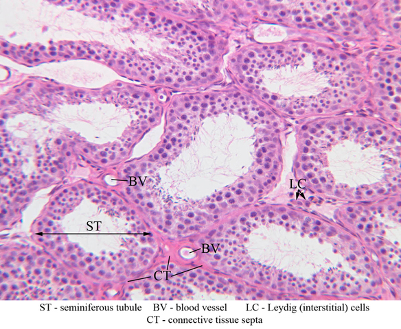


Testis Table of Identifications
| Row | Structure | Abbreviation | Optimal Stain | Representative Section | Note |
|---|---|---|---|---|---|
| 1 | Cauda (tail of) Epididymis | (none) | H&E | |
|
| 2 | Corpus (body of) Epididymis | (none) | H&E | |
|
| 3 | Tunica Vaginalis | (none) | H&E, PAS | |
|
| 4 | Tunica Albuginea | (none) | H&E, PAS | |
|
| 5 | Seminiferous Tubules | (none) | H&E, PAS | |
|
| 6 | Epididymis | (none) | H&E, PAS | |
|
| 7 | Septa | S | H&E | |
|
| 8 | Blood Vessel | BV | H&E | |
|
| 9 | Leydig (Interstitial) Cells | LC | H&E | |
|
| 10 | Connective Tissue Septa | CT | H&E | |
|
| 11 | Seminiferous Epithelium | SE | H&E | |
|
| 12 | Lamina Propria | LP | H&E | |
|
| 13 | Myoid Cell | MC | H&E | |
|
| 14 | Fibroblast | F | H&E | |
|
| 15 | Endothelial Cell | En | H&E | |
|
| 16 | Spermatocytes | Sc | H&E | |
|
| 17 | Spermatids | Sp | H&E | |
|
| 18 | Sertoli Nuclei | Sn | H&E | |
|
| 19 | Spermatogonia | Sg | H&E | |
Top of page
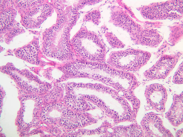
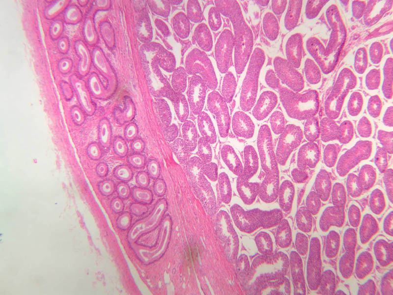
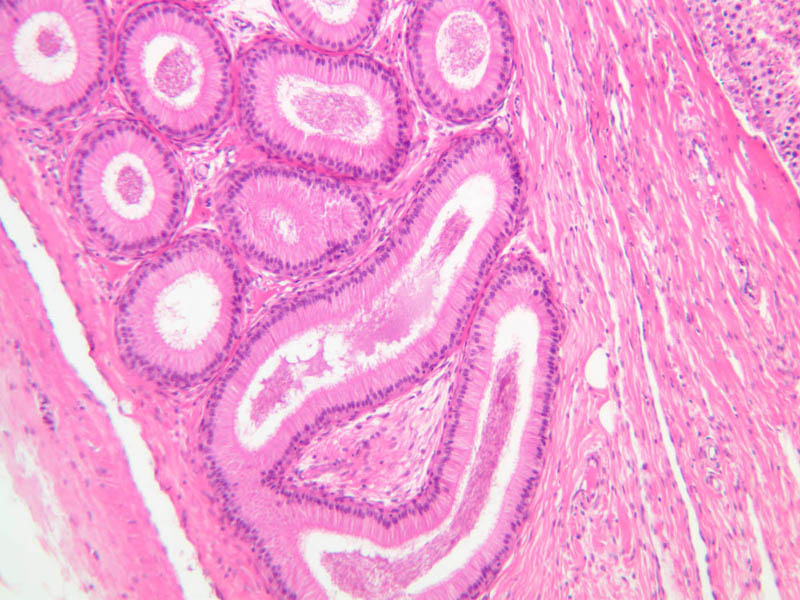
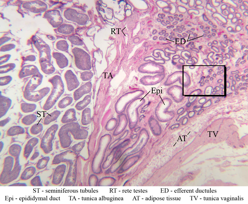
Excurrent Ducts
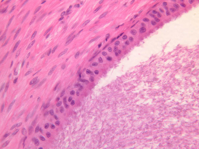
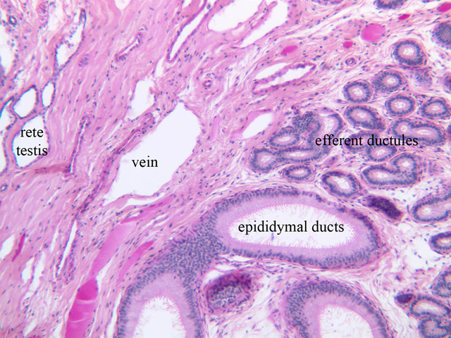
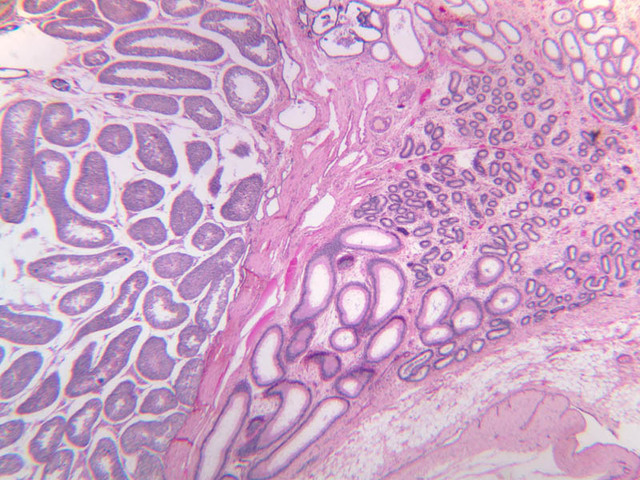
Spermiogenesis is concluded when immature (non-motile) spermatozoa detach from the Sertoli cells and lie free in the lumen of the seminiferous tubule. In the course of their passage through the intratesticular ducts and the ductus epididymis, sperm acquire motility and become fully capable of fertilization. This maturation process requires the presence of androgens as well as glycoprotein secretions of cells that line the excurrent ducts--especially the epididymis. Three mechanisms account for the movement of newly released sperm from the seminiferous tubule into and through the excurrent duct system: (1) contraction of myoid cells, (2) propulsion by ciliated cells in some portions of the duct system and (3) a physical current produced by net secretion of fluid proximally and net resorption of fluid distally. In proximodistal sequence, the excurrent duct system consists of the following regions: rete testis, ductuli efferentes, epididymis, vas deferens, urethra. Top of pageRete testis
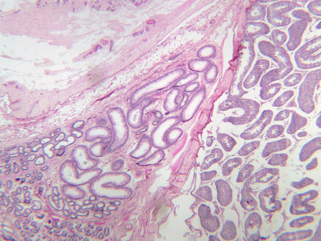
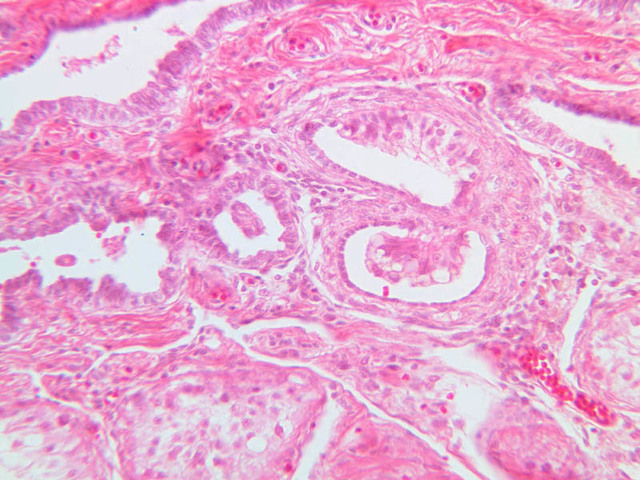
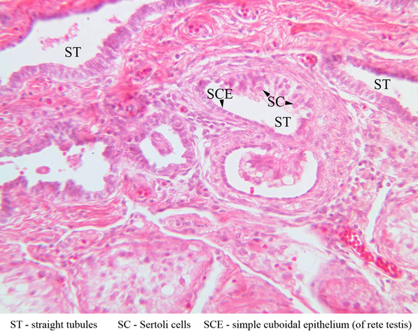
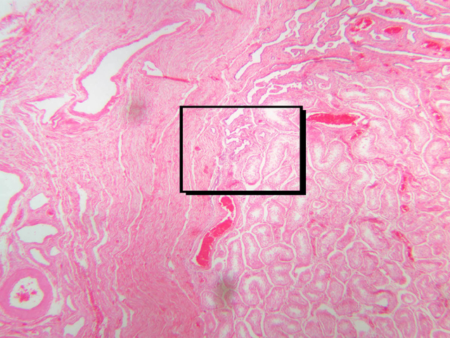
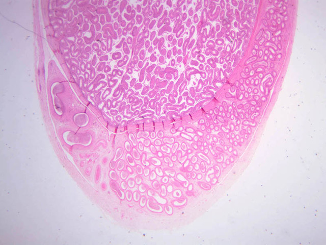
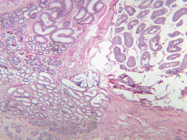
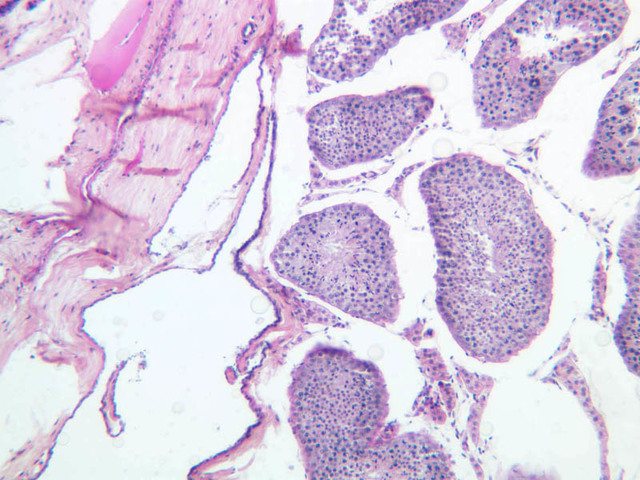
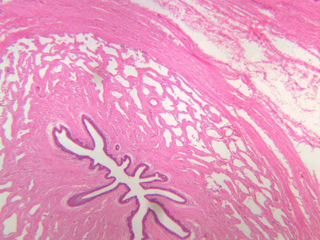
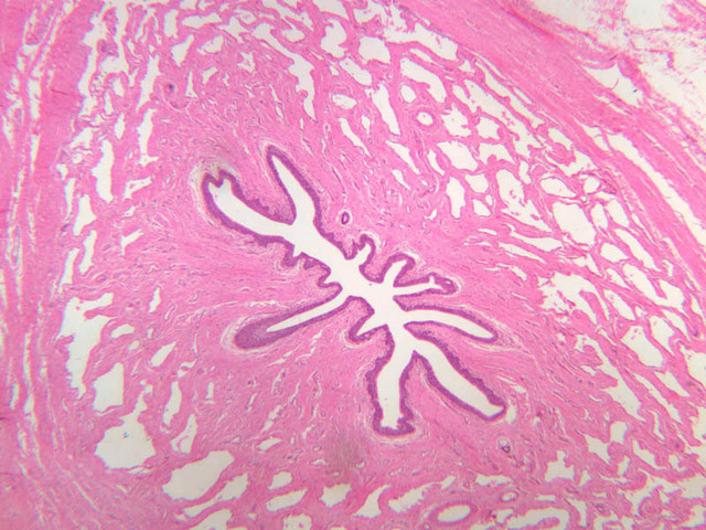
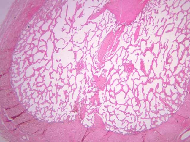
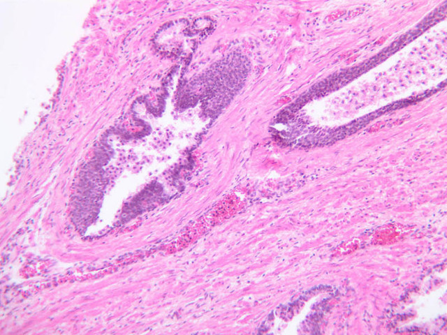
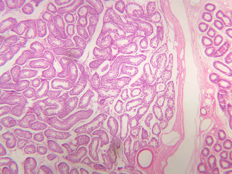
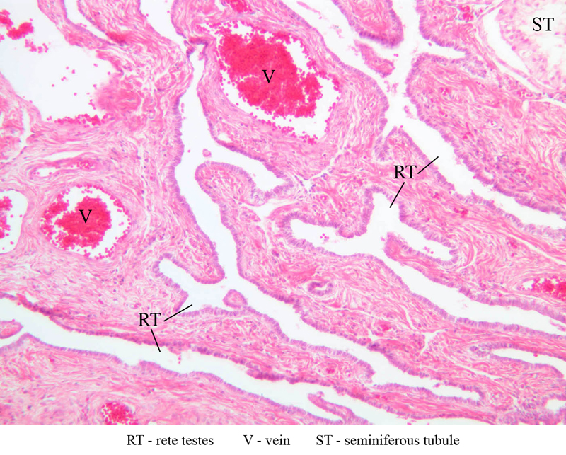
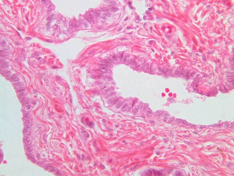
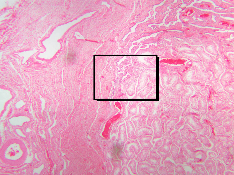
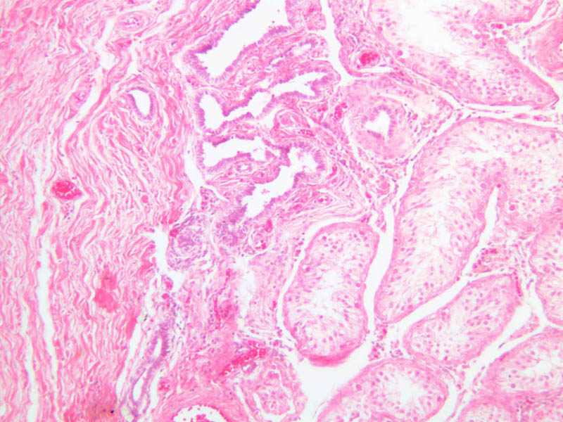
The rete testis is an anastomosing network of irregular, epithelium-lined spaces located in the mediastinum testis. The mediastinum testis is an inward extension of the tunica albuginea located on the posterior aspect of the testis. Just before it joins the rete testis, each end of the seminiferous tubule tapers and its epithelial lining consists entirely of Sertoli cells. These tapered, germ cell-free tubules are known as tubuli recti, or straight tubules (B-79, H&E [10x, 20x-labeled, 40x]; B-81, PAS [10x, 20x, 40x]). Locate the rete spaces, deep to the tunica albuginea, at one pole of the testicular section. Note that the epithelium lining these spaces varies from cuboidal to squamous (slide B- 79, H&E [2.5x-labeled, 10x-labeled, 20x, 40x] [2.5x-labeled, 10x, 20x, 40x]).Rete Testis Image Gallery


Rete Testis Table of Identifications
| Row | Structure | Abbreviation | Optimal Stain | Representative Section | Note |
|---|---|---|---|---|---|
| 1 | Straight Tubules | ST | H&E | |
|
| 2 | Sertoli Cells | SC | H&E | |
|
| 3 | Simple Cuboidal Epithelium (of Rete Testis) | SCE | H&E | |
|
| 4 | Tunica Albuginea | TA | H&E | |
|
| 5 | Rete Testis | RT | H&E | |
|
| 6 | Seminiferous Tubules | ST | H&E | |
|
| 7 | Vein | V | H&E | |
Top of page
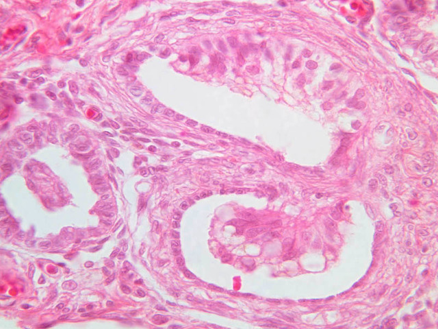
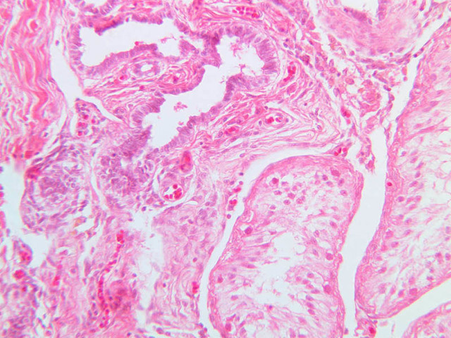
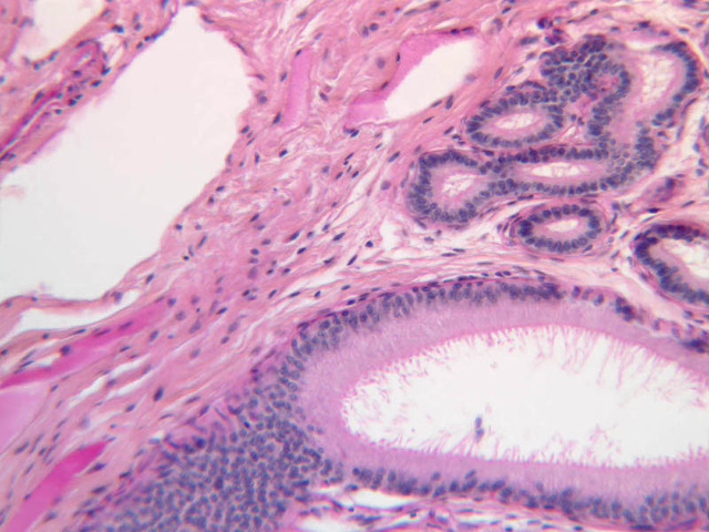
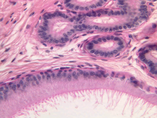
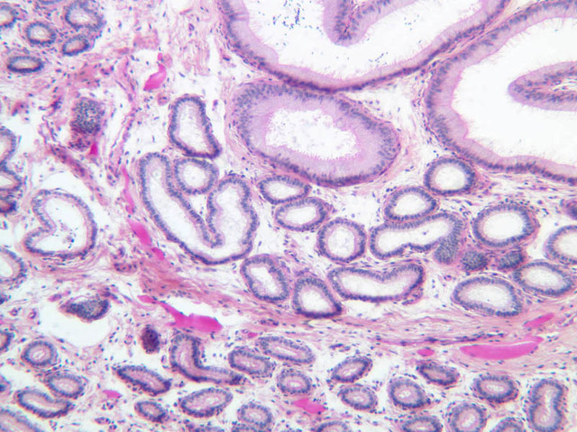
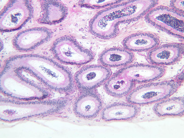
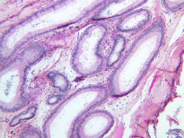
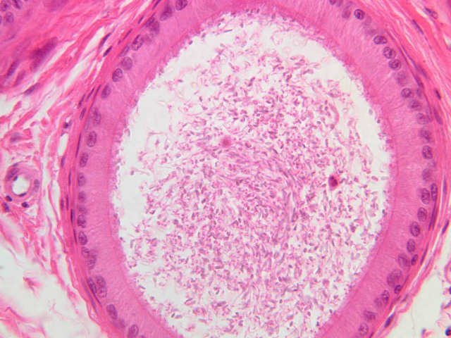
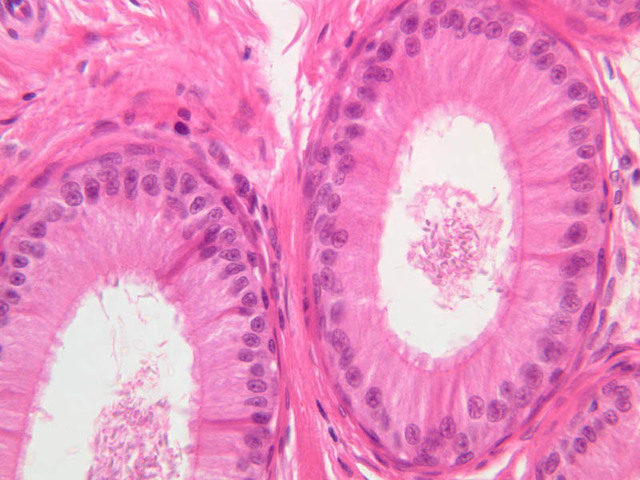
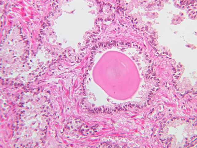
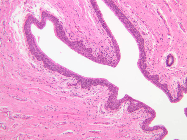
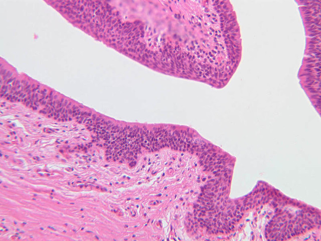
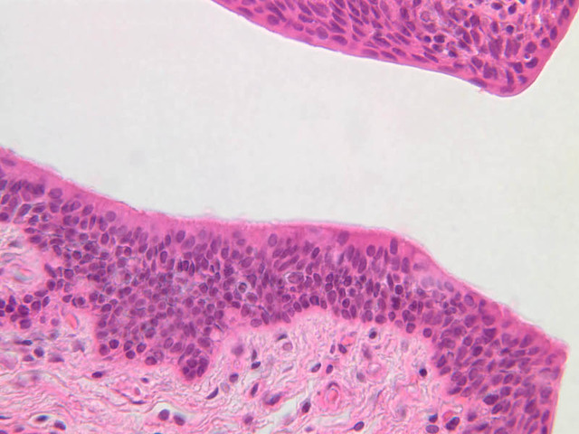
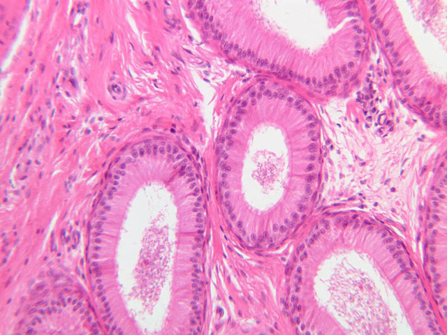
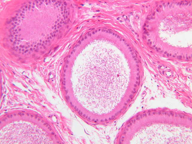
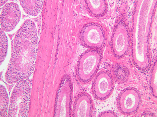
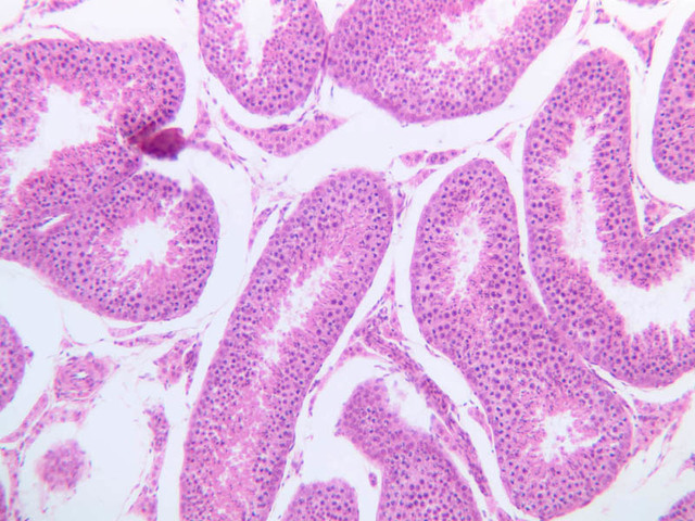
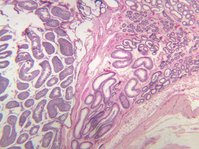
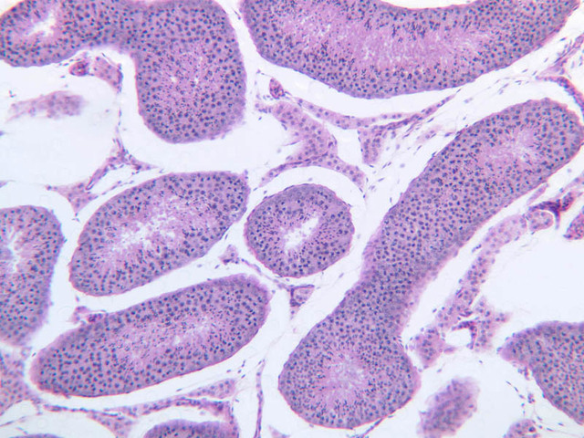
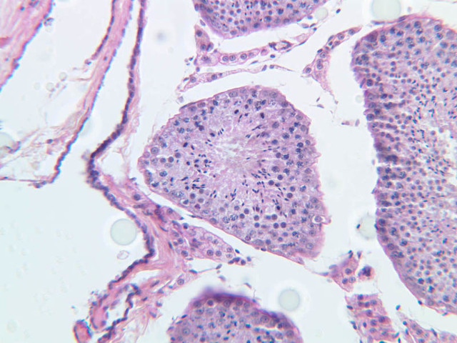
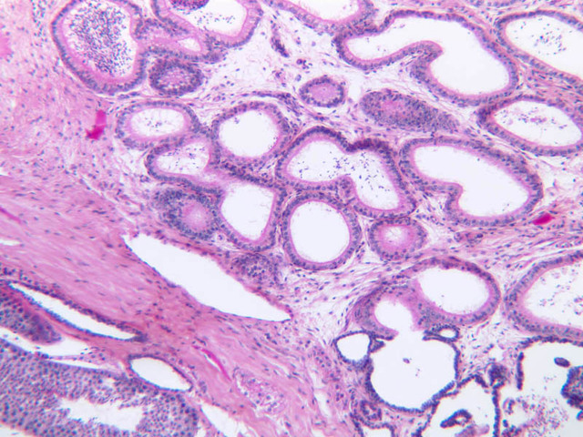
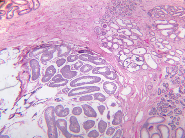
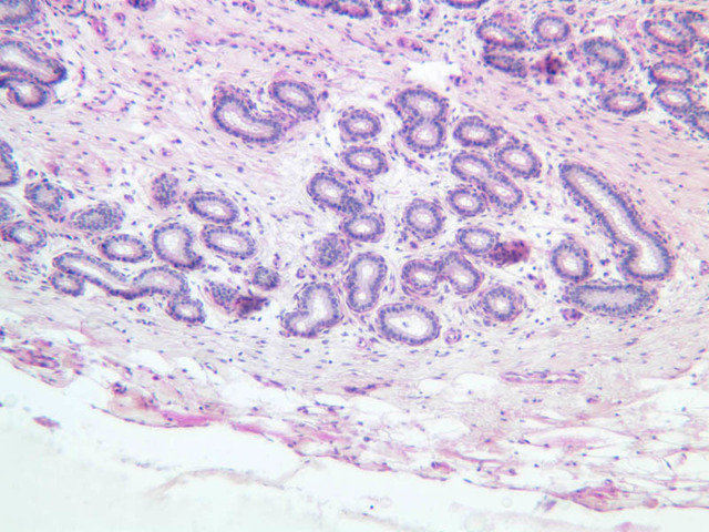
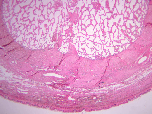
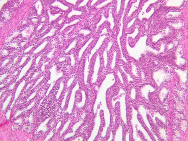
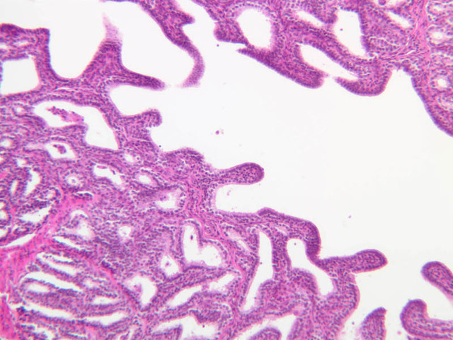
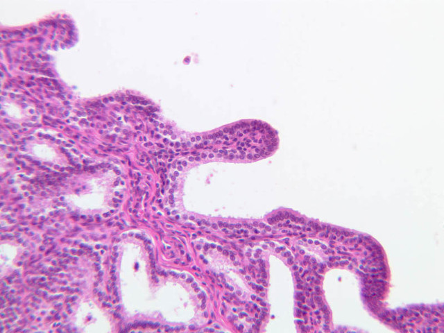
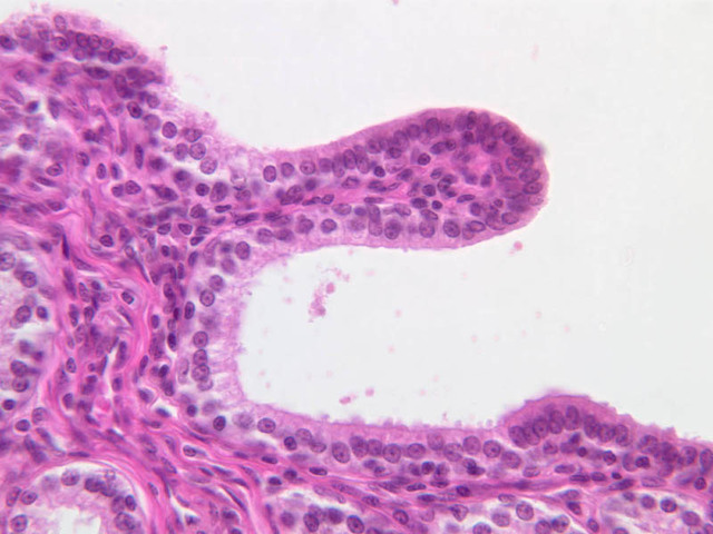
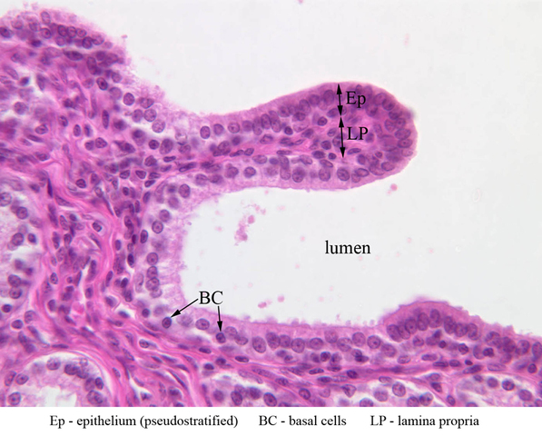
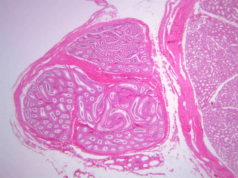
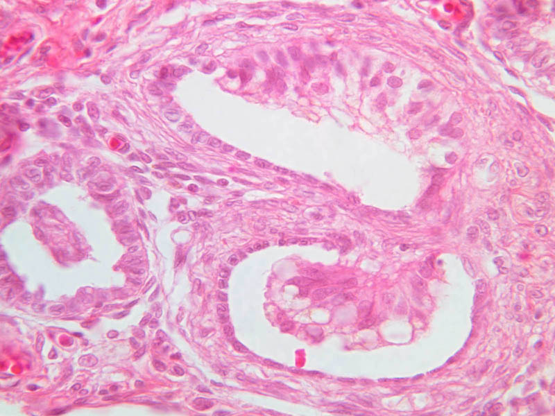
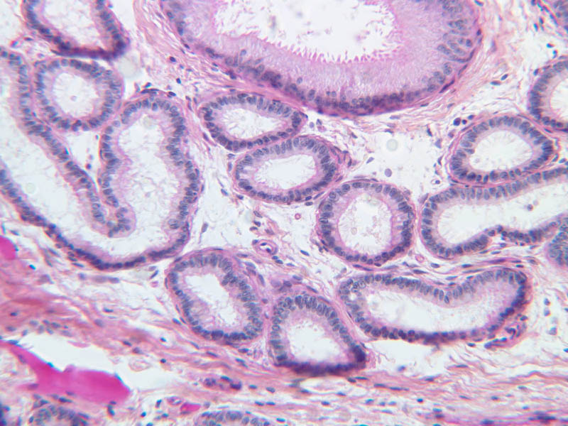
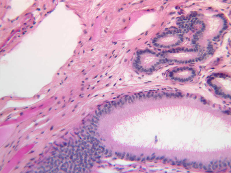
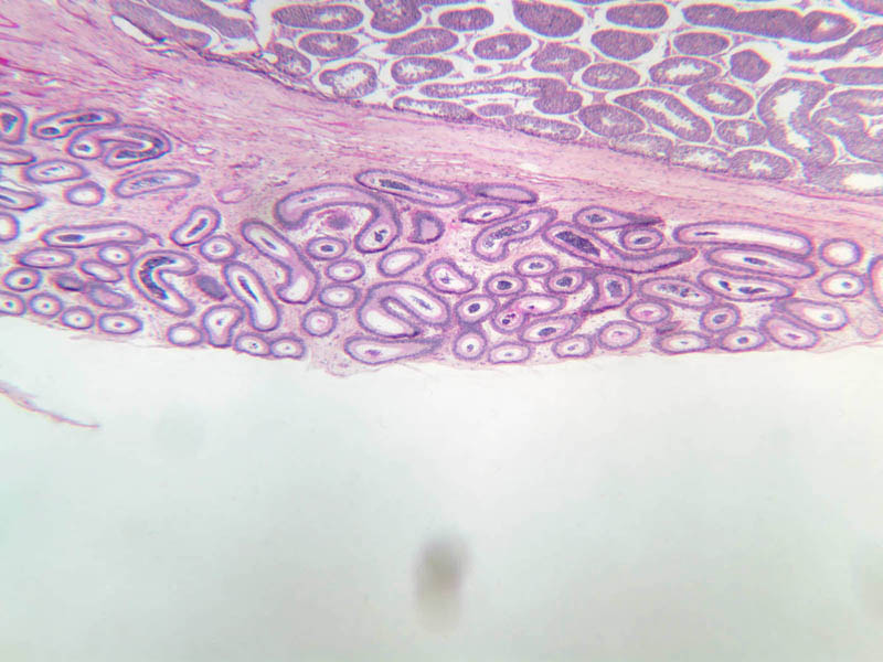
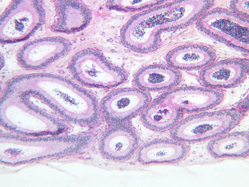
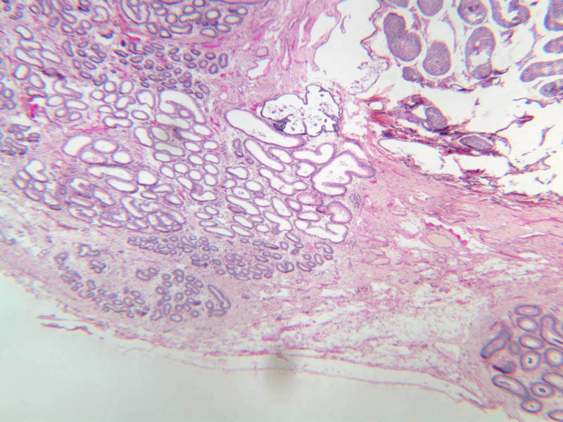
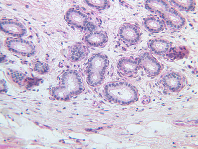
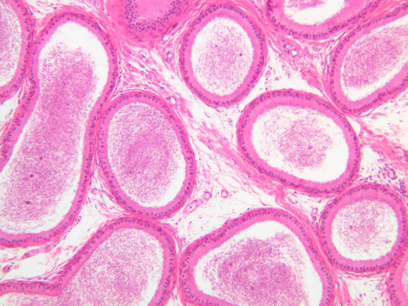
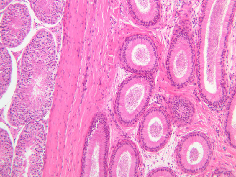
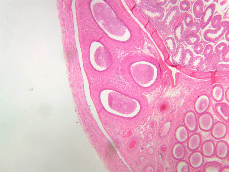
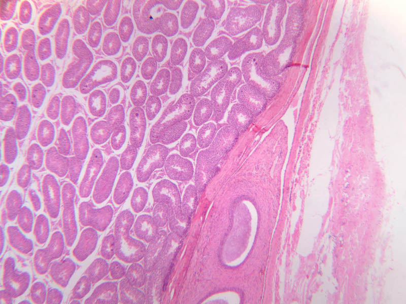
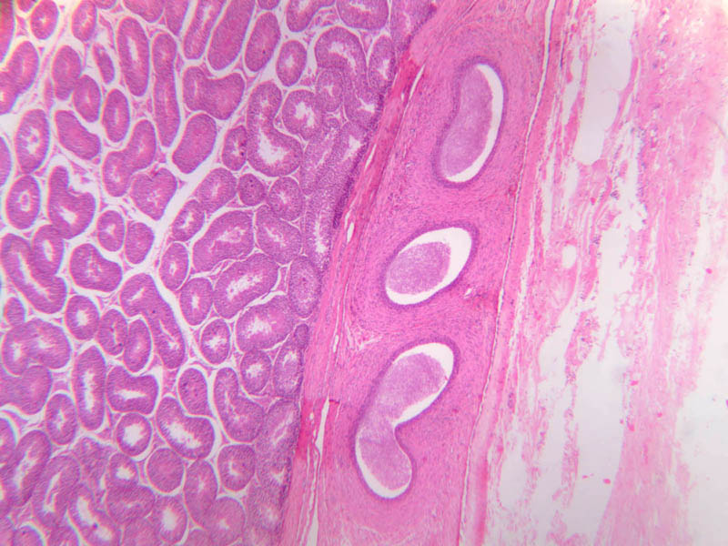
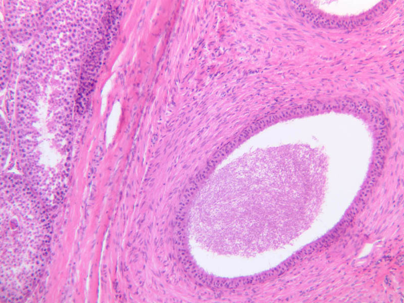
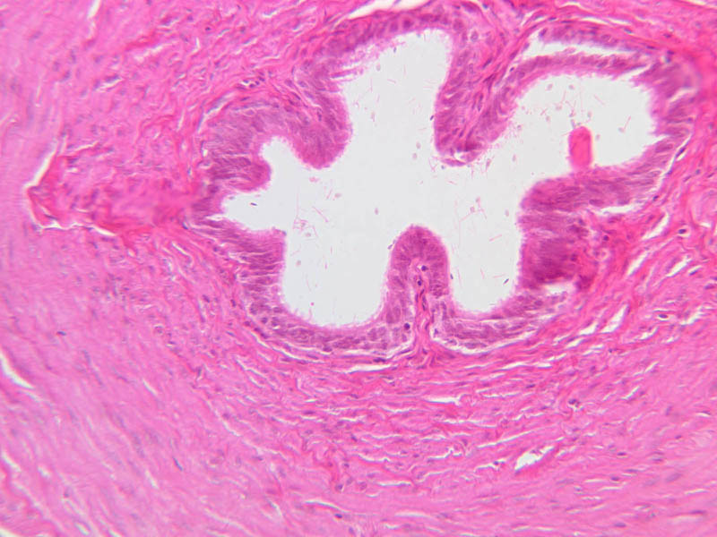
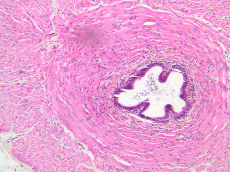
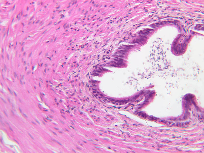
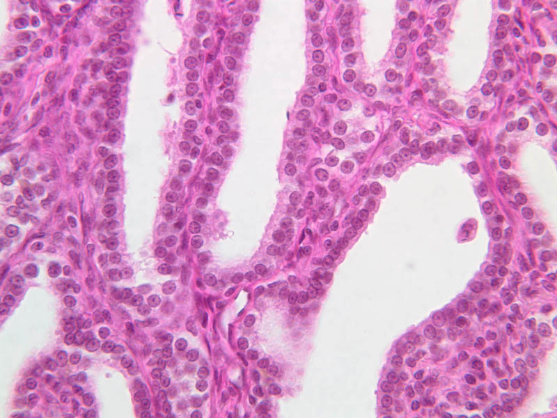
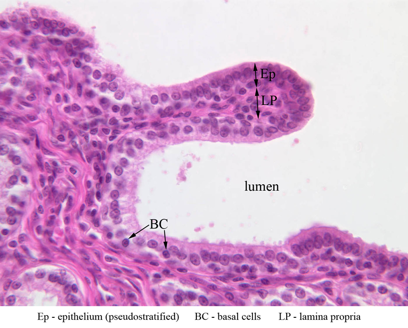
Ductuli efferentes (efferent ductules)
The rete testis is linked to the epididymis by 12 or more highly convoluted ductuli efferentes. A pseudostratified columnar epithelium of variable thickness lines these structures. In addition to basal cells, the epithelium exhibits a patchy arrangement of tall columnar ciliated cells and low columnar or cuboidal cells with brush borders. You should be able to find a thin layer of circularly arranged smooth muscle cells in the tubule wall. Ductuli efferentes are classically described as having a scalloped appearance (i.e. when cut in cross section, the hight of the epithelium varies circumferencially). This scalloped appearance may be difficult to identify in your slide set (B-81, PAS [2.5x-labeled, 10x, 20x-labeled, 40x] [2.5x, 10x-labeled, 20x, 40x] [10x, 20x, 40x]).Efferent Duct Image Gallery


Efferent Duct Table of Identifications
| Row | Structure | Abbreviation | Optimal Stain | Representative Section | Note |
|---|---|---|---|---|---|
| 1 | Seminiferous Tubules | ST | PAS | |
|
| 2 | Rete Testis | RT | PAS | |
|
| 3 | Efferent Ductules | ED | PAS | |
|
| 4 | Epididymis | Epi | PAS | |
|
| 5 | Tunica Albuginea | TA | PAS | |
|
| 6 | Adipose Tissue | AT | PAS | |
|
| 7 | Tunica Vaginalis | TV | PAS | |
|
| 8 | Smooth Muscle | SM | PAS | |
|
| 9 | Connective Tissue | CT | PAS | |
|
| 10 | Blood Vessels | BV | PAS | |
|
| 11 | Vein | (none) | PAS | |
Top of page
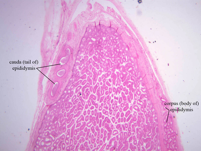
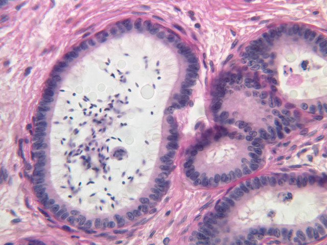
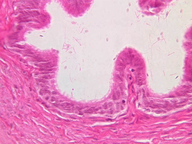
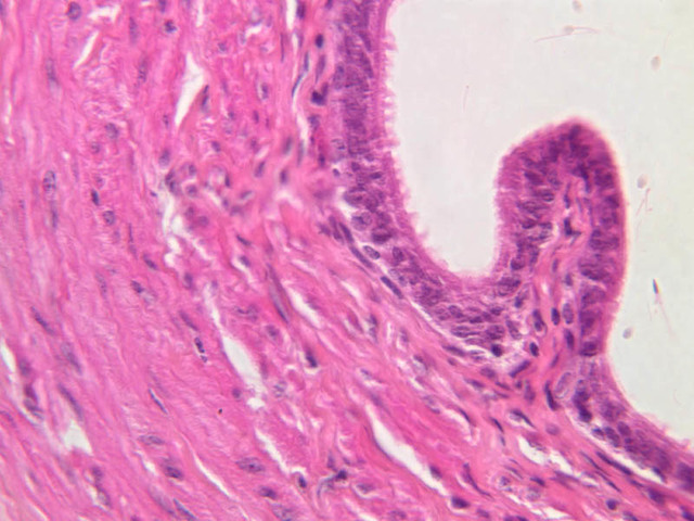
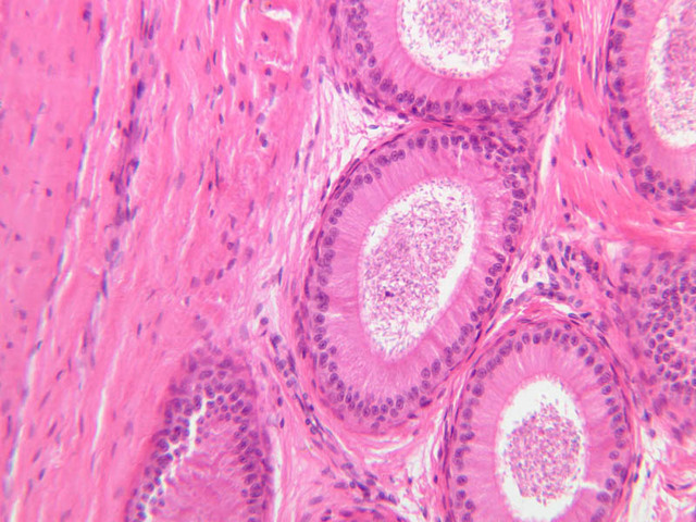
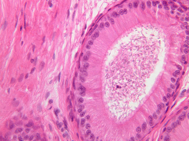
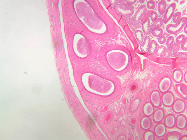
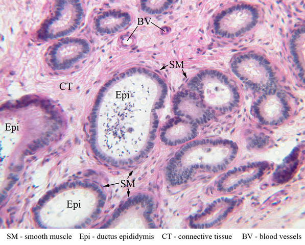
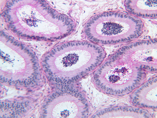
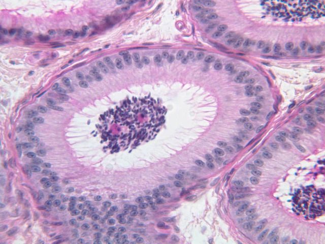
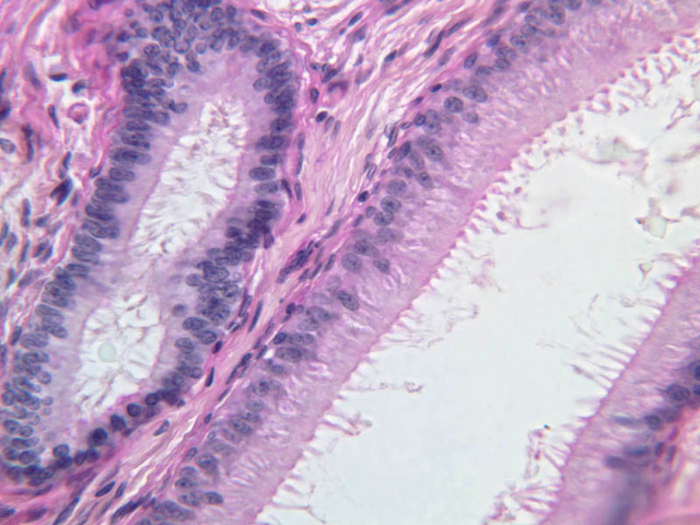
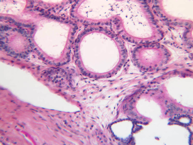
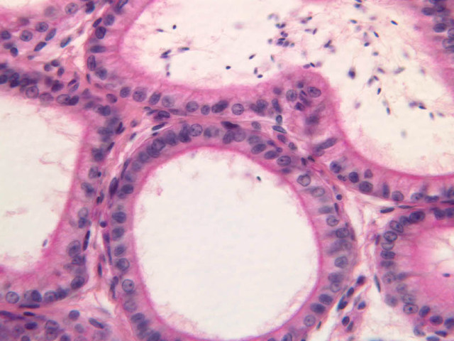
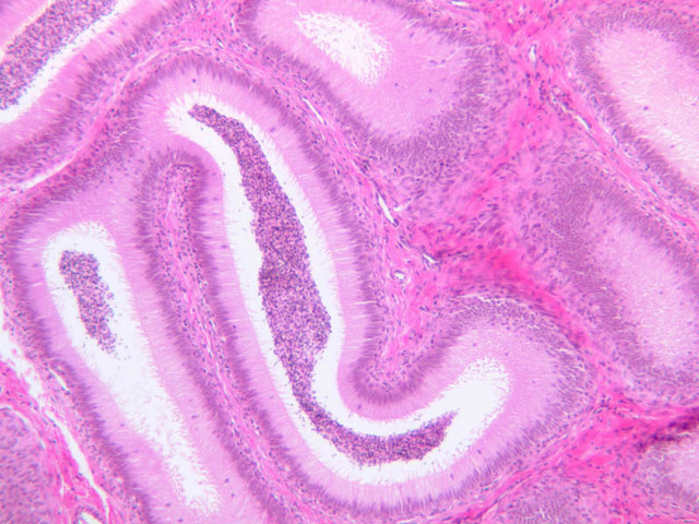
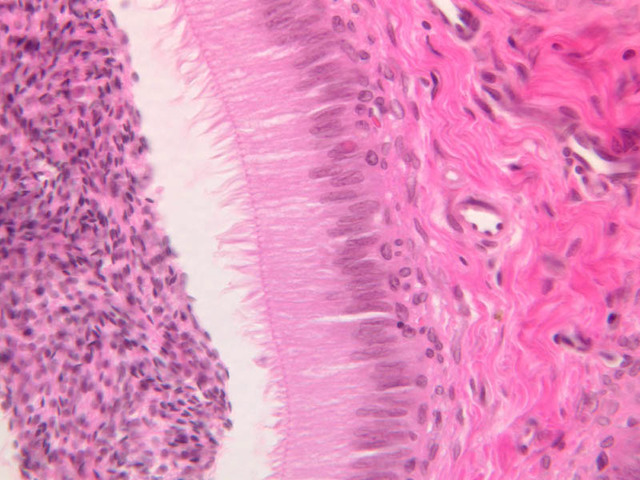
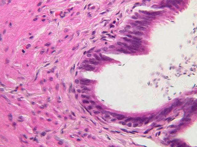
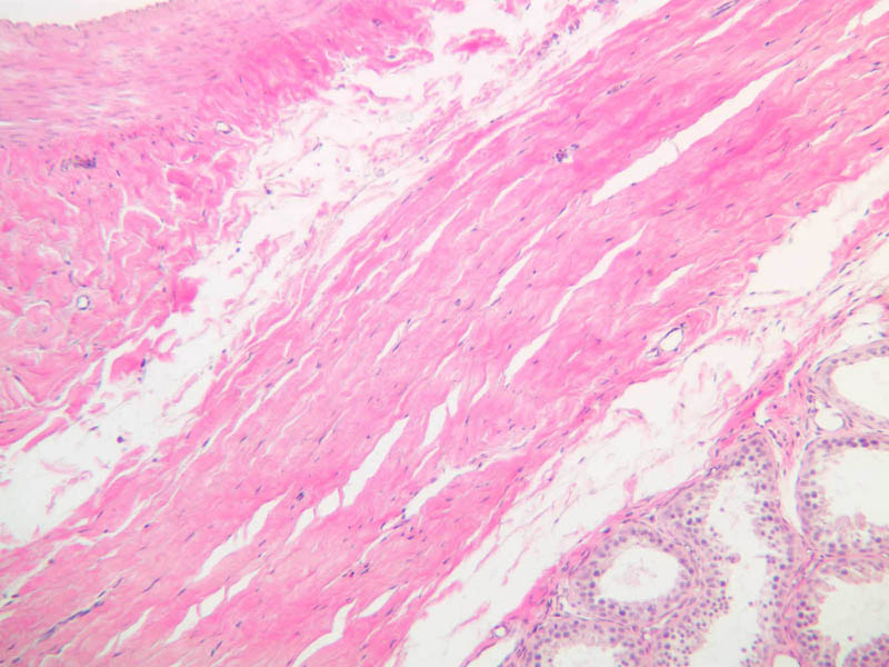
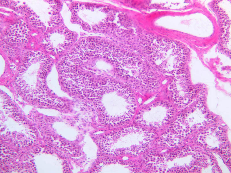
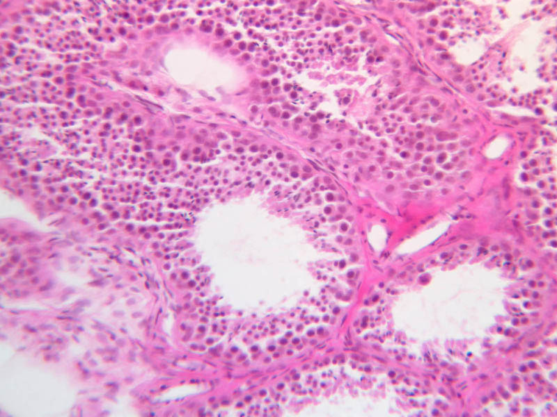
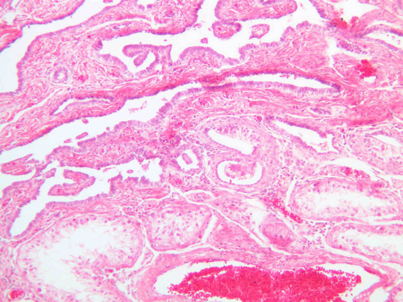
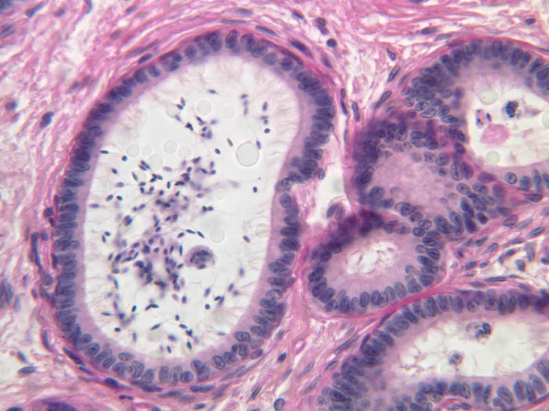
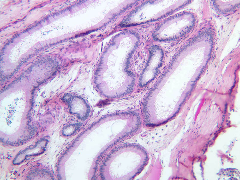
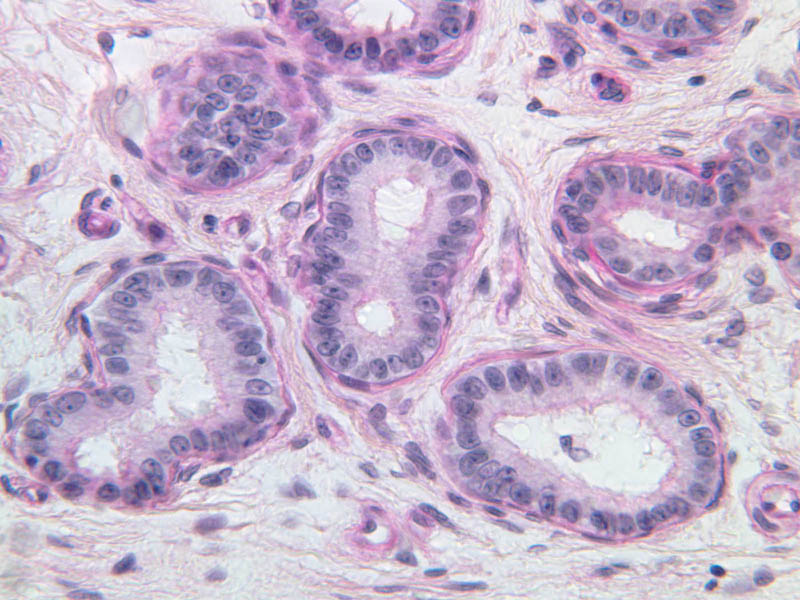
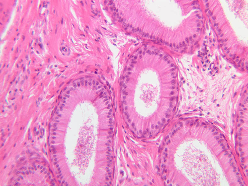
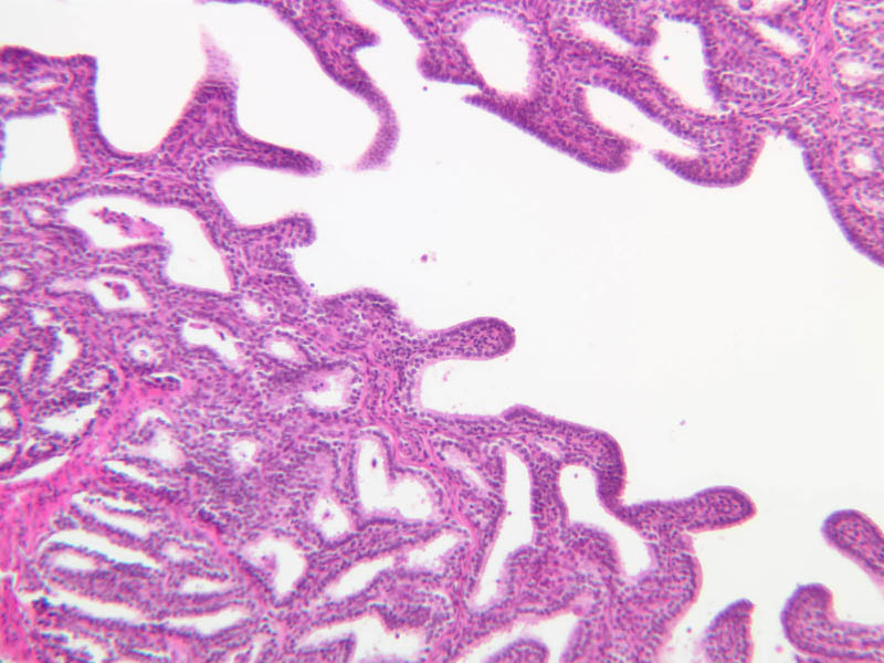
Epididymis
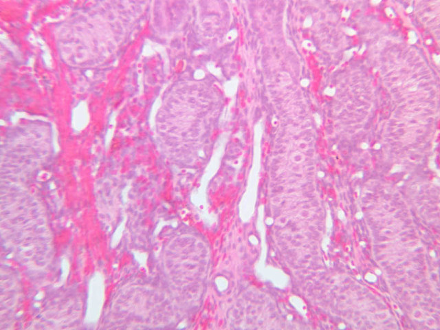
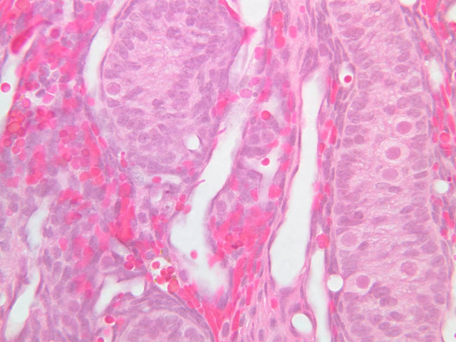
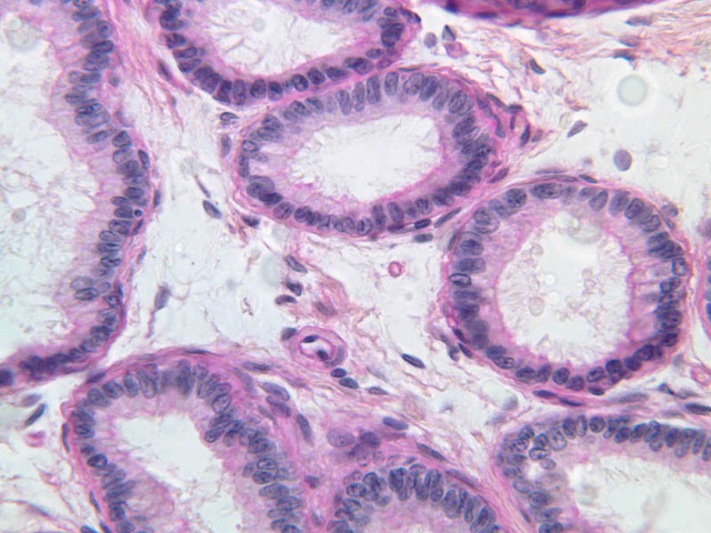
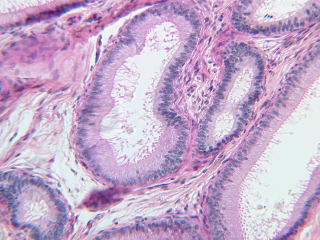
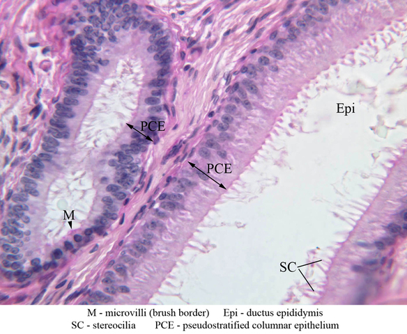
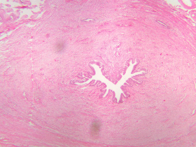
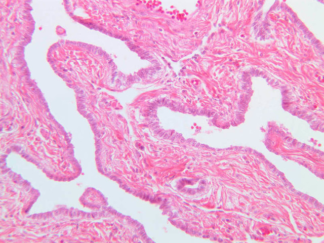
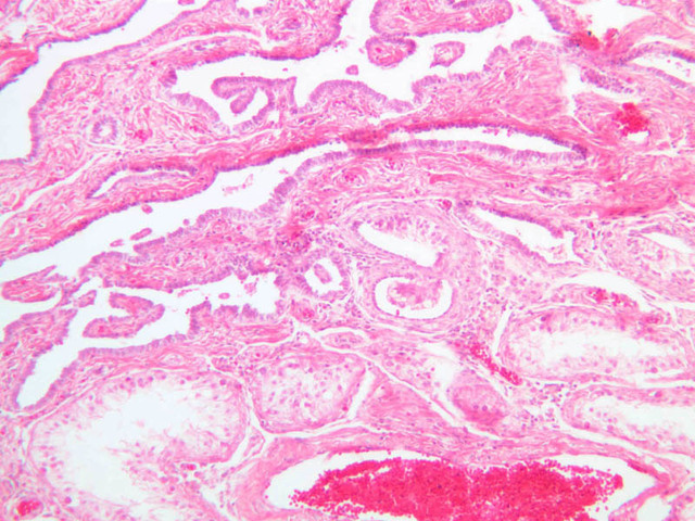
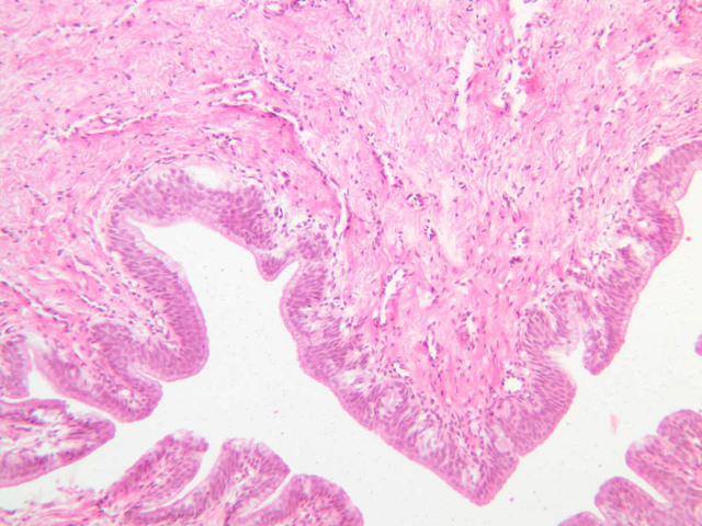
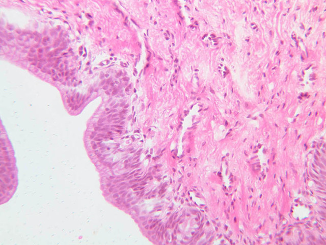
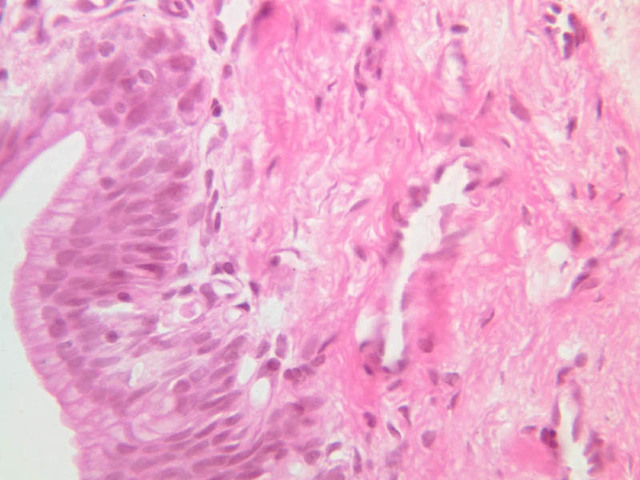
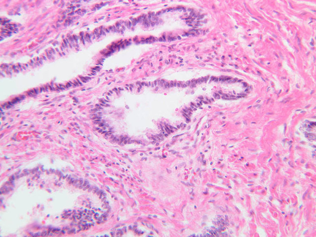
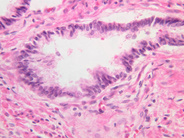
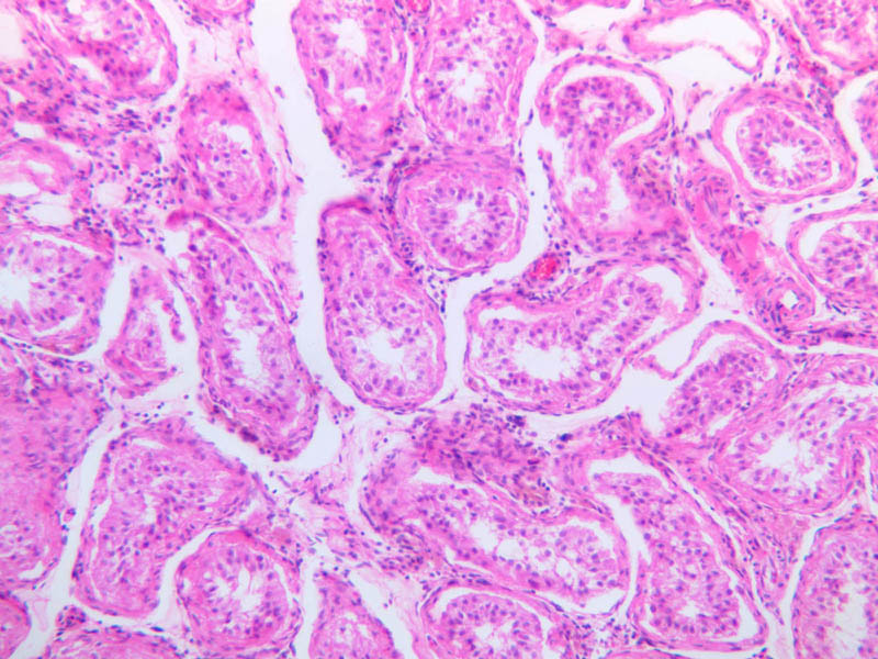
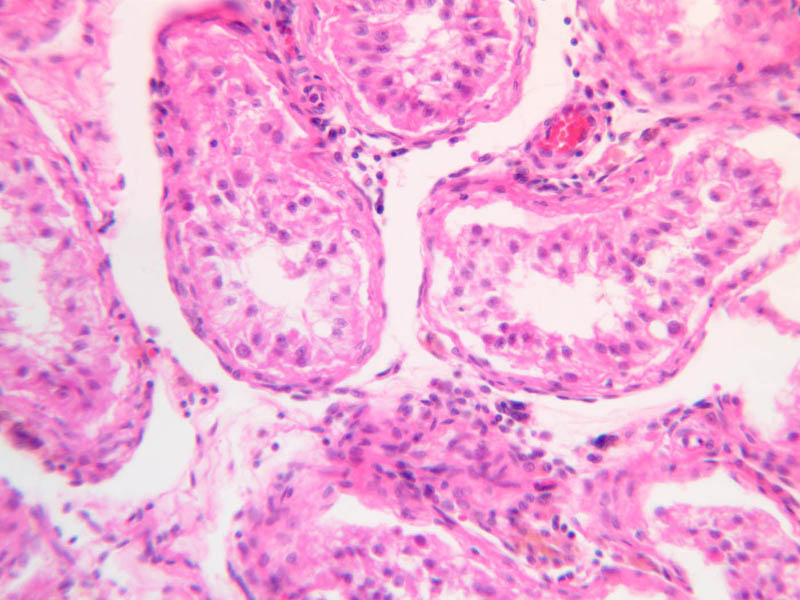
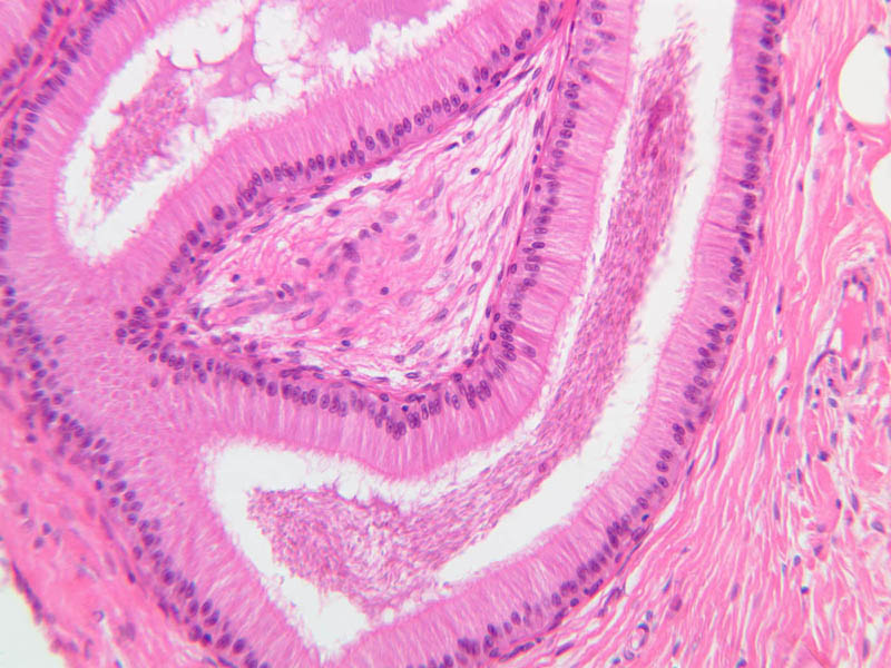
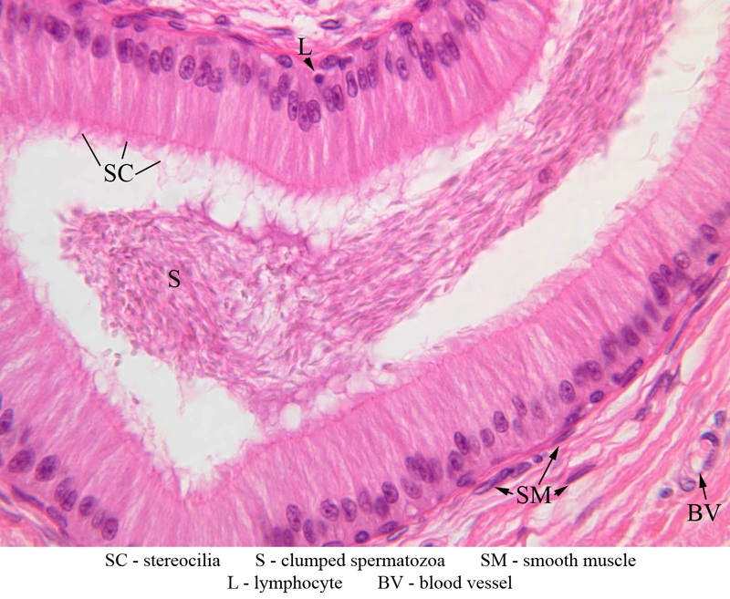
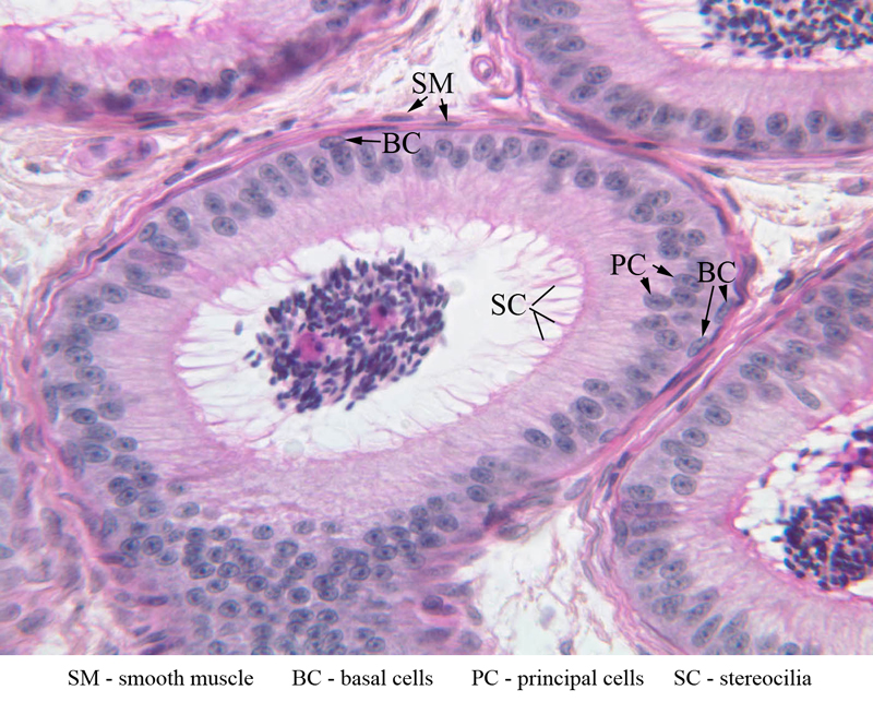
The epididymis is an organ that is composed of the efferent ductules and the epididymal duct. The epididymal duct consists of a single highly coiled tube that is closely applied to the posterior aspect of the testis. Locate areas of epididymal tissue at the periphery of the testis (slide B-80, H&E [2.5x, 10x, 20x, 40x-labeled] [10x, 20x, 40x]; B-81, PAS [2.5x, 10x, 20x, 40x-labeled] [2.5x, 10x, 20x, 40x-labeled] [2.5x, 10x, 20x, 40x]; B-82, H&E [2.5x, 10x, 20x, 40x]). A pseudostratified columnar epithelium that bears long tufts of microvilli, misleadingly called stereocilia, lines the entire length of the epididymal duct. The epithelium is exceptionally tall in the head of the epididymis (B-80 [2.5x, 10x, 20x, 40x] [10x, 20x, 40x] [2.5x, 10x, 20x, 40x]) and becomes progressively shorter (B-80 [10x, 20x, 40x]) towards the distal end of the duct (B-80 [2.5x] [2.5x, 10x, 20x, 40x]). As you study the epithelium, you should identify principal cells, with their prominent tufts of stereocilia, and basal cells. You should also be able to find occasional lymphocytes (sometimes called "halo" cells) that are migrating through the epithelium. By studying a number of different profiles of epididymal duct, you should find that both the principal cells and the stereocilia are only about half as tall in the tail of the epididymis as they are in its head. Study the muscular layers of the wall of the epididymis. The most proximal portion of the epididymis has a single layer of circularly arranged smooth muscle; in the intermediate segment an outer longitudinal layer of muscle is added; in the distal portion (tail) an inner longitudinal muscle layer is added. You should be able to find profiles of epididymal duct with muscular layers of conspicuously different thickness; however, it is unlikely that you will be able to find profiles with one, two and three muscle layers in the same section.Epididymis Image Gallery


Epididymis Table of Identifications
| Row | Structure | Abbreviation | Optimal Stain | Representative Section | Note |
|---|---|---|---|---|---|
| 1 | Stereocilia | SC | H&E | |
|
| 2 | Slumped Spermatozoa | S | H&E | |
|
| 3 | Smooth Muscle | SM | H&E | |
|
| 4 | Lymphocyte | L | H&E | |
|
| 5 | Blood Vessel | BV | H&E | |
|
| 6 | Basal Cells | BC | PAS | |
|
| 7 | Principal Cells | PC | PAS | |
|
| 8 | Microvilli (Brush Border) | M | PAS | |
|
| 9 | Pseudostratified Columnar Epithelium | PCE | PAS | |
Top of page
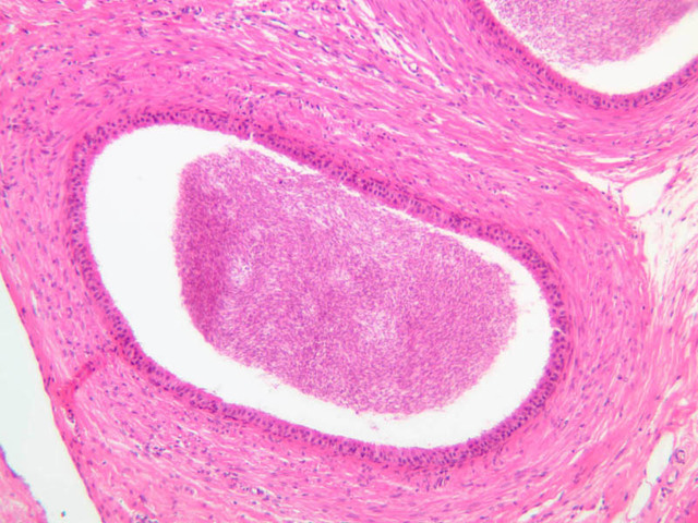
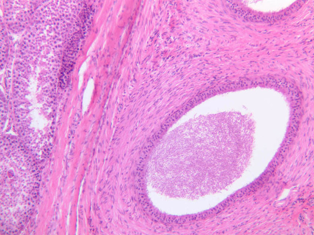
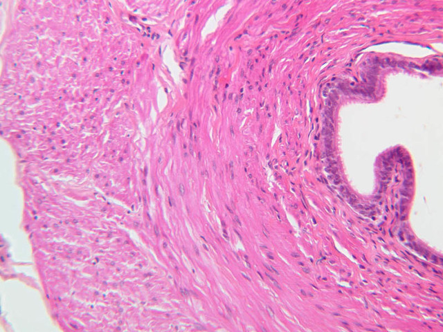
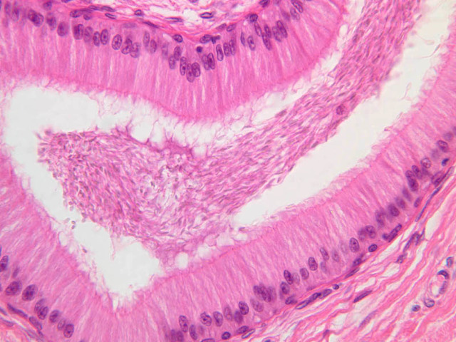
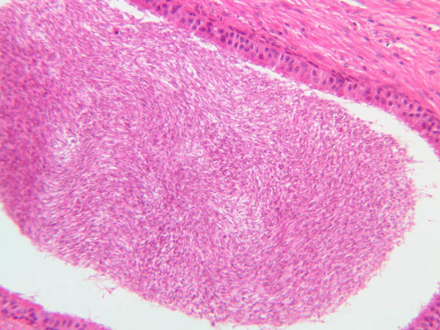
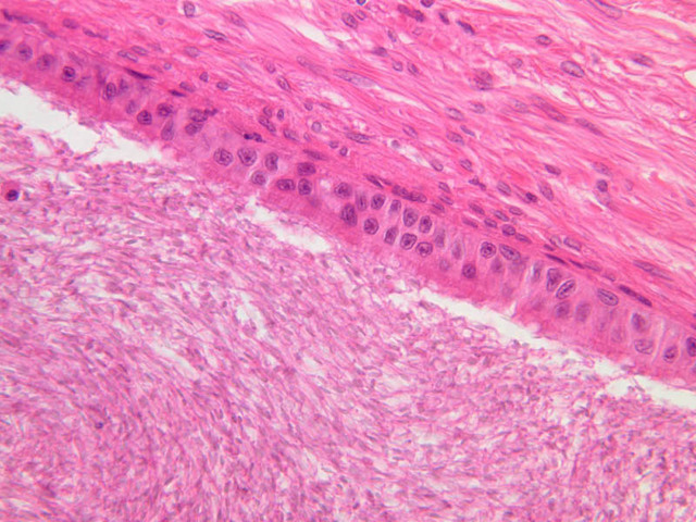
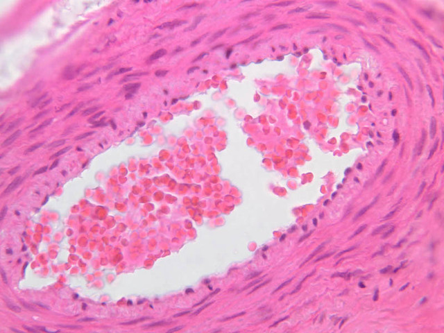
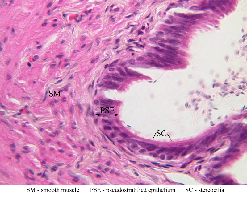
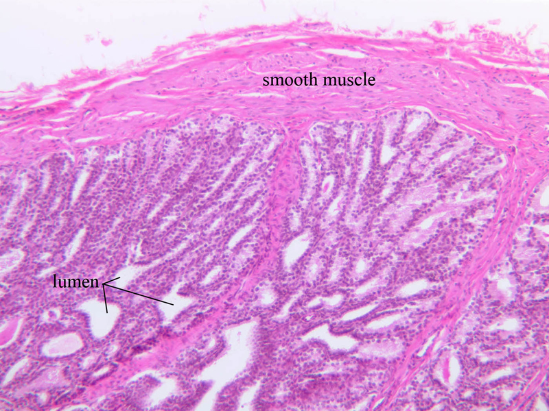
Vas Deferens (ductus deferens)
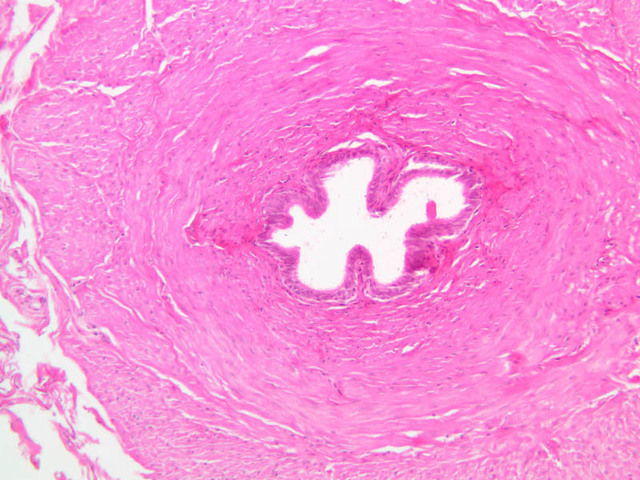
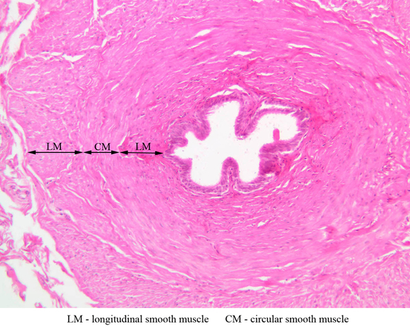
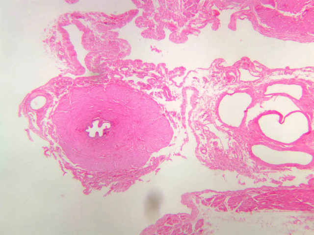
Locate the ductus deferens in section B-83 (spermatic cord, H&E [2.5x, 10x-labeled, 20x, 40x] [20x, 40x] [10x, 20x, 40x-labeled]). Avoid confusing it with the testicular artery. The duct is a thick, muscular tube lined by a pseudostratified epithelium of varying height, some of the cells of which may bear stereocilia. Examine the muscular coat. Can you distinguish muscle fiber groups in different orientations? Why is such a prominent muscular coat necessary? From the ductus deferens, sperm pass into the prostatic and penile urethrae. At ejaculation, viscid secretions from the prostate and seminal vesicles are added to the sperm to form semen.Vas Deferens Image Gallery


Vas Deferens Table of Identifications
| Row | Structure | Abbreviation | Optimal Stain | Representative Section | Note |
|---|---|---|---|---|---|
| 1 | Longitudinal Muscle | LM | H&E | |
|
| 2 | Circular Muscle | CM | H&E | |
|
| 3 | Smooth Muscle | SM | H&E | |
|
| 4 | Pseudostratified Epithelium | PSE | H&E | |
|
| 5 | Stereocilia | SC | H&E | |
Top of page
Accessory Glands
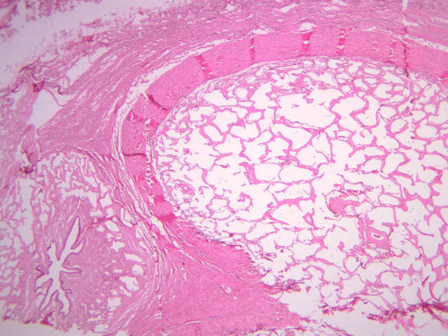
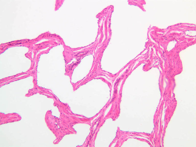
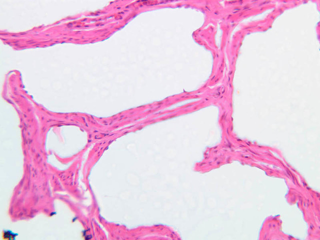
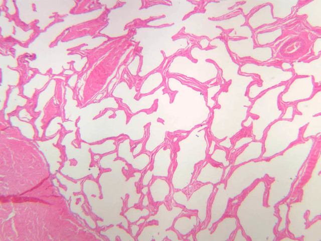
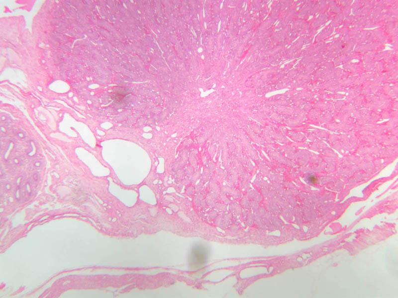
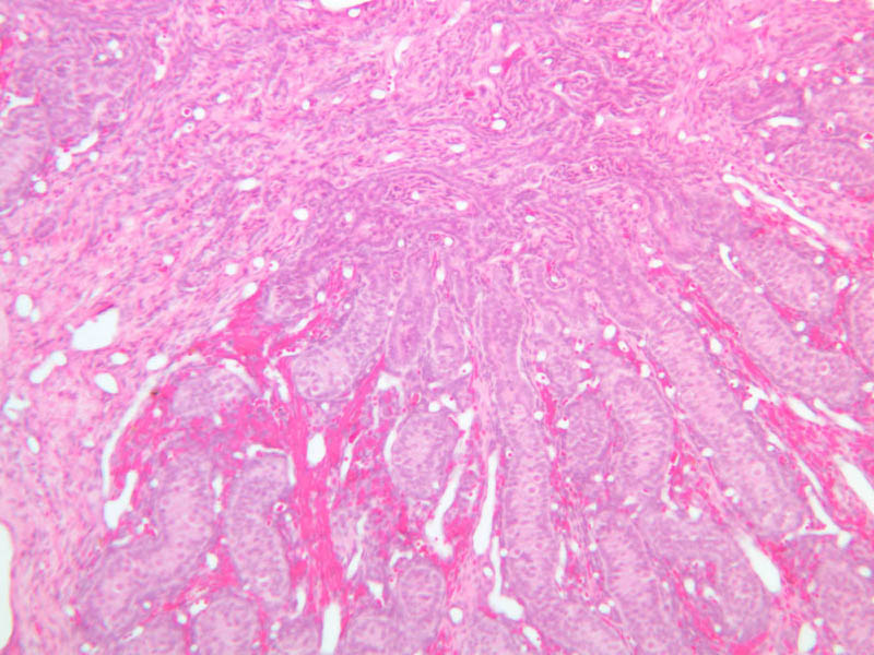
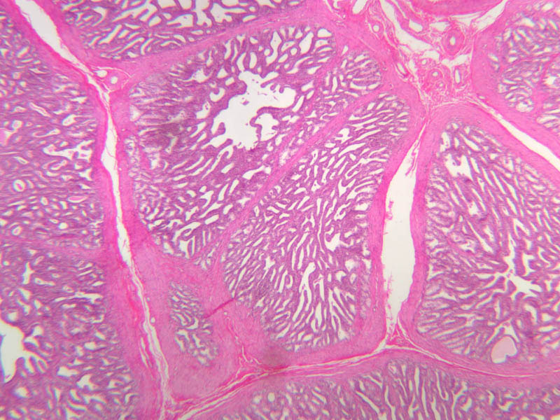
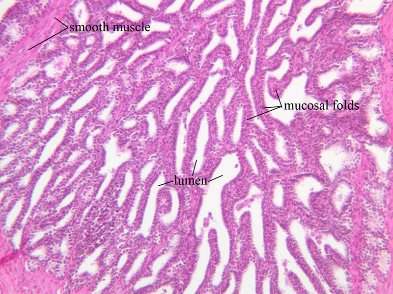
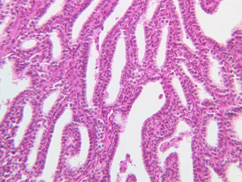
Seminal Vesicle
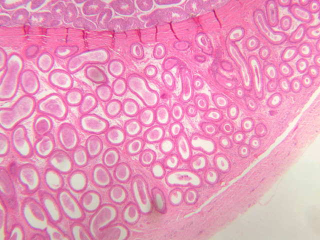
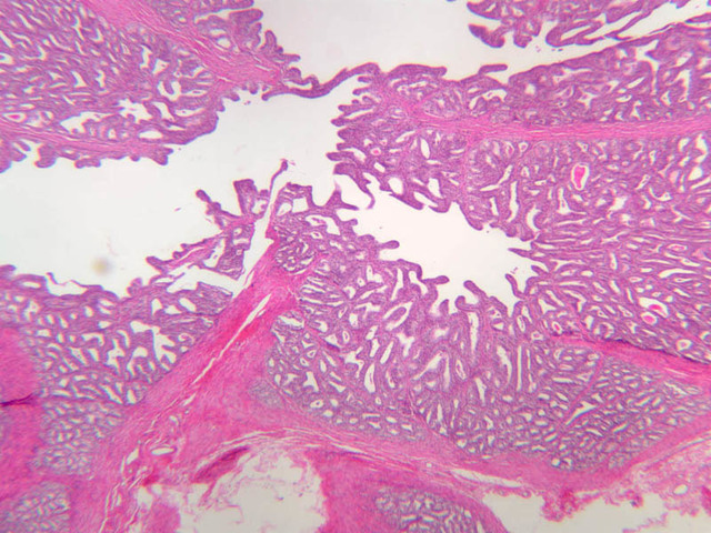
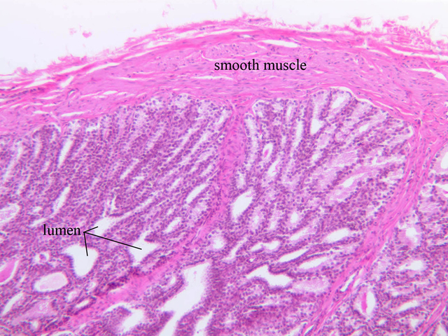
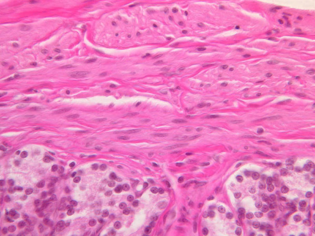
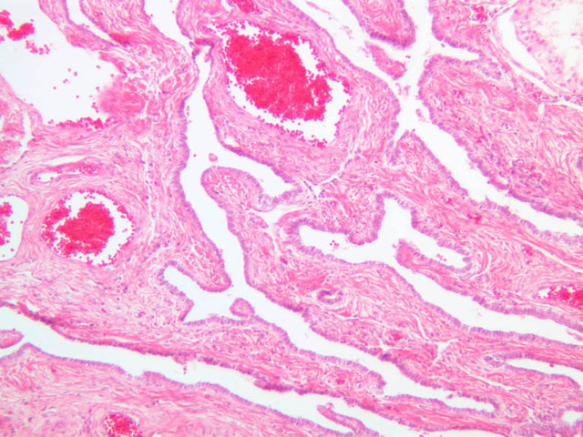
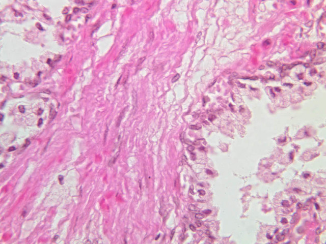
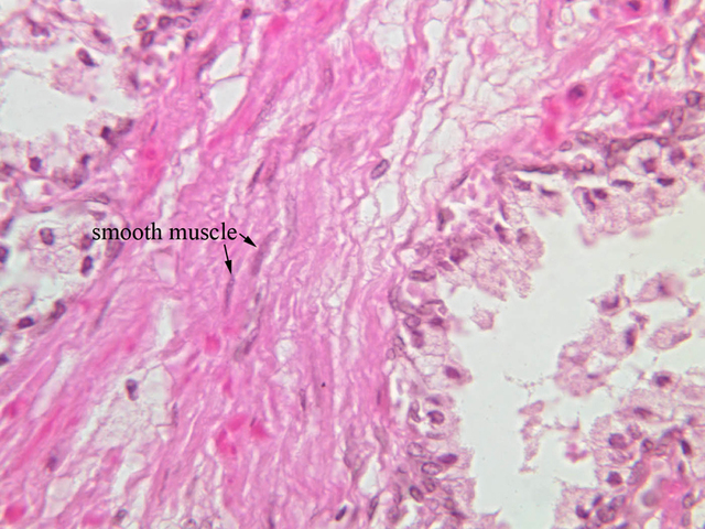
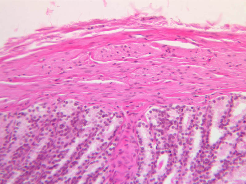
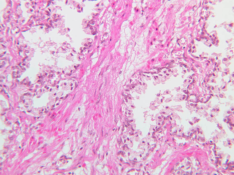
The seminal vesicles originate as diverticula of the ductuli deferentia. The duct of each seminal vesicle joins the associated ductus deferens a short distance before the latter empties into the prostatic urethra. With the scanning objective, note that the seminal vesicle appears to consist of several thick walled tubules lined with a secretory epithelium. Although the luminal lining is thrown into many folds and often appears cavitated, the cavities are not tubular glands, but freely connect with the central lumen. The epithelium, which rests on a vascular lamina propria, varies from low columnar to pseudostratified. The outer wall of the seminal vesicle is made up largely of an inner circular and an outer longitudinal layer of smooth muscle (slide B-89, H&E [2.5x, 10x-labeled, 20x, 40x] [2.5x, 10x, 20x, 40x-labeled] [2.5x, 10x-labeled, 20x, 40x]).Seminal Vesicle Image Gallery


Seminal Vesicle Table of Identifications
| Row | Structure | Abbreviation | Optimal Stain | Representative Section | Note |
|---|---|---|---|---|---|
| 1 | Smooth Muscle | (none) | H&E | |
|
| 2 | Lumen | (none) | H&E | |
|
| 3 | Mucosal Folds | (none) | H&E | |
|
| 4 | Epithelium (Pseudostratified) | Ep | H&E | |
|
| 5 | Basal Cells | BC | H&E | |
|
| 6 | Lamina Propria | LP | H&E | |
Top of page
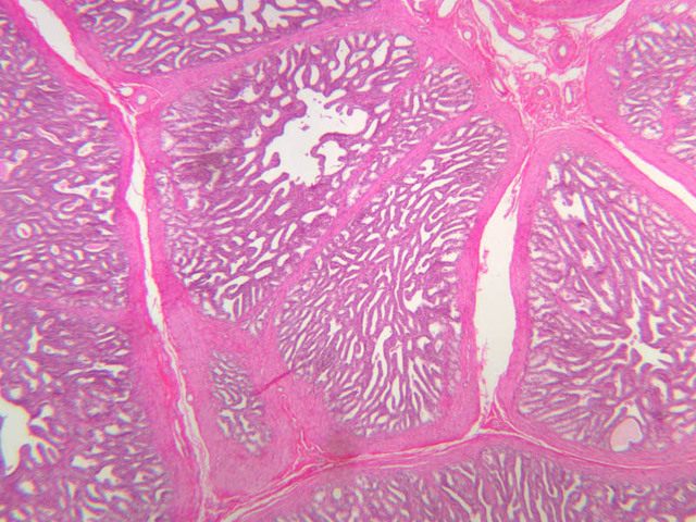
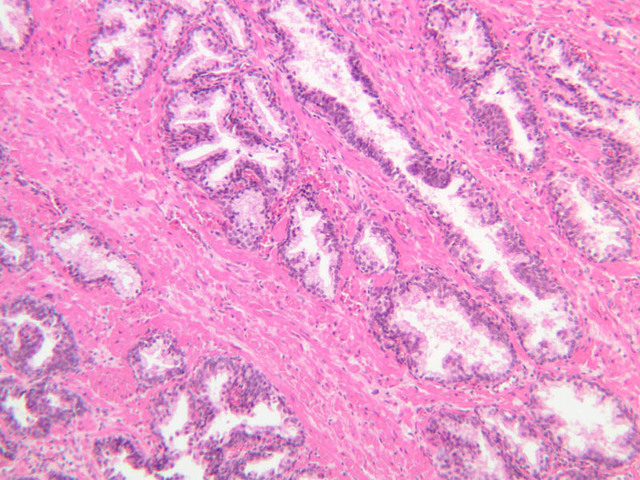
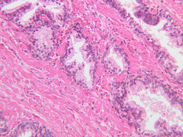
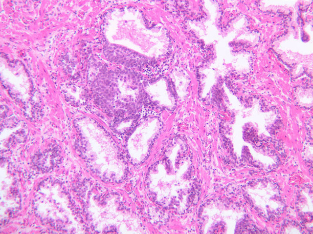
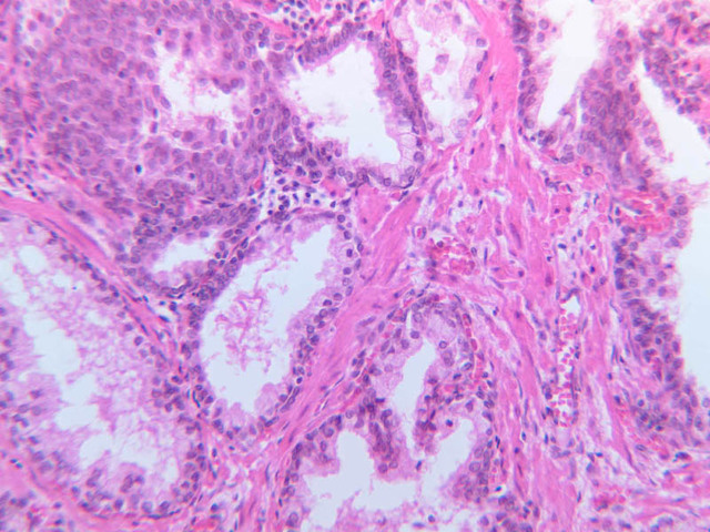
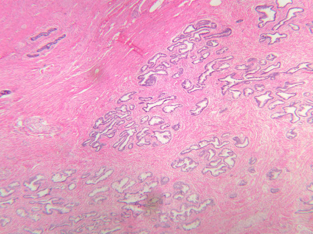
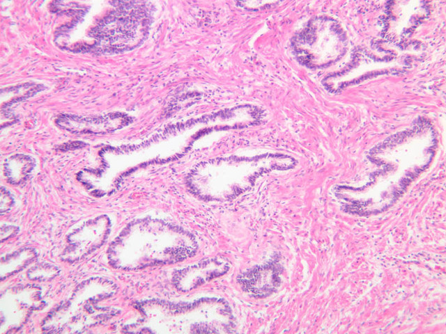
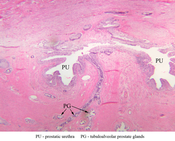
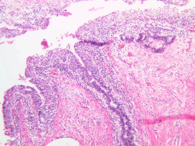
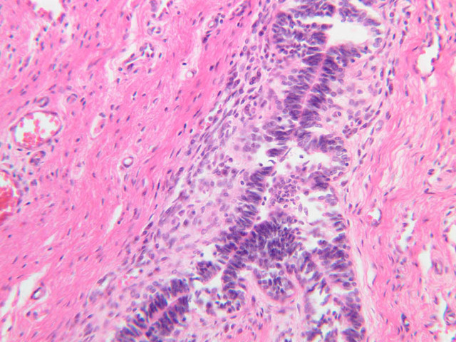
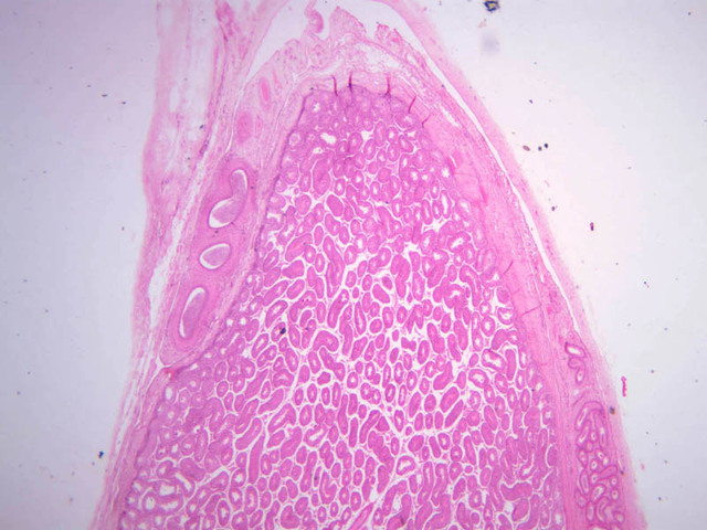
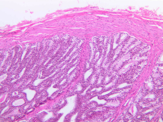
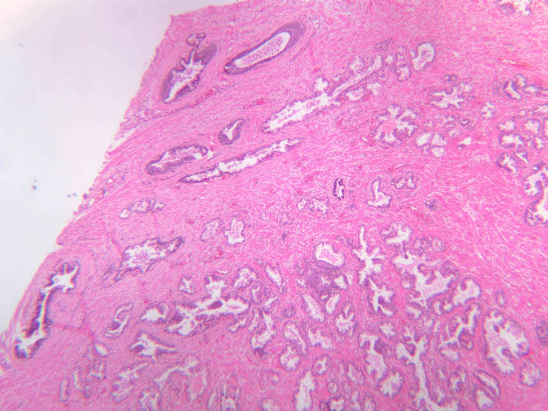
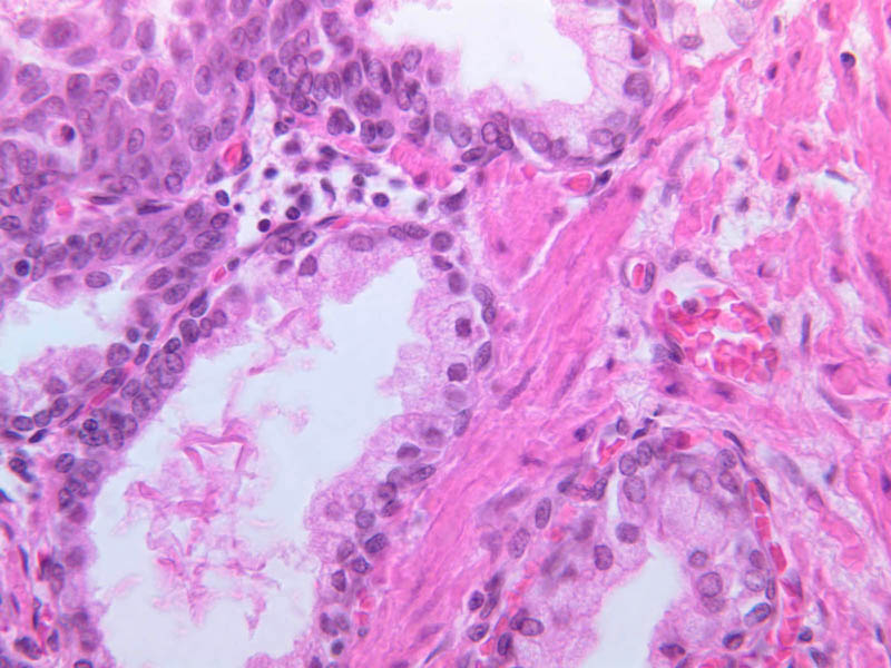
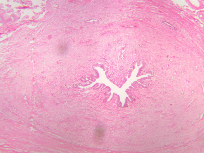
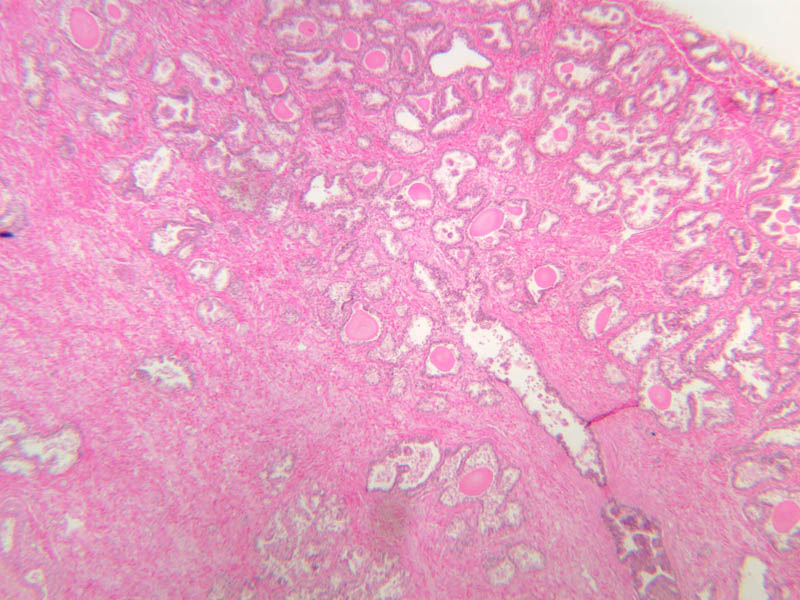
Prostate Gland
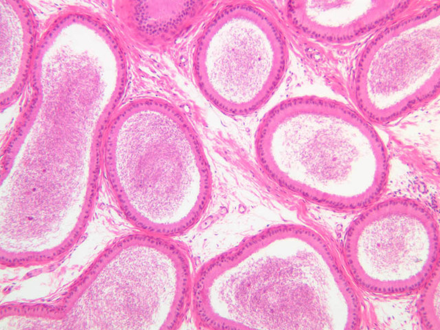
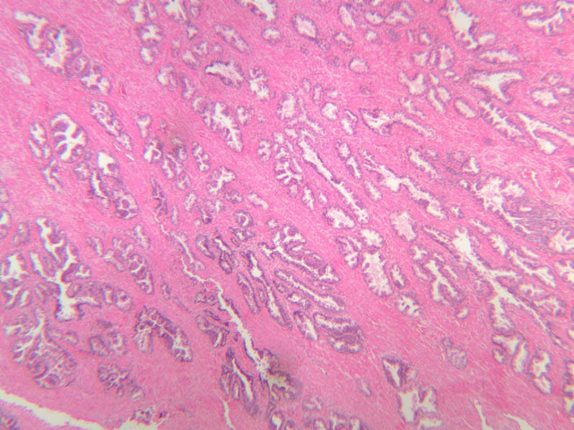
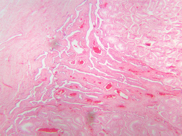
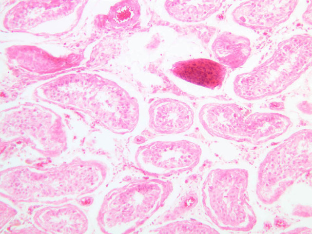
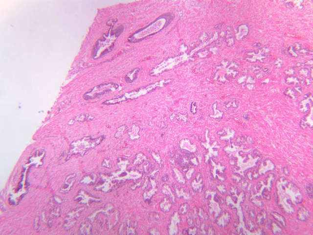
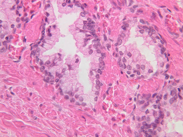
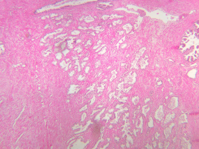
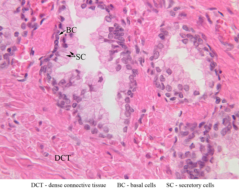
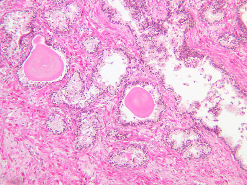
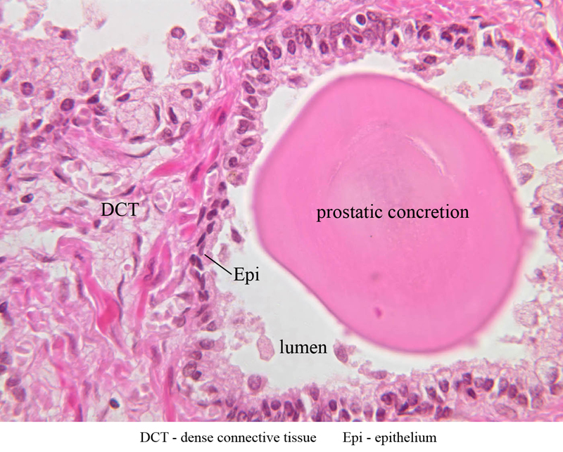
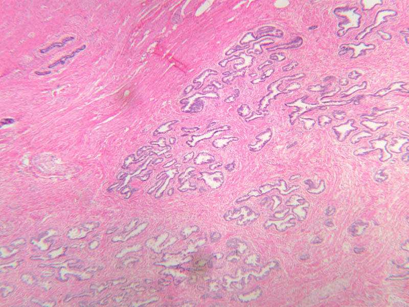
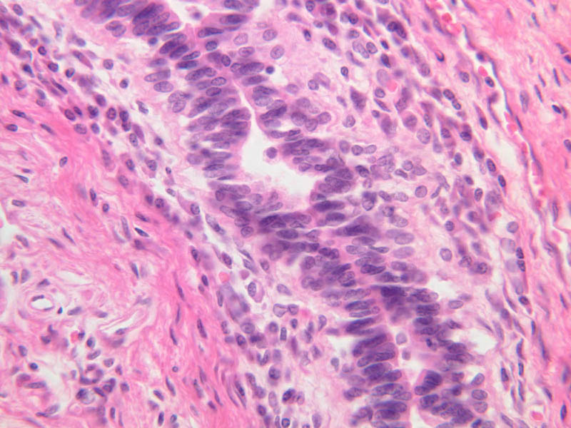
The prostate is an aggregate of numerous tubulo-alveolar glands embedded in a dense stroma of collagen fibers and smooth muscle (B-90 [2.5x, 10x, 20x, 40x-labeled]; B-91 [2.5x, 10x, [[http://www.medicalhistology.us/wiki/bin/viewfile/Main/ProstateImages?rev=1;filename=b91_prostate_adult_20x_he.jpg[20x]], 40x-labeled] [2.5x, 10x, 10x, 20x, 40x]). Note that the prostatic urethra, the portion of the urethra which runs through the prostate gland, is also present (B-91 [2.5x, 10x, 20x, 40x]). Under high dry magnification, it is obvious that the glandular epithelium varies from simple cuboidal to pseudostratified columnar. Prostatic acini often contain eosinophilic corpora amalacea (prostatic concretions) (B-90 [2.5x, 10x, 20x, 40x-labeled]). The number and degree of calcification of these spherical, lamellated structures increase with age. In some sections, the prostatic urethra can be identified. Functional, as well as morphological, maintenance of the adult prostate and seminal vesicles depends on adequate levels of circulating testosterone. Examine the prepubertal prostate (B-92 [2.5x, 10x, 20x, 40x] [2.5x-labeled, 10x, 20x, 40x]). How does this specimen differ from that on slide B-90?Prostate Image Gallery


Prostate Vesicle Table of Identifications
| Row | Structure | Abbreviation | Optimal Stain | Representative Section | Note |
|---|---|---|---|---|---|
| 1 | Smooth Muscle | (none) | H&E | |
|
| 2 | Dense Connective Tissue | DCT | H&E | |
|
| 3 | Basal Cells | BC | H&E | |
|
| 4 | Secretory Cells | SC | H&E | |
|
| 5 | Epithelium | Epi | H&E | |
|
| 6 | Lumen | (none) | H&E | |
|
| 7 | Prostatic Concretion | (none) | H&E | |
|
| 8 | Prostatic Urethra | PU | H&E | |
|
| 9 | Tubuloalveolar Prostate Glands | PG | H&E | |
Top of page
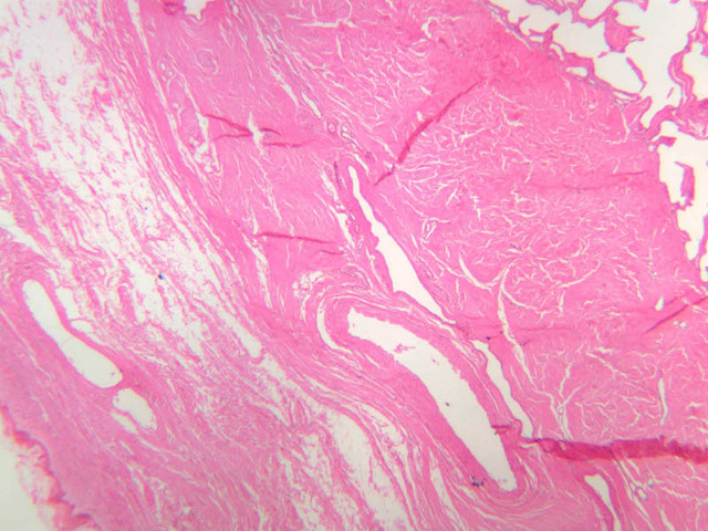
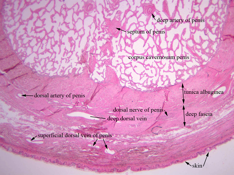
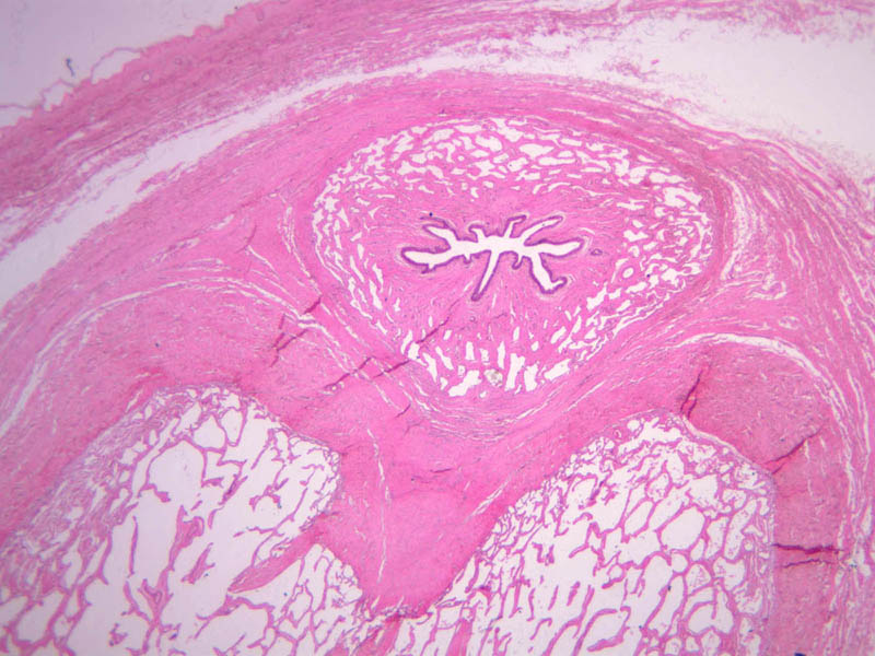
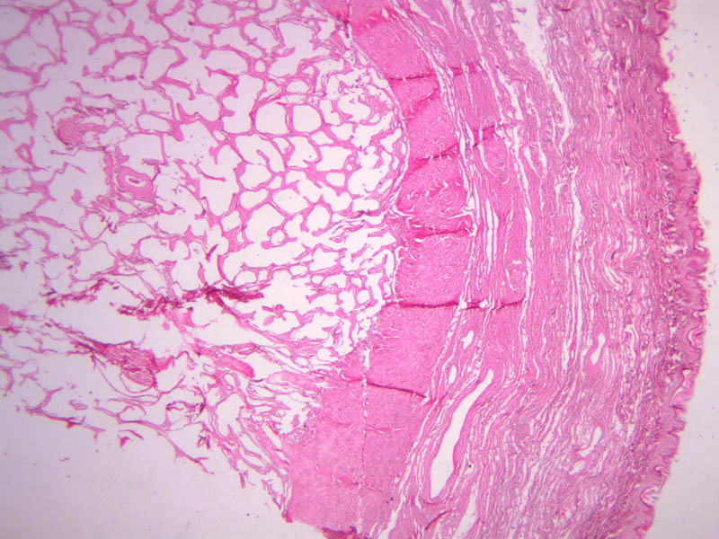
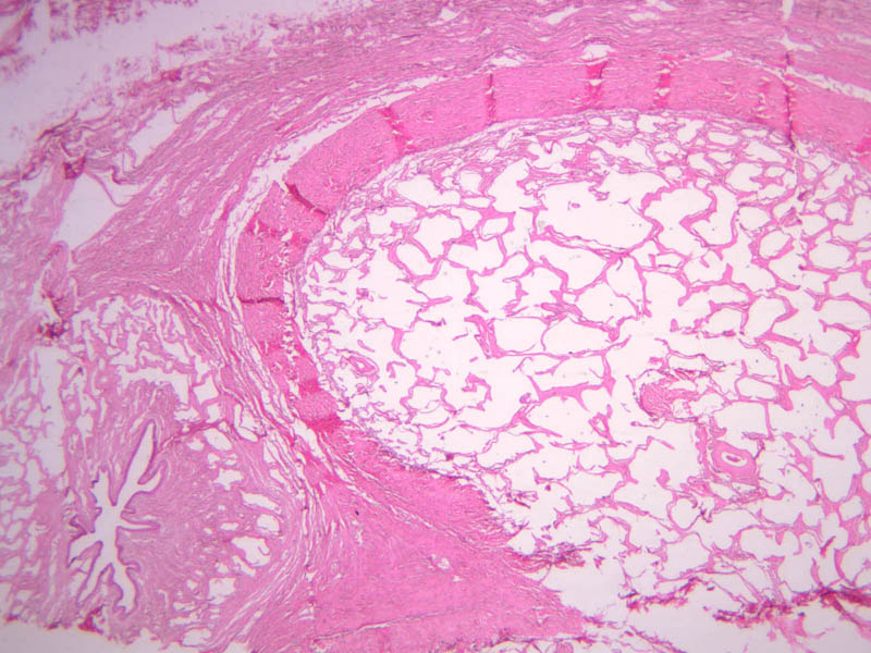
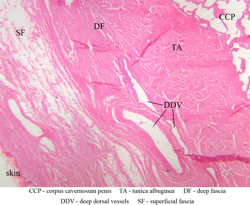
Penis
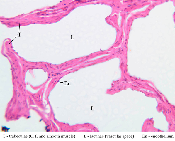
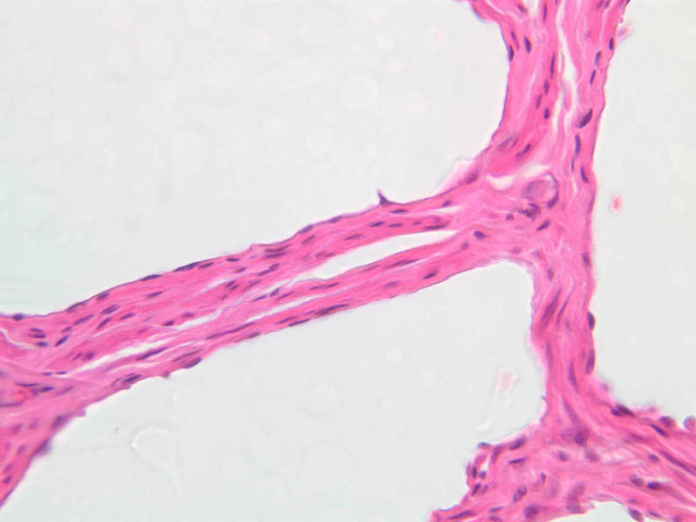
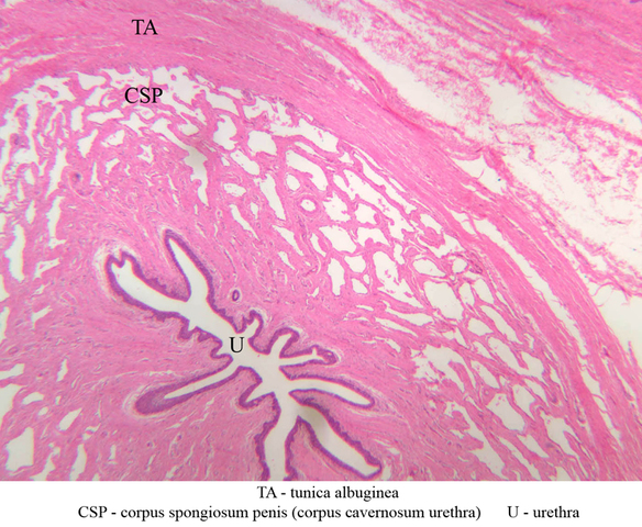
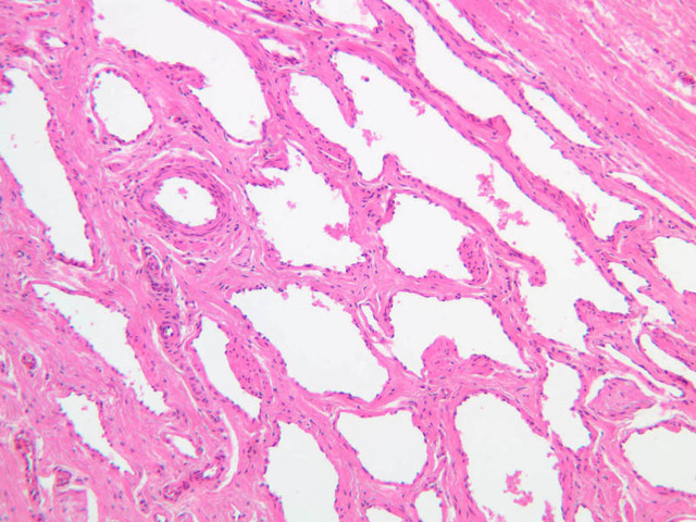
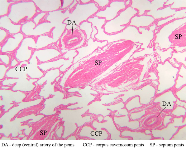
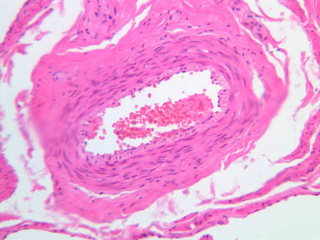
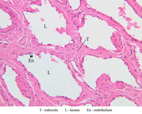
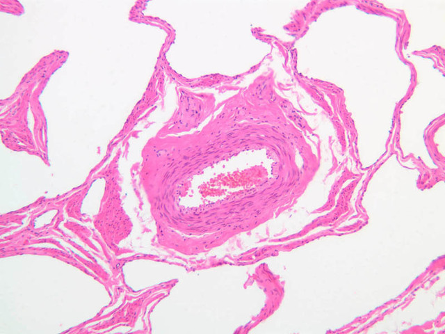
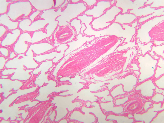
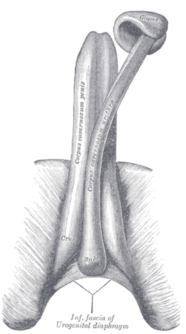
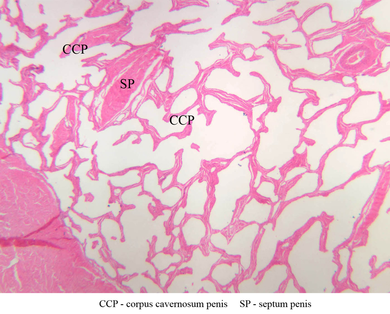
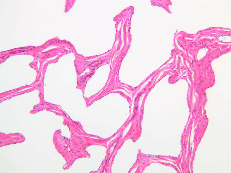
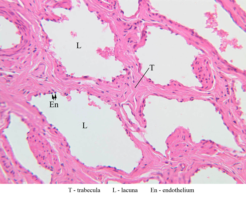
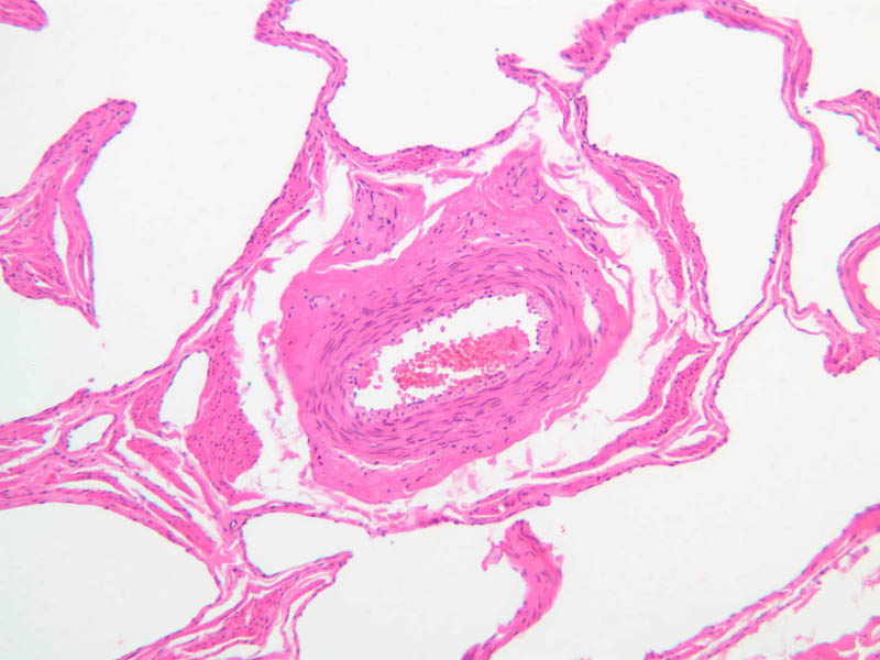
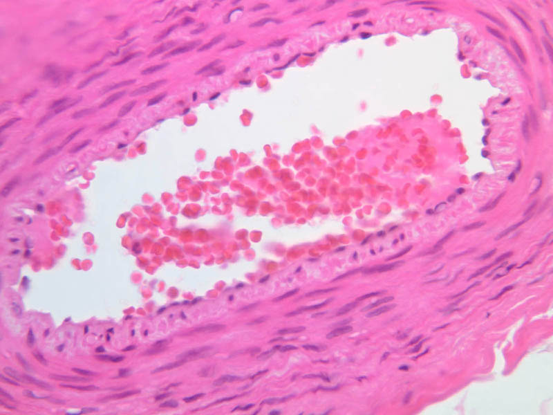
Examine the section on slide B-85 with your naked eye. Locate the paired, dorsally situated, corpora cavernosa penis ([1x-labeled, 1x] [1x, 1x, 1x] [2.5x-labeled, 10x, 20x-labeled, 40x] [2.5x]) and ventrally situated corpus spongiosum (aka corpus cavernosum urethrae) ([2.5x-labeled, 10x, 20x-labeled, 40x]), which surrounds the penile (spongy) urethra ([2.5x, 10x, 20x, 40x]). With the low power objective, note the dense collagenous connective tissue (tunica albuginea penis) that surrounds and separates the corpora cavernosa ([2.5x-labeled]). The corpus spongiosum is also surrounded by a connective tissue sheath, but its tunic is very much thinner and contains a greater proportion of elastic fibers than that of the corpora cavernosa. What is the consequence of the thinner tunica albuginea of the corpus spongiosum? The median septum between the corpora cavernosa is incomplete distally so that there is communication between the vascular spaces of these paired structures. What is the practical consequence of this feature? The erectile tissue of the penis consists of trabeculae of connective tissue and smooth muscle, which are covered with endothelium and divide the cavernous bodies into a sponge-like systems of irregular lacunae (vascular spaces). Overall, the trabeculae are thinner and the lacunae larger in the corpus cavernosum penis than in the corpus spongiosum. Look near the center of each corpus cavernosum penis for the deep (or central) artery of the penis ([2.5x-labeled, 10x, 20x, 40x] [40x]). Examine the trabeculae near the deep artery of the penis in search of small, highly coiled branches of the deep artery, known as helicine arteries. The helicine arteries open directly into the cavernous spaces and are notable for a thick tunica intima of longitudinally oriented smooth muscle fibers which serve to keep the arteries closed when the penis is flaccid. Thin-walled veins drain the lacunae through the connective sheath.Penis Image Gallery


Penis Table of Identifications
| Row | Structure | Abbreviation | Optimal Stain | Representative Section | Note |
|---|---|---|---|---|---|
| 1 | Deep Artery of the Penis | (none) | H&E | |
|
| 2 | Septum of the Penis | (none) | H&E | |
|
| 3 | Corpus Cavernosum Penis | (none) | H&E | |
|
| 4 | Dorsal Artery of the Penis | (none) | H&E | |
|
| 5 | Dorsal Nerve of the Penis | (none) | H&E | |
|
| 6 | Deep Dorsal Vein | (none) | H&E | |
|
| 7 | Superficial Dorsal Vein | (none) | H&E | |
|
| 8 | Tunica Albuginea | (none) | H&E | |
|
| 9 | Deep Fascia | (none) | H&E | |
|
| 10 | Skin | (none) | H&E | |
|
| 11 | Trabeculae (Connective Tissue and Smooth Muscle) | T | H&E | |
|
| 12 | Lacunae (Vascular Space) | L | H&E | |
|
| 13 | Endothelium | En | H&E | |
|
| 14 | Corpus Spongiosum | CSP | H&E | |
|
| 15 | Urethra | U | H&E | |
Top of page
Chapter Fifteen Review
Review of Slides


Review of Identifications
| Row | Structure | Abbreviation | Optimal Stain | Representative Section | Note |
|---|---|---|---|---|---|
| 1 | Cauda (tail of) Epididymis | (none) | H&E | |
|
| 2 | Corpus (body of) Epididymis | (none) | H&E | |
|
| 3 | Tunica Vaginalis | (none) | H&E, PAS | |
|
| 4 | Tunica Albuginea | (none) | H&E, PAS | |
|
| 5 | Seminiferous Tubules | (none) | H&E, PAS | |
|
| 6 | Epididymis | (none) | H&E, PAS | |
|
| 7 | Septa | S | H&E | |
|
| 8 | Blood Vessel | BV | H&E | |
|
| 9 | Leydig (Interstitial) Cells | LC | H&E | |
|
| 10 | Connective Tissue Septa | CT | H&E | |
|
| 11 | Seminiferous Epithelium | SE | H&E | |
|
| 12 | Lamina Propria | LP | H&E | |
|
| 13 | Myoid Cell | MC | H&E | |
|
| 14 | Fibroblast | F | H&E | |
|
| 15 | Endothelial Cell | En | H&E | |
|
| 16 | Spermatocytes | Sc | H&E | |
|
| 17 | Spermatids | Sp | H&E | |
|
| 18 | Sertoli Nuclei | Sn | H&E | |
|
| 19 | Spermatogonia | Sg | H&E | |
|
| 20 | Straight Tubules | ST | H&E | |
|
| 21 | Simple Cuboidal Epithelium (of Rete Testis) | SCE | H&E | |
|
| 22 | Rete Testis | RT | H&E | |
|
| 23 | Vein | V | H&E | |
|
| 24 | Efferent Ductules | ED | PAS | |
|
| 25 | Adipose Tissue | AT | PAS | |
|
| 26 | Smooth Muscle | SM, (none) | PAS, H&E | |
|
| 27 | Connective Tissue | CT | PAS | |
|
| 28 | Blood Vessels | BV | PAS | |
|
| 29 | Stereocilia | SC | H&E | |
|
| 30 | Slumped Spermatozoa | S | H&E | |
|
| 31 | Lymphocyte | L | H&E | |
|
| 32 | Basal Cells | BC | PAS | |
|
| 33 | Principal Cells | PC | PAS | |
|
| 34 | Microvilli (Brush Border) | M | PAS | |
|
| 35 | Pseudostratified Columnar Epithelium | PCE | PAS | |
|
| 36 | Longitudinal Muscle | LM | H&E | |
|
| 37 | Circular Muscle | CM | H&E | |
|
| 38 | Pseudostratified Epithelium | PSE, Ep | H&E | |
|
| 39 | Lumen | (none) | H&E | |
|
| 40 | Mucosal Folds | (none) | H&E | |
|
| 41 | Dense Connective Tissue | DCT | H&E | |
|
| 42 | Secretory Cells | SC | H&E | |
|
| 43 | Epithelium | Epi | H&E | |
|
| 44 | Prostatic Concretion | (none) | H&E | |
|
| 45 | Prostatic Urethra | PU | H&E | |
|
| 46 | Tubuloalveolar Prostate Glands | PG | H&E | |
|
| 47 | Deep Artery of the Penis | (none) | H&E | |
|
| 48 | Septum of the Penis | (none) | H&E | |
|
| 49 | Corpus Cavernosum Penis | (none) | H&E | |
|
| 50 | Dorsal Artery of the Penis | (none) | H&E | |
|
| 51 | Dorsal Nerve of the Penis | (none) | H&E | |
|
| 52 | Deep Dorsal Vein | (none) | H&E | |
|
| 53 | Superficial Dorsal Vein | (none) | H&E | |
|
| 54 | Tunica Albuginea (of Penis) | (none) | H&E | |
|
| 55 | Deep Fascia | (none) | H&E | |
|
| 56 | Skin | (none) | H&E | |
|
| 57 | Trabeculae (Connective Tissue and Smooth Muscle) | T | H&E | |
|
| 58 | Lacunae (Vascular Space) | L | H&E | |
|
| 59 | Endothelium | En | H&E | |
|
| 60 | Corpus Spongiosum | CSP | H&E | |
|
| 61 | Urethra | U | H&E | |
Top of page
Comments
- My bad. Nevertheless, I am a little confused about the testis table compared to the review table. Why are the EDs omitted from the testis table? Are they not part of the testis? I realized that clicking on the "structure" column was handy. I imagine that row sorting reflects order of discussion. I expected, incorrectly, alphabetization of IDs and then did not notice they were not sorted. Thus, I immediately went to the "E's" and did not find ED. Plus, ED is not in the testis table proper. Perhaps a discreet note could remind the user that the columns are sortable. -- LorenEvey - 09 Jul 2007
- See, now you are just toying with me. Look here : EDs -- AshleyLPistorio - 09 Jul 2007
- Hey, while you are at it, let's label an efferent ductule. -- LorenEvey - 09 Jul 2007
- Oh, they are not equal as far as I know. The helicine aa are like curly little offshoots of the deep aa that feed the spaces of the trabeculae. I'll probably deal with this during my editing process. You said the text equated them? Which text - here or HH? You know, Dr. H is not infallible. -- AshleyLPistorio - 07 Jul 2007
- The text equates them with trabeculae. I am just getting back on board with your efforts. This is looking great. What caught my attention was the relationships between Bucks f., Superf. Dorsal vv., Deep Dorsal v., Dorsal n., and Dorsal a. I can foresee that this atlas might, on occasion, get referenced from the gross course. Perhaps from the message boards. I was merely thinking ahead. Particular affinity? -- LorenEvey - 07 Jul 2007
- Don't know that I see one in the 1x slides. Did you find one? You have a particular affinity for the helicine aa.?
 -- AshleyLPistorio - 07 Jul 2007
- Hey, can we (you ) include a label for the helicine aa? -- LorenEvey - 07 Jul 2007
Edit | Attach | Print version | History: r2 < r1 | Backlinks | View wiki text | More topic actions
Topic revision: r2 - 20 Jun 2015, LorenEvey

- Epithelium
- Connective Tissue
- Muscle
- Nervous Tissue
- Cardiovascular System
- Skin Appendages and Sensory Receptors
- Lymphatic System
- Cartilage and Bone
- Respiratory System
- Peripheral Blood and Bone Marrow
- Oral Cavity and Salivary Glands
- Esophagus and Gastrointestinal Tract
- Pancreas, Liver, and Gall Bladder
- Endocrine Organs
- Male Reproductive System
- Female Reproductive System
- Urinary System
Ideas, requests, problems regarding Medical Histology? Send feedback