 |
|
You are here: Medical Histology>Main Web>AtlasContents>EsophagusAndGastrointestinalAtlas12 (20 Jun 2015, LorenEvey)Edit Attach
Chapter Twelve: Esophagus and Gastrointestinal Tract


Introduction
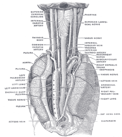
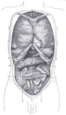
Prior to learning the microarchitecture of Esophagus and Gastrointestinal Tract, use the table below to review some of the gross anatomy of these tissues:| Structure | Image |
|---|---|
| The Esophagus | |
| The Stomach | |
| Gross Anatomical Location of Much of the GI Tract in the Thoracic Abdomen | |
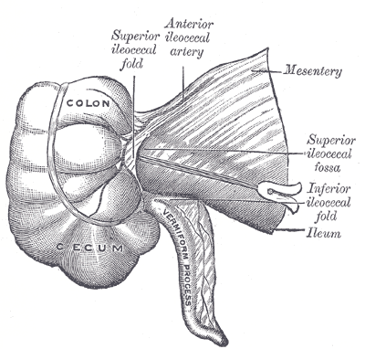
| The Vermiform Appendix | |
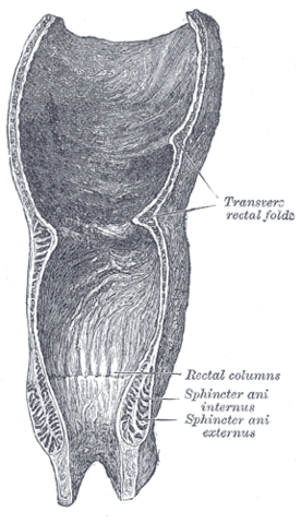
| The Rectum and Anal Canal | |
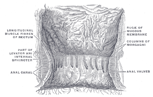
| Close-up View of the Anal Canal | |
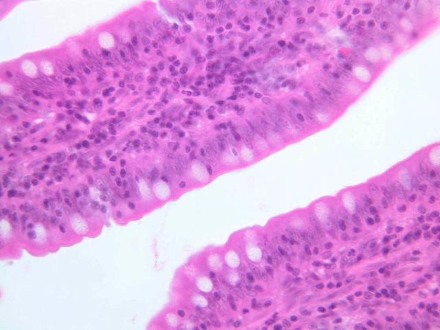
Esophagus
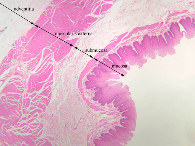
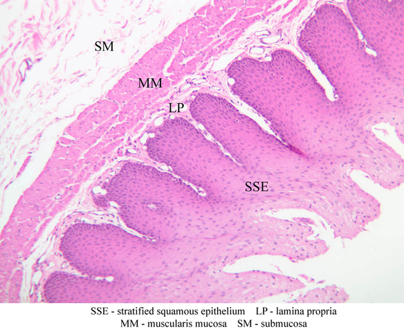
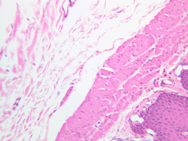
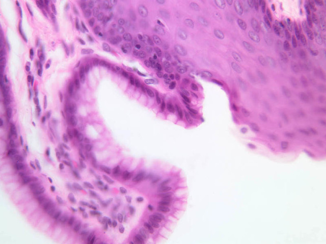
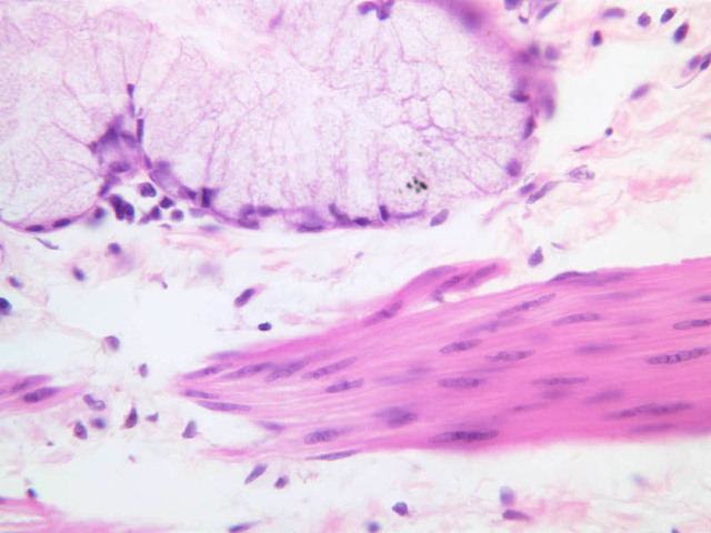
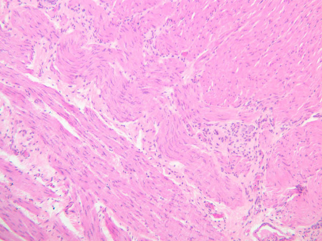
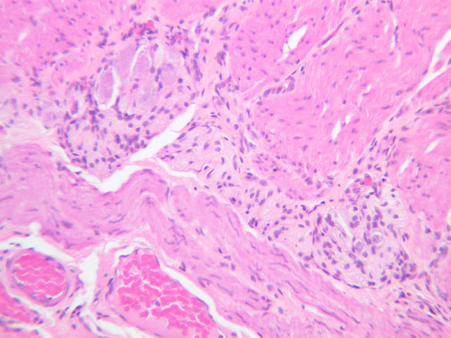
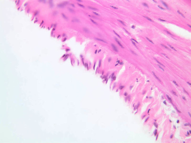
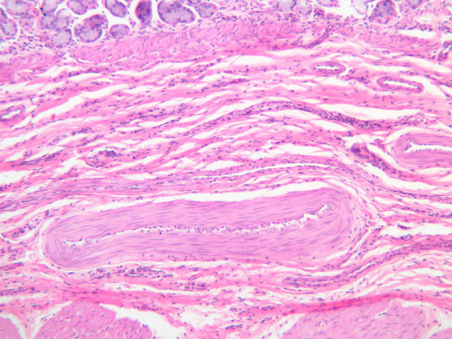
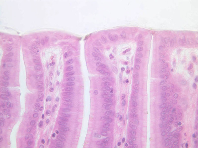
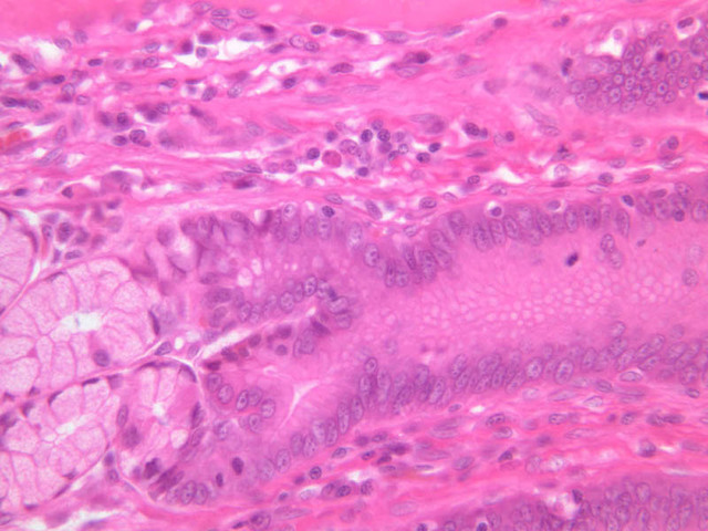
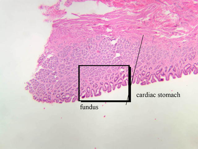
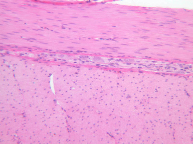
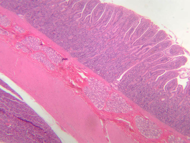
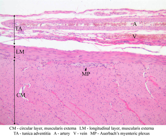
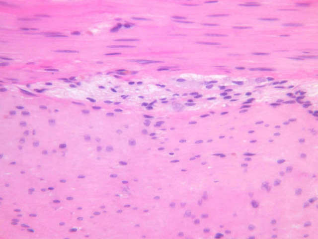
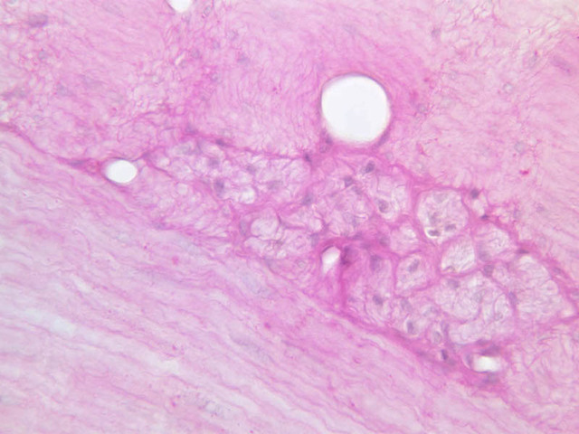
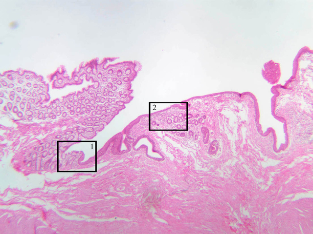
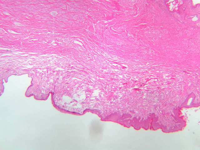
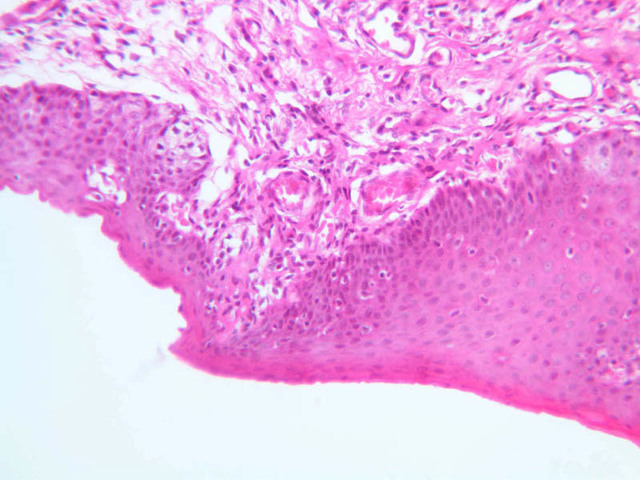
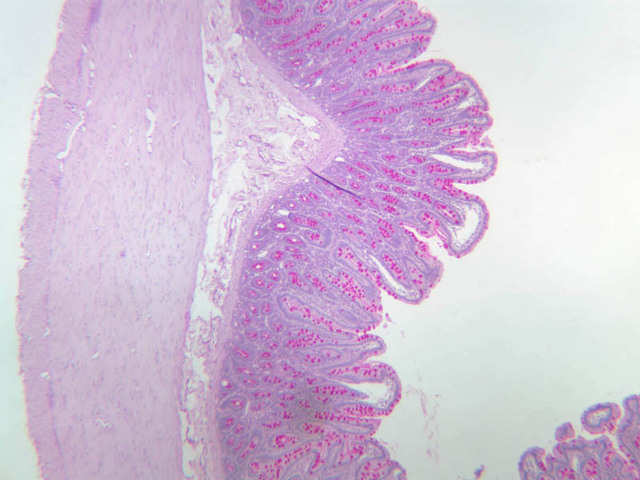
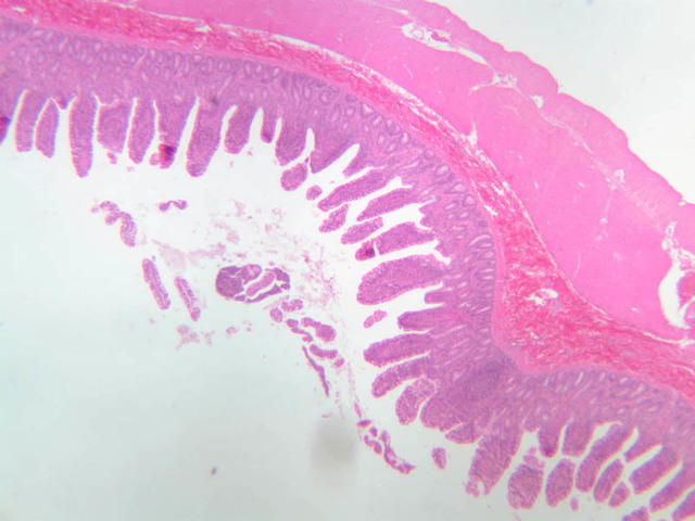
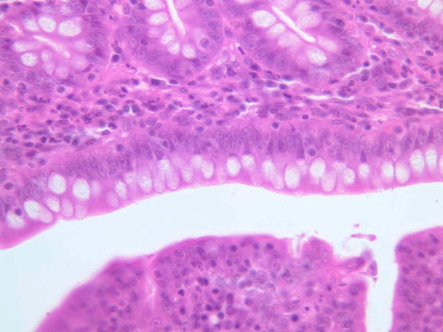
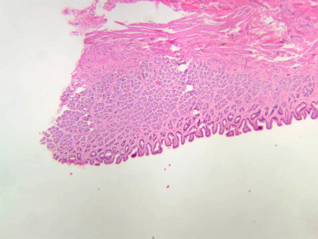
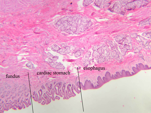
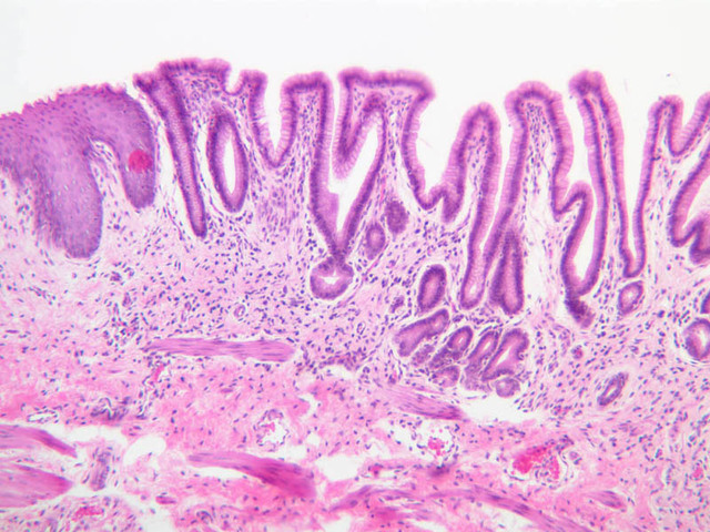
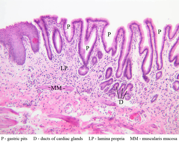
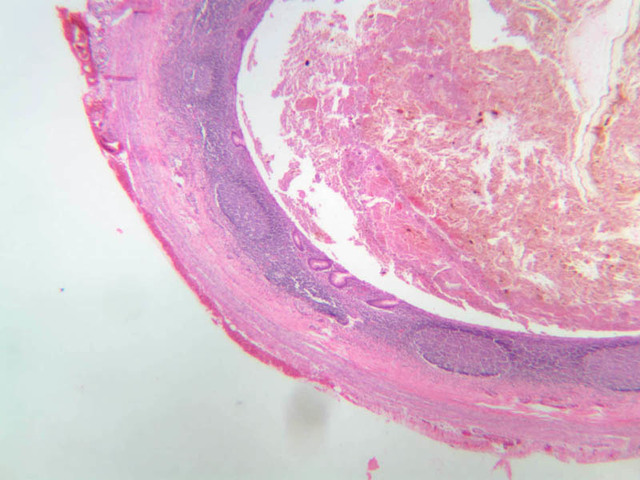
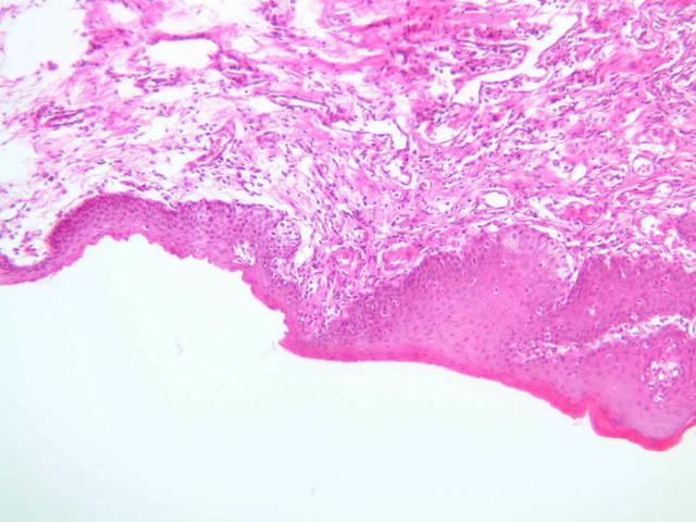
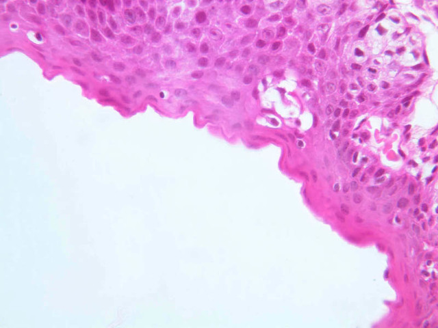
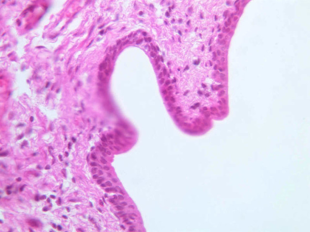
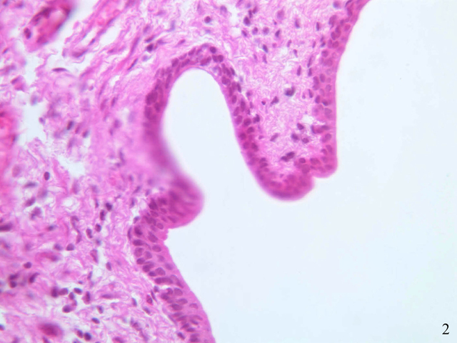
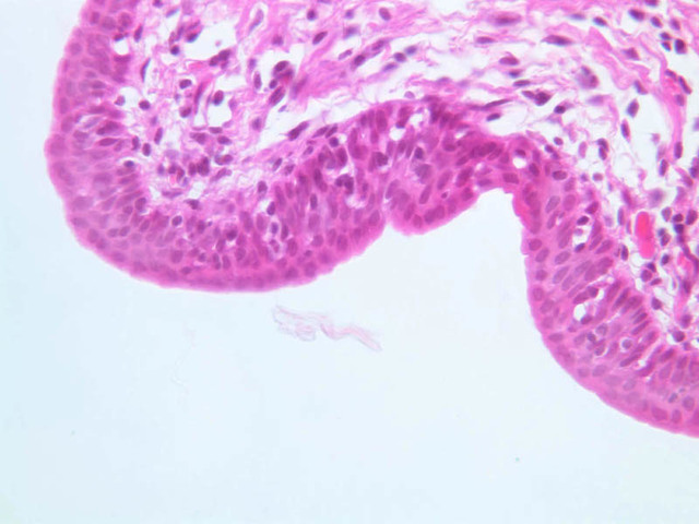
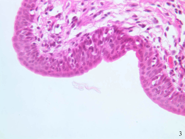
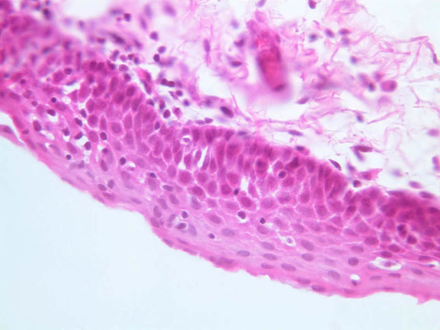
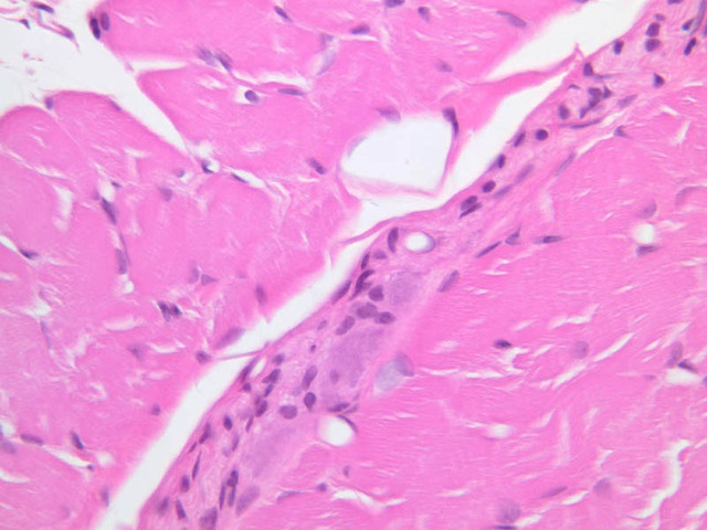
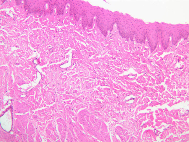
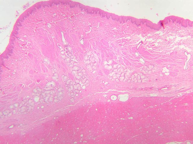
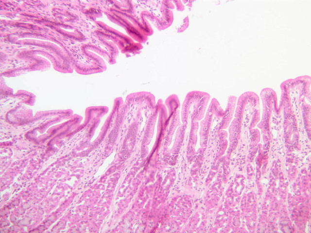
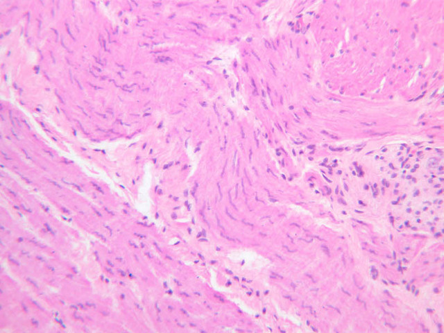
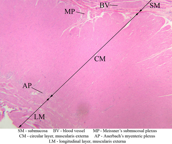
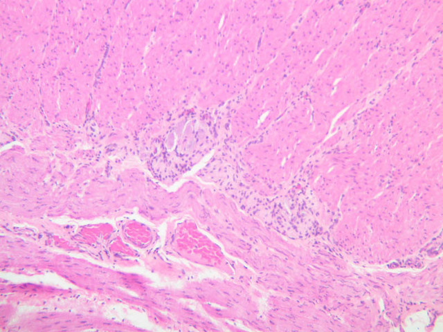
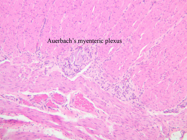
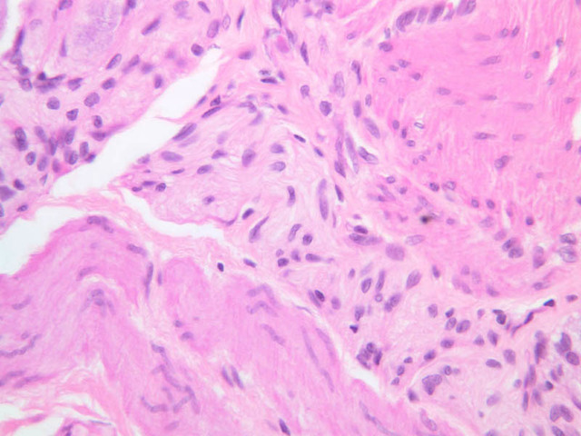
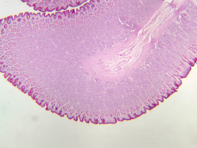
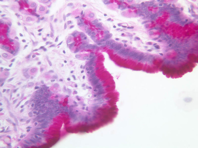
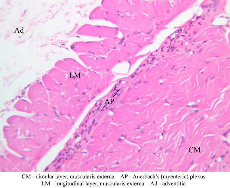
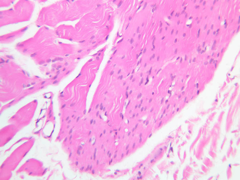
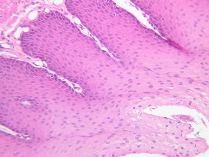
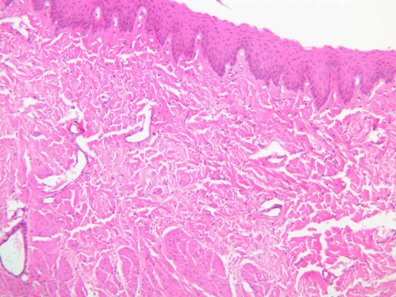
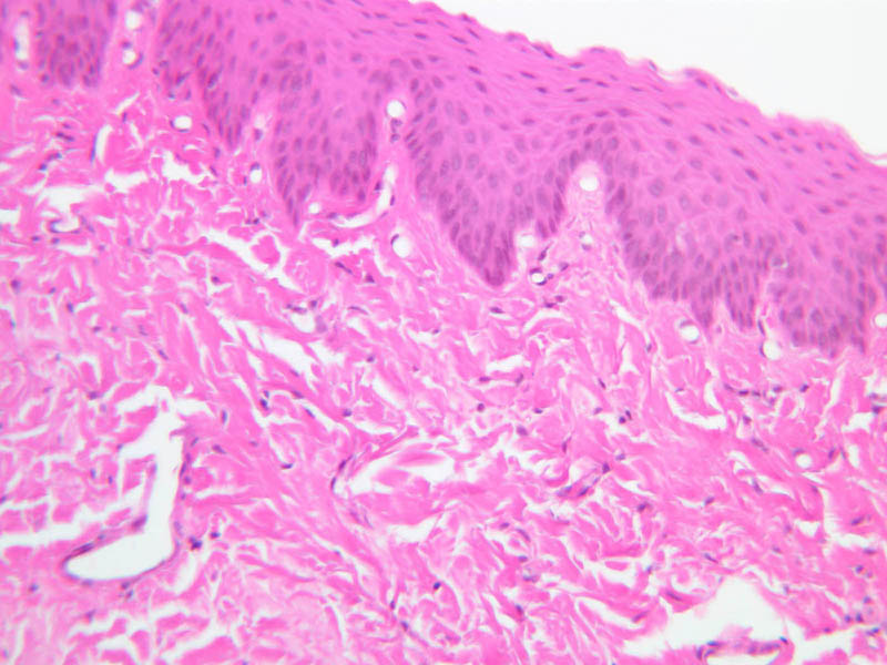
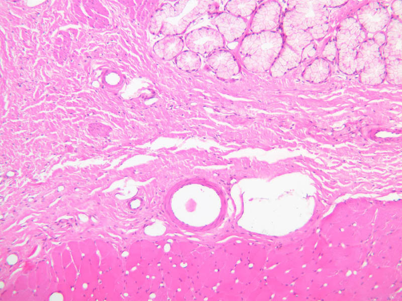
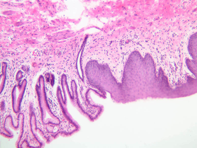
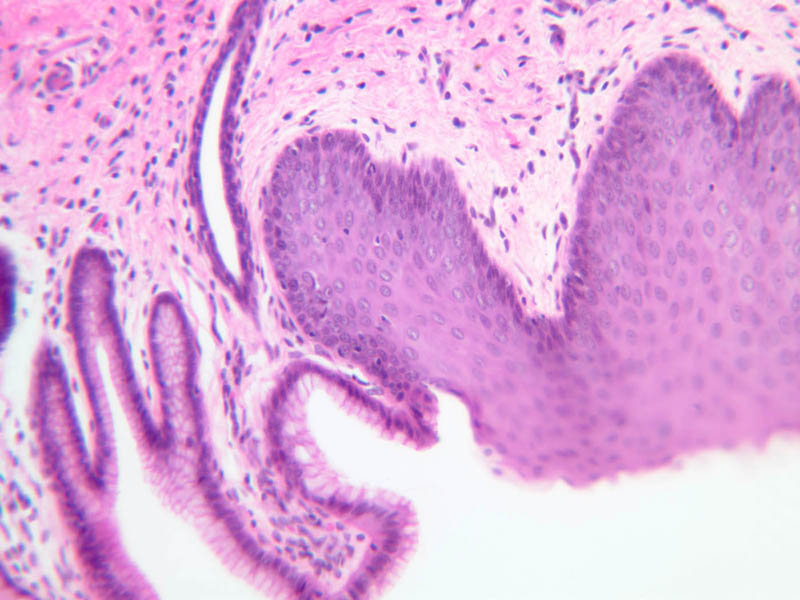
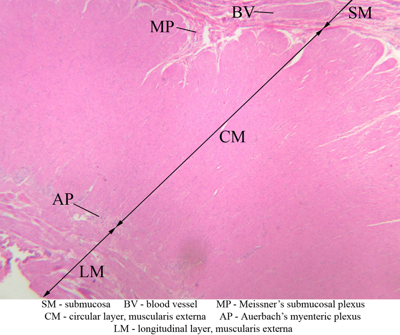
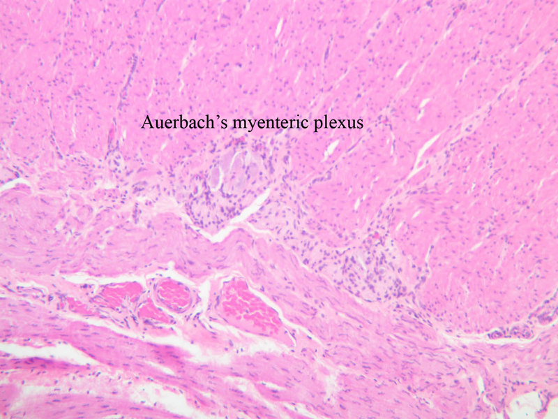
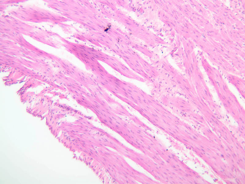
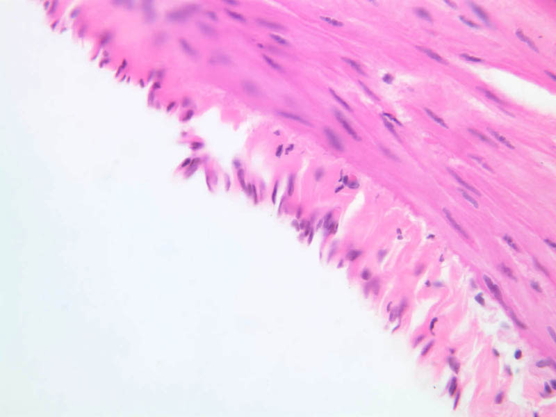
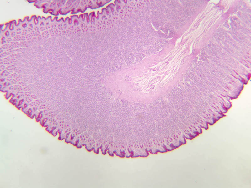
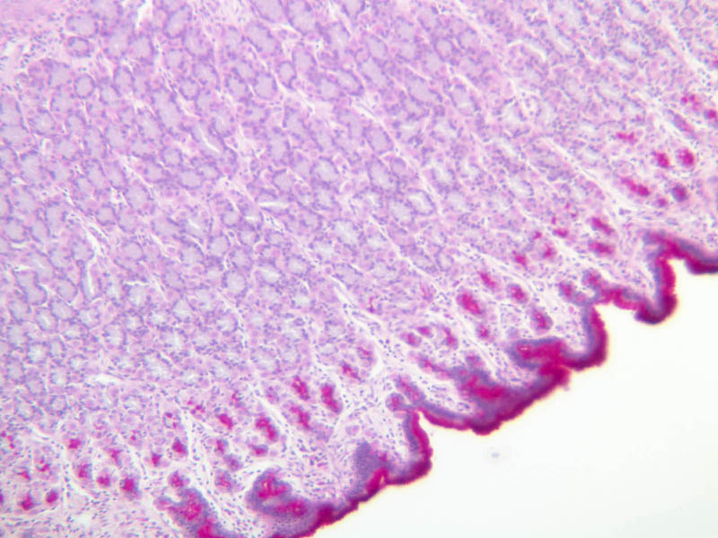
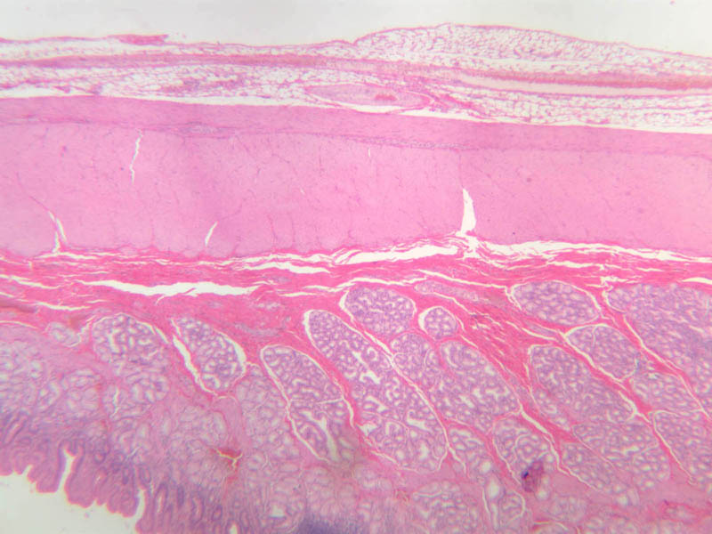
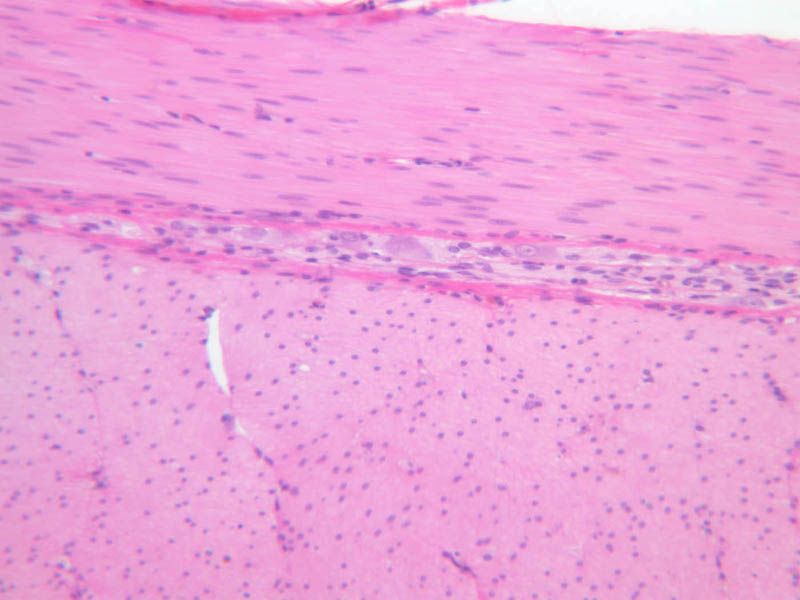
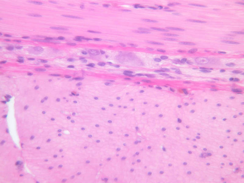
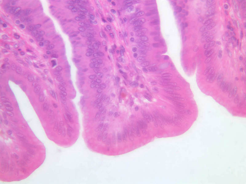
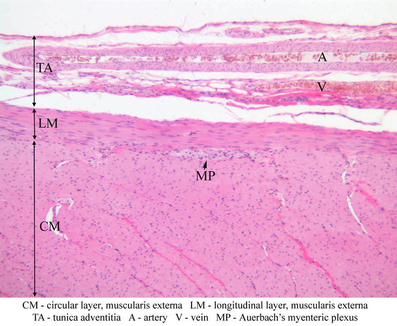
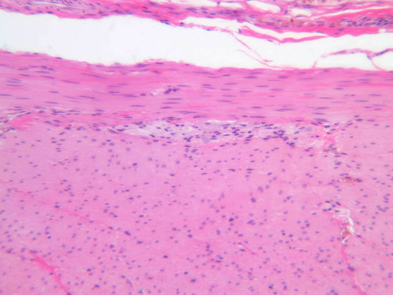
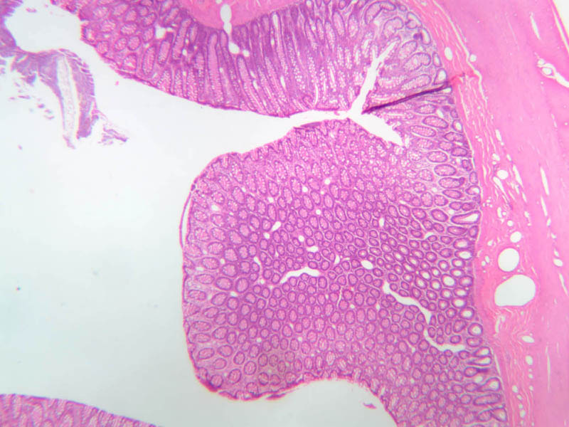
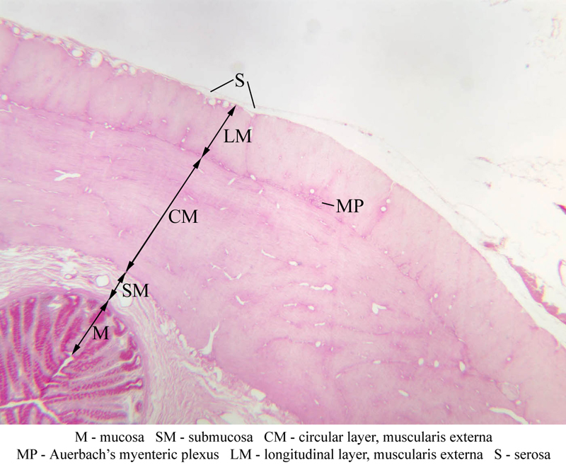
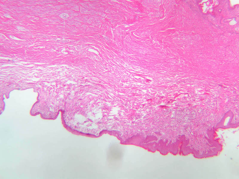
Sections of esophagus appear on slides B-2 (H&E [2.5x-labeled, 10x-labeled, 20x-labeled, 40x] [20x, 20x. 20x]) and B-3 (H&E [2.5x-labeled, 20x, 20x, 10x]). By studying these sections, you will be able to familiarize yourself with the basic structural plan of the entire digestive tube. Find the esophageal lumen and identify the three components of the mucosa (mucous membrane): a stratified squamous (nonkeratinized) epithelium, a layer of loose connective tissue (lamina propria) and a thin double layer (inner circular/outer longitudinal) of smooth muscle, known as the muscularis mucosae. The submucosa, which consists largely of dense irregular connective tissue, lies beneath the mucosa. Carefully scan the submucosa to find ganglion cells and bundles of nerve fibers that comprise Meissner's plexus (submucosal plexus). You may also find mucous glands (esophageal glands proper) in the submucosa. The muscularis externa lies beneath the submucosa. In the esophagus, the muscularis externa consists of an inner circular and an outer longitudinal layer of muscle. Note that at the mid-esophageal levels represented by these slides, the muscularis externa consists of a mixture of smooth and skeletal muscle. What kind of muscle would you expect to find in the upper esophagus? In the lower esophagus? Between the two layers of the muscularis externa you should be able to find ganglion cells and nerve fibers of Auerbach's plexus (myenteric plexus). External to the muscularis externa is a layer of connective tissue, the tunica adventitia, which binds the esophagus to surrounding structures. A section through the esophageocardiac junction appears on slide B-1 (H&E [2.5x-labeled, 10x, 20x, 40x] [2.5x, 10x, 20x, 40x]). As you examine the epithelial surface of this section, you should be able to find a region of rather abrupt transition from the stratified squamous epithelium typical of the esophagus to the simple columnar epithelium of the gastric cardia. There may be a narrow zone of stratified cuboidal or stratified columnar epithelium between the definitive epithelia of esophagus and stomach.Esophagus Image Gallery


Esophagus Table of Identifications
| Row | Structure | Abbreviation | Optimal Stain | Representative Section | Note |
|---|---|---|---|---|---|
| 1 | Adventitia | (none), Ad | H&E | |
|
| 2 | Muscularis Externa | (none) | H&E | |
|
| 3 | Submucosa | (none) | H&E | |
|
| 4 | Mucosa | (none) | H&E | |
|
| 5 | Stratified Squamous Epithelium | SSE | H&E | |
|
| 6 | Lamina Propria | LP | H&E | |
|
| 7 | Muscularis Mucosa | MM | H&E | |
|
| 8 | Circular Layer, Muscularis Externa | CM | H&E | |
|
| 9 | Auerbach's (Myenteric) Plexus | AP | H&E | |
|
| 10 | Longitudinal Layer, Muscularis Externa | LM | H&E | |
|
| 11 | Mucous Glands | (none) | H&E | |
|
| 12 | Esophagus | (none) | H&E | |
|
| 13 | Cardiac Stomach | (none) | H&E | |
|
| 14 | Fundus | (none) | H&E | |
Back to Top of Page
Stomach
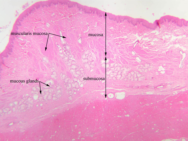
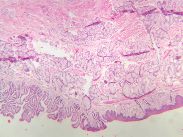
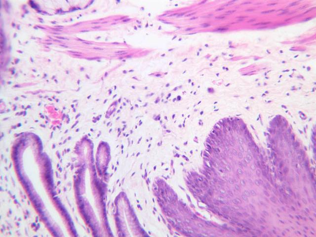
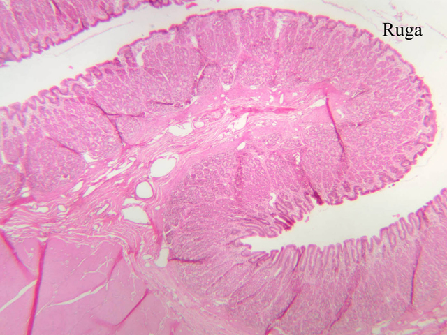
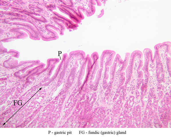
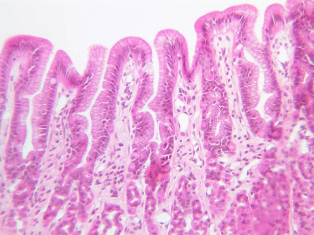
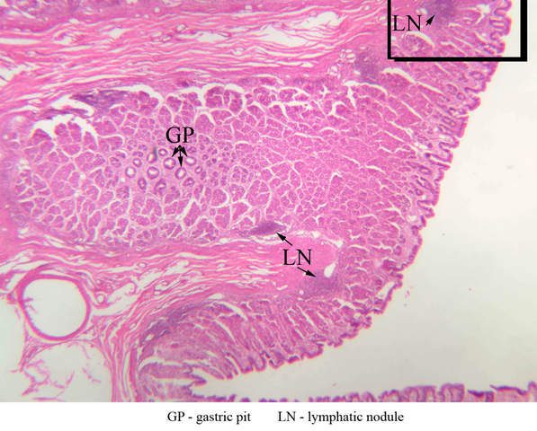
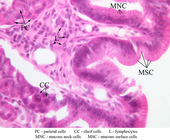
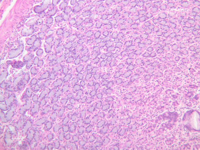
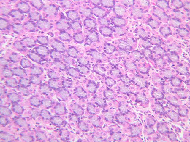
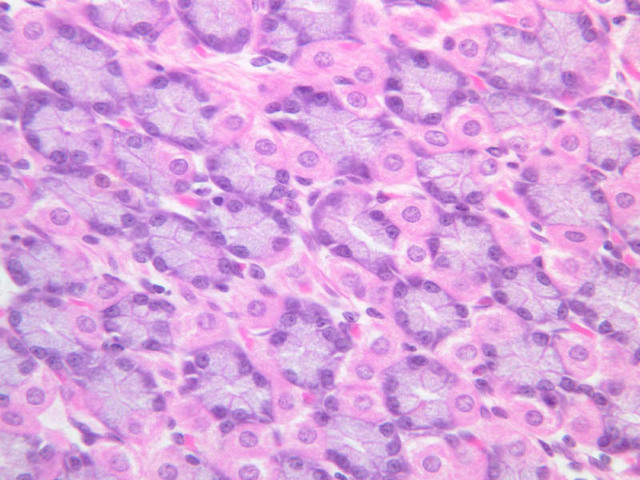
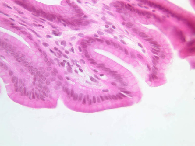
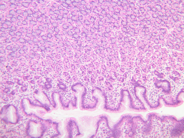
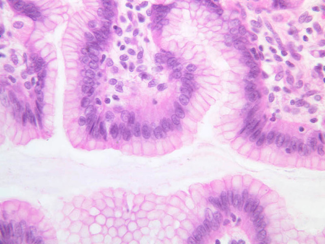
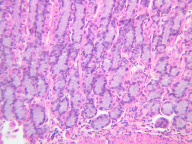
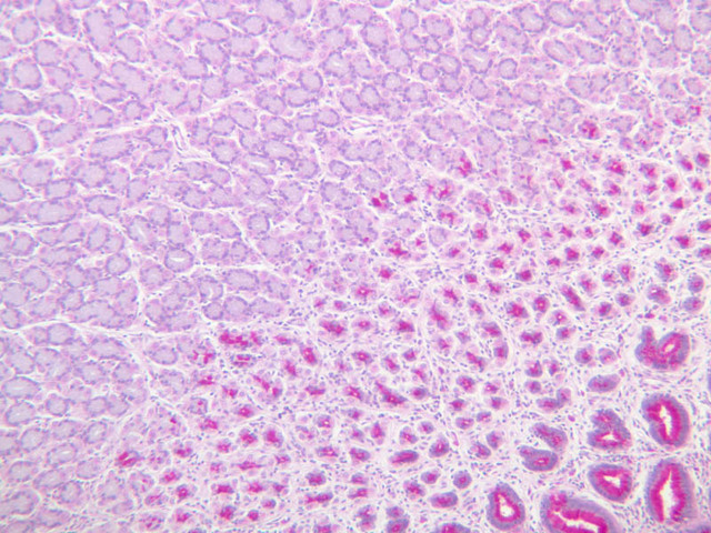
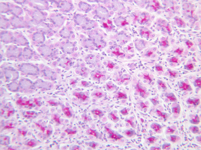
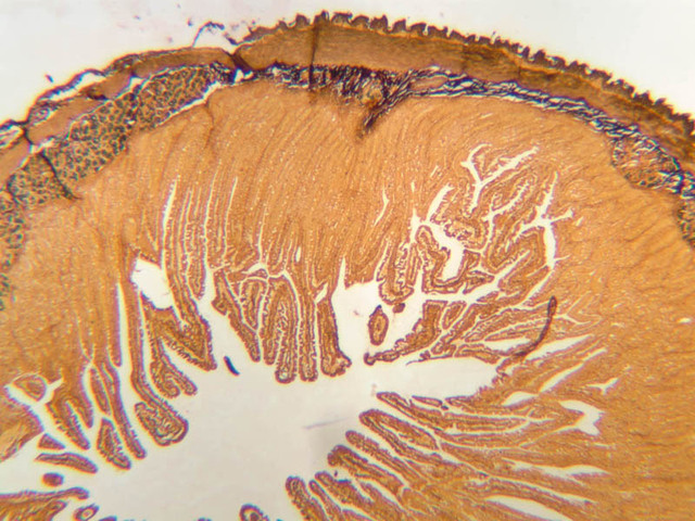
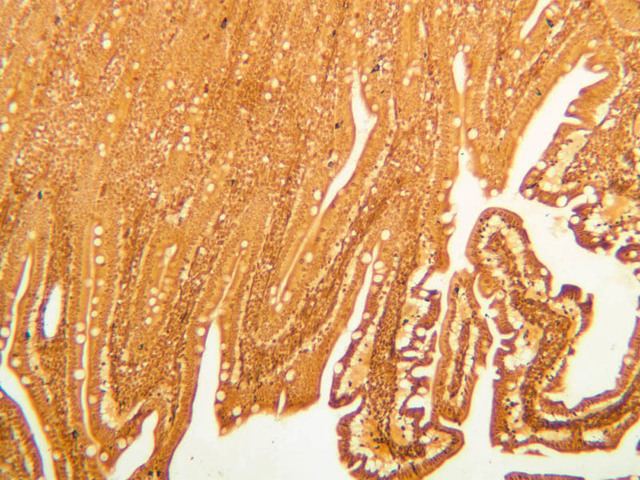
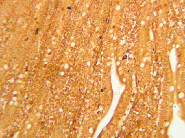
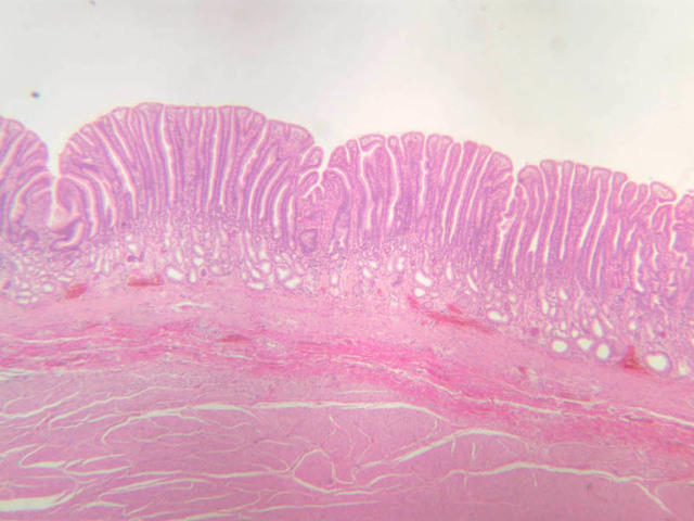
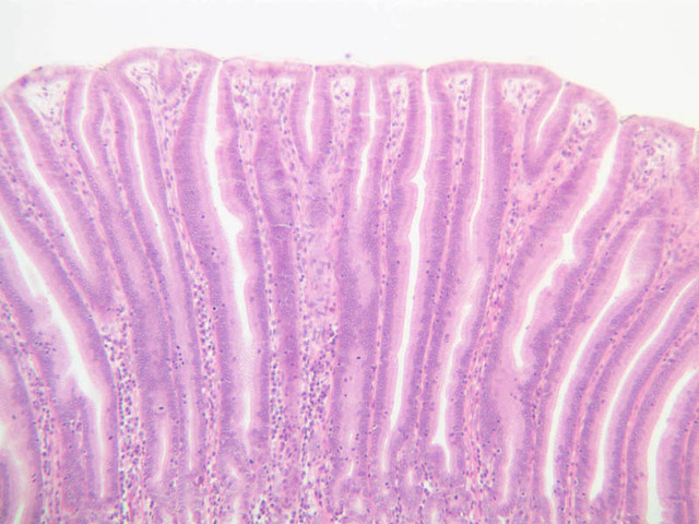
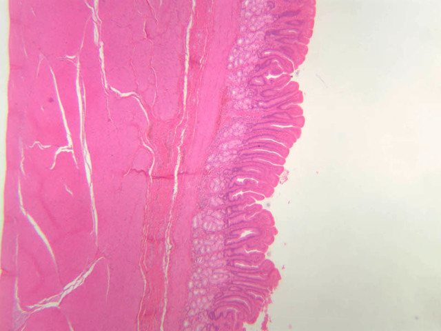
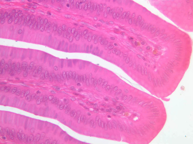
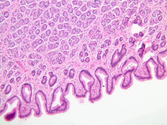
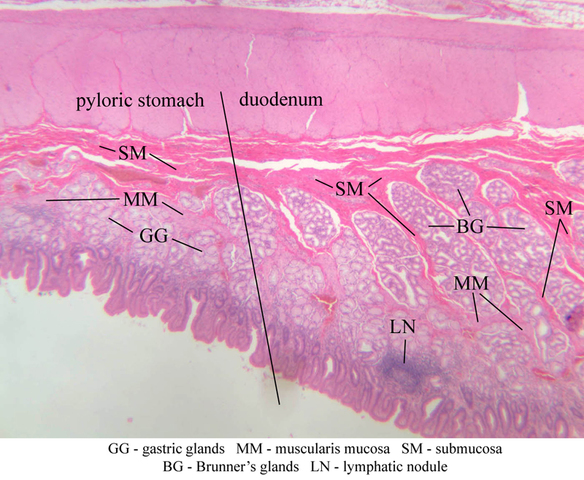
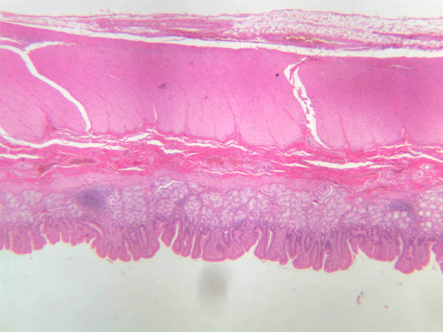
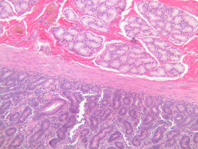
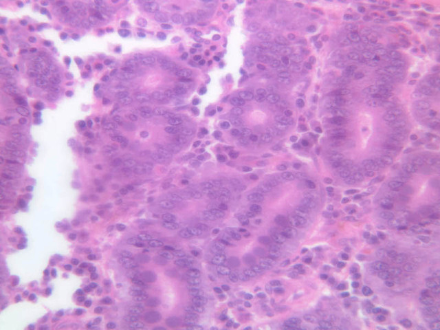
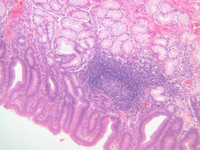
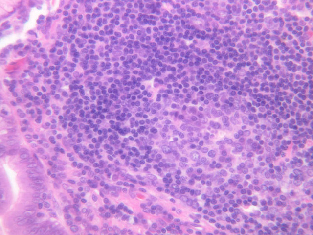
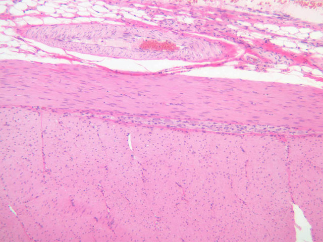
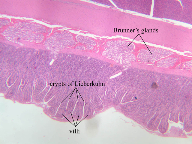
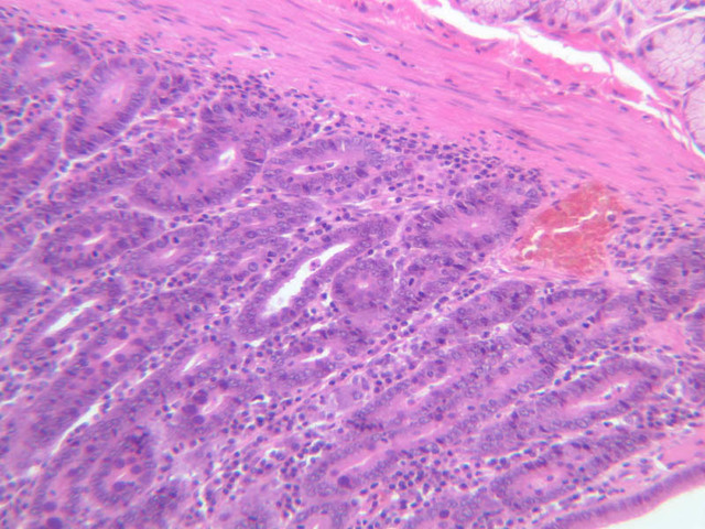
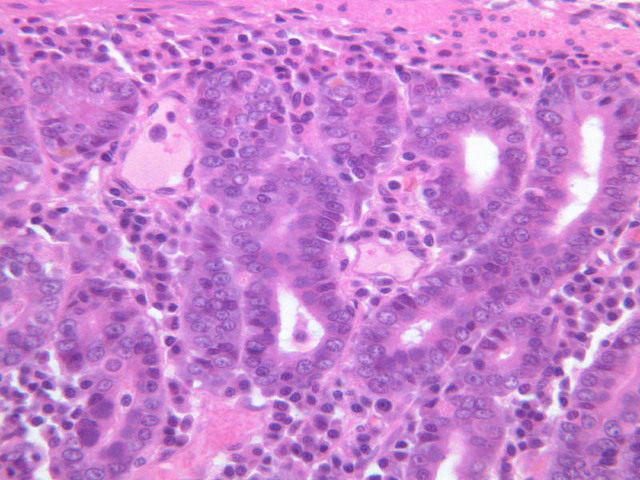
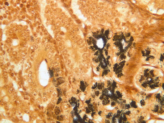
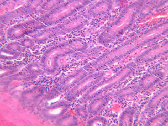
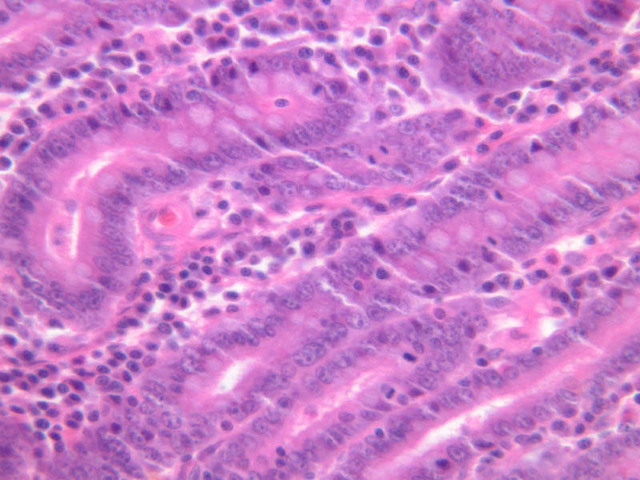
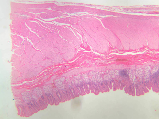
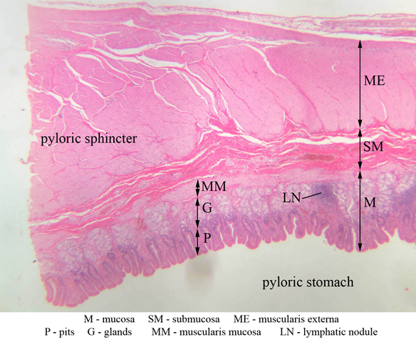
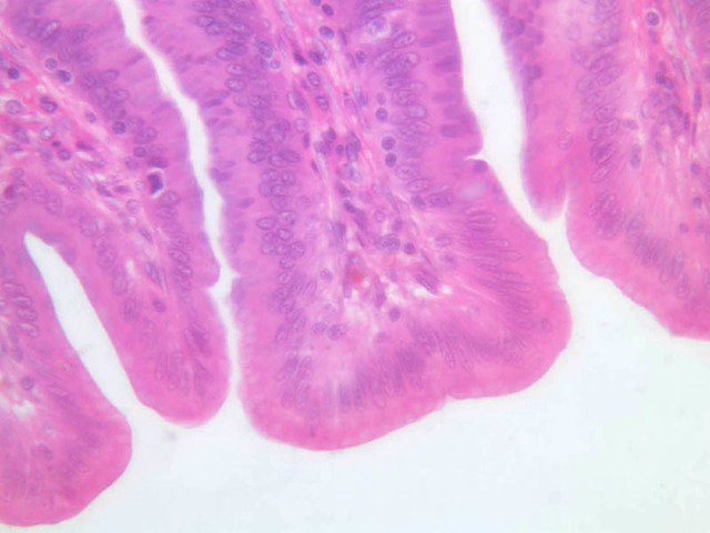
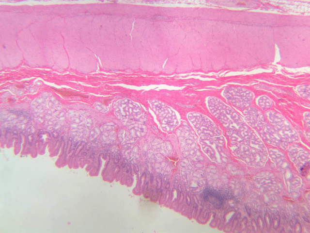
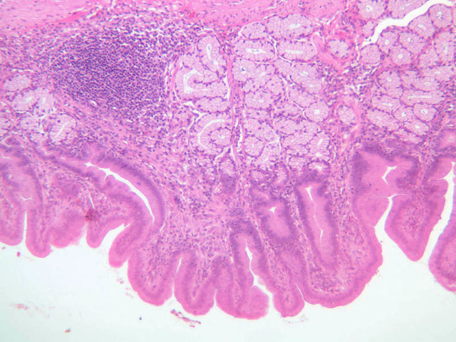
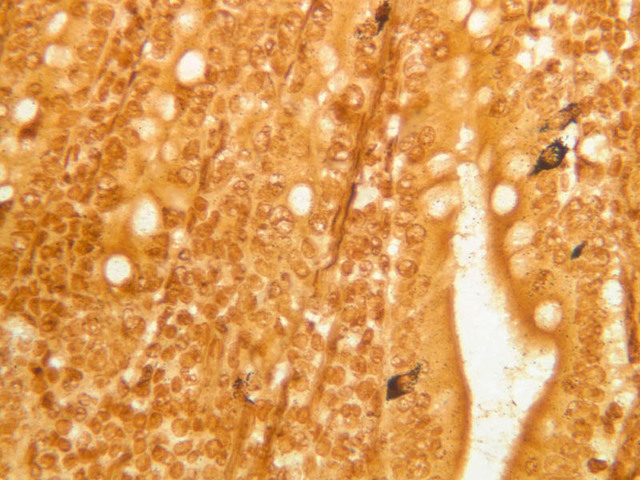
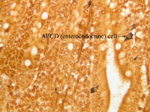
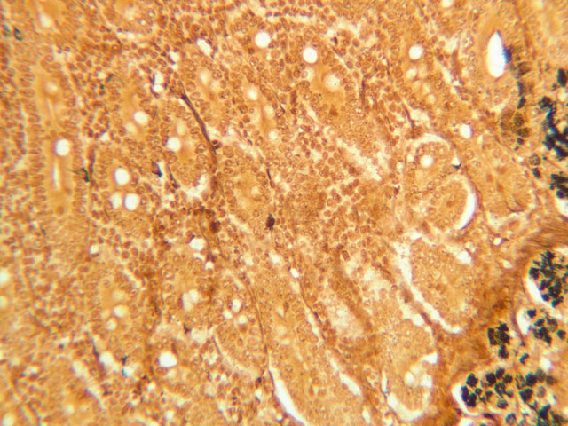
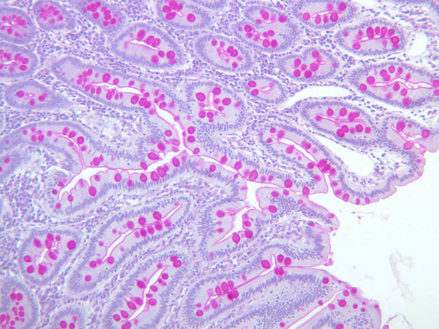
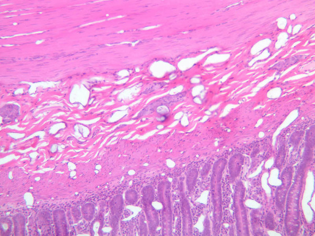
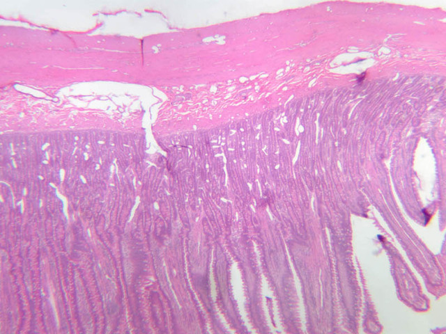
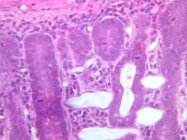
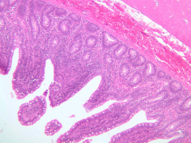
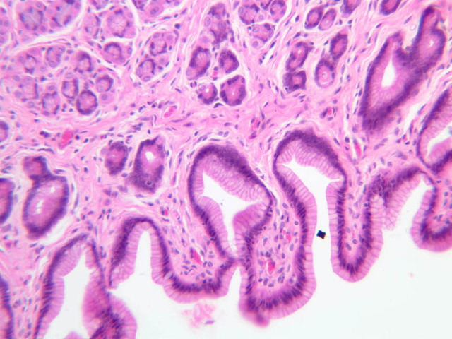
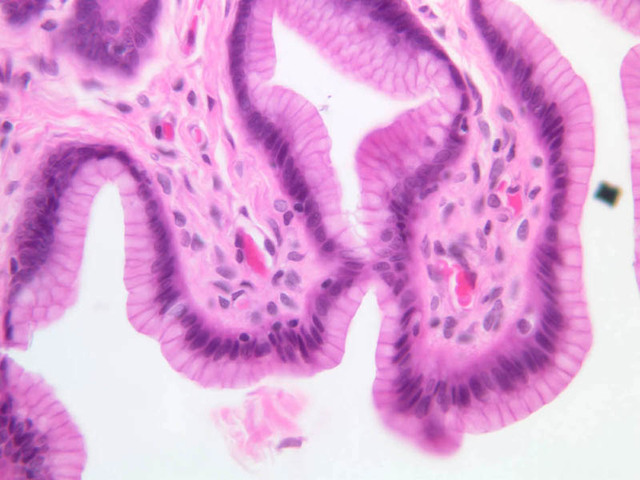
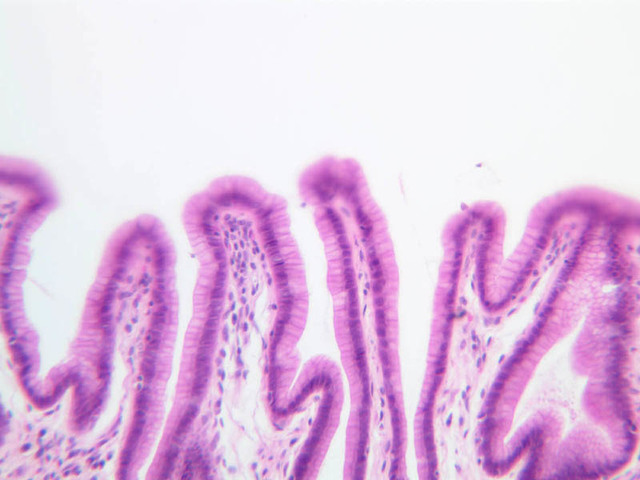
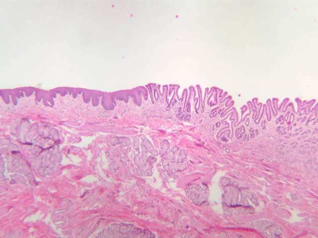
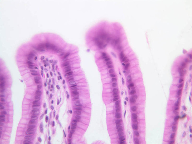
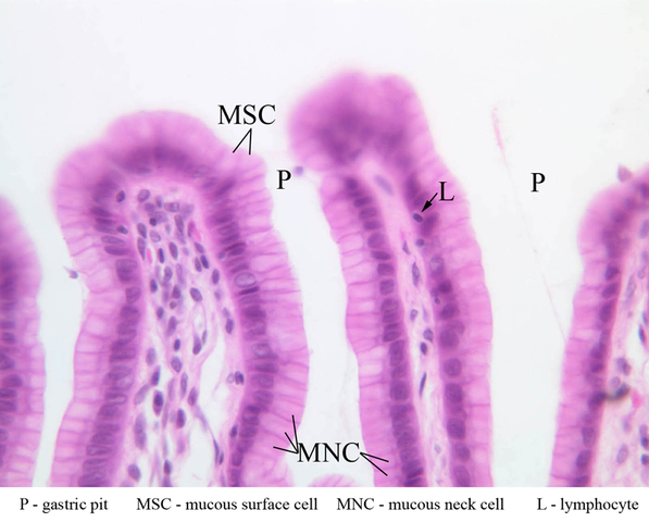
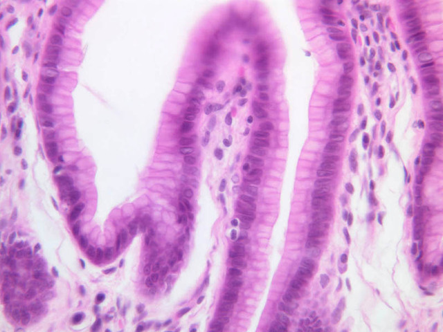
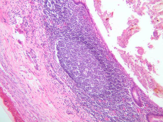
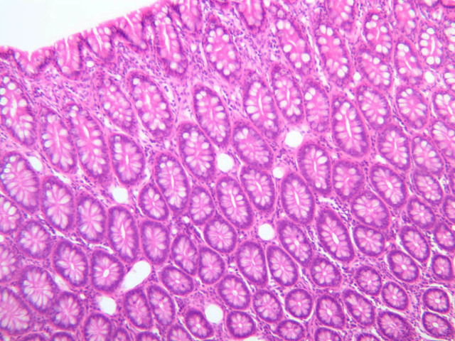
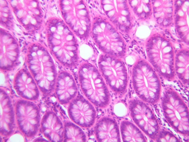
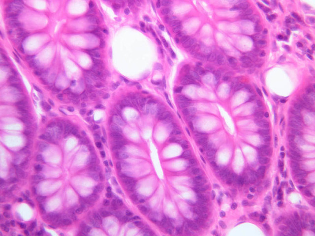
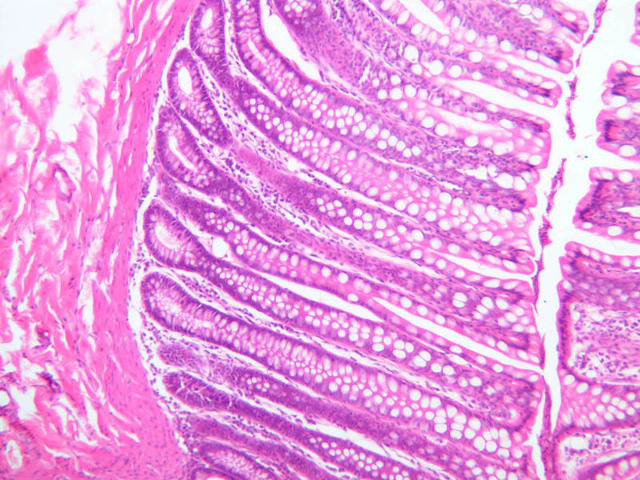
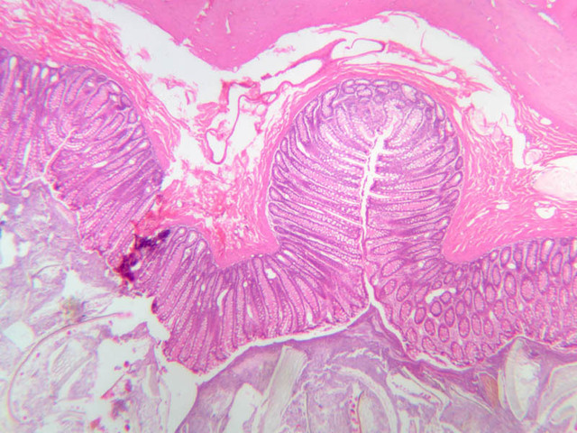
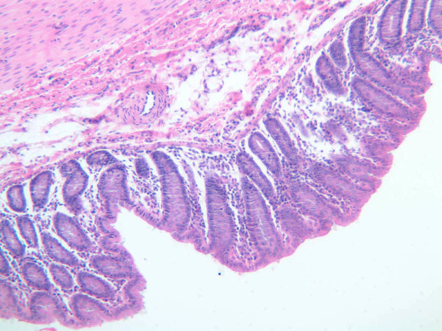
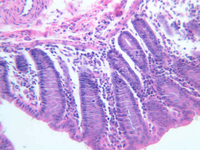
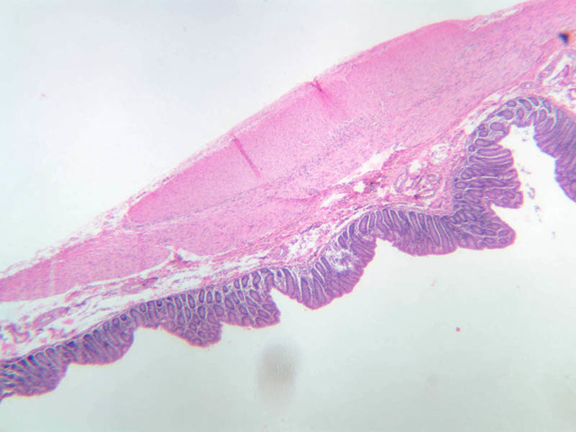
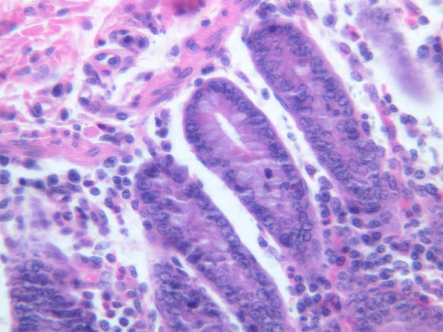
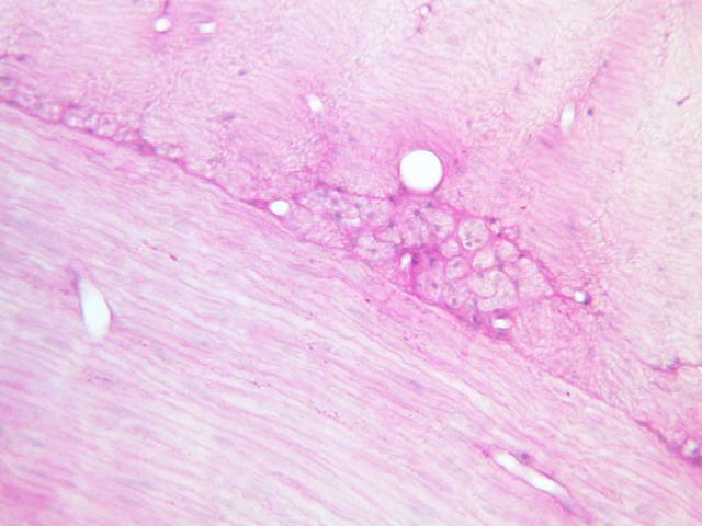
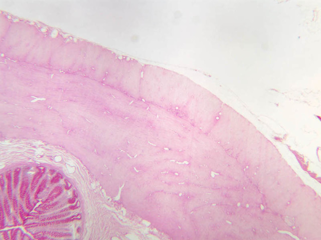
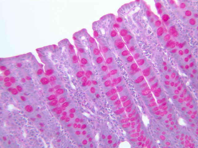
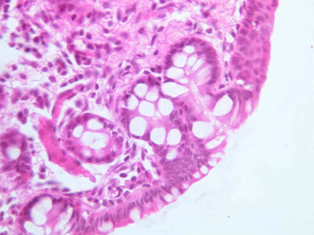
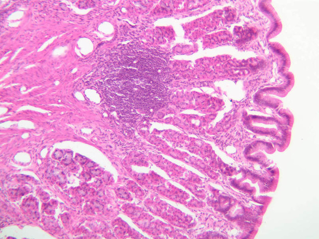
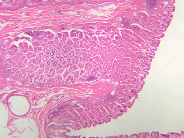
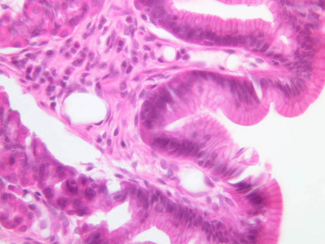
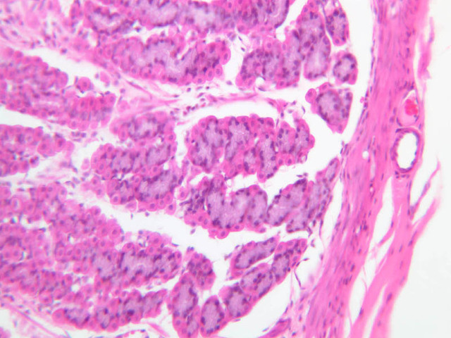
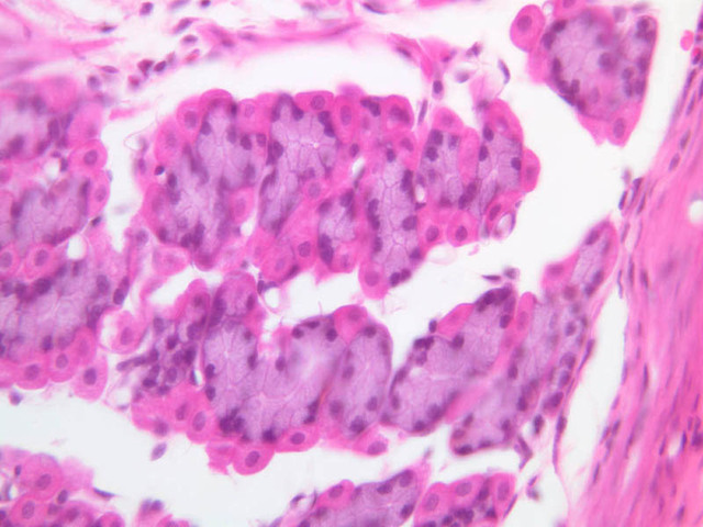
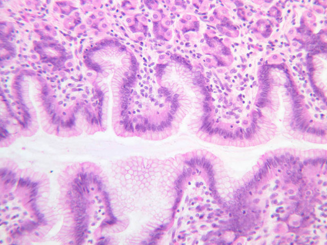
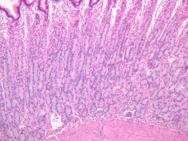
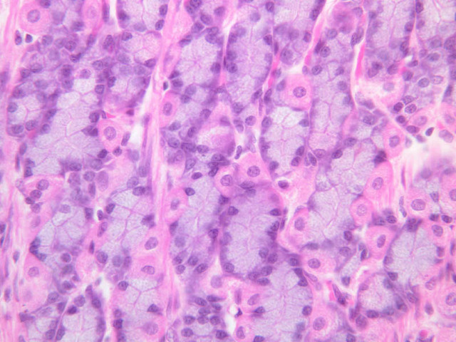
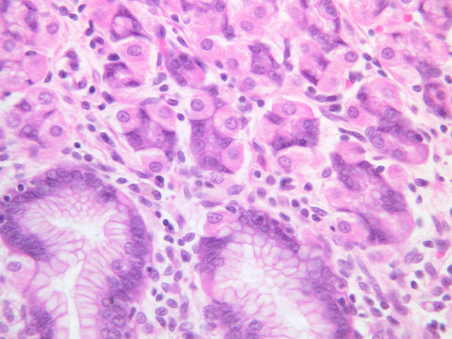
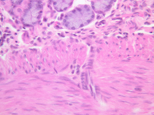
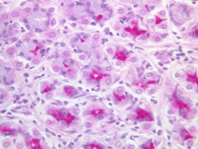
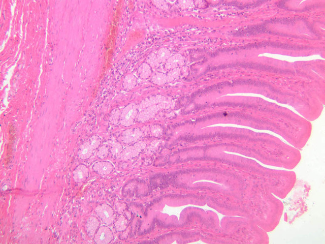
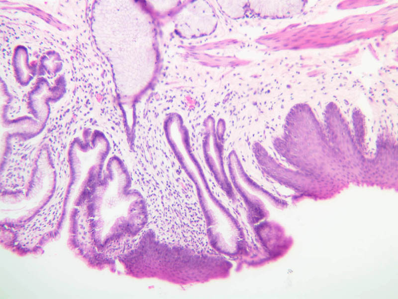
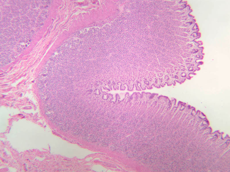
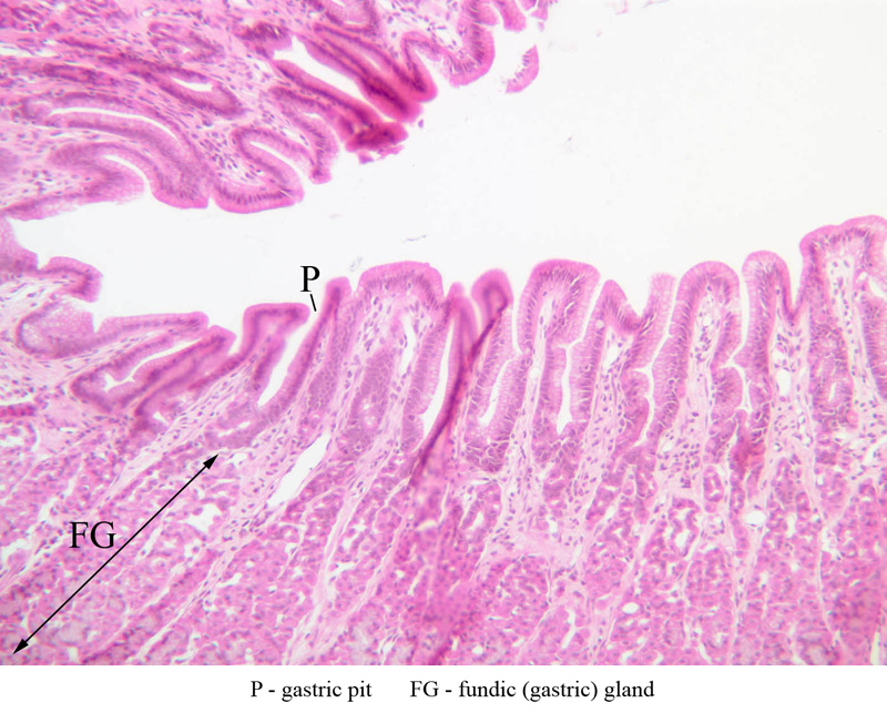
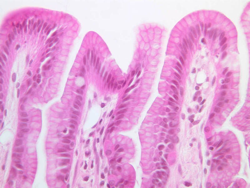
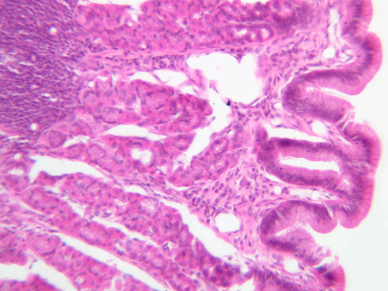
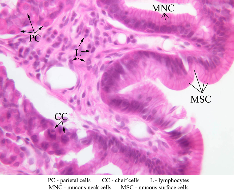
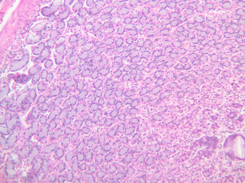
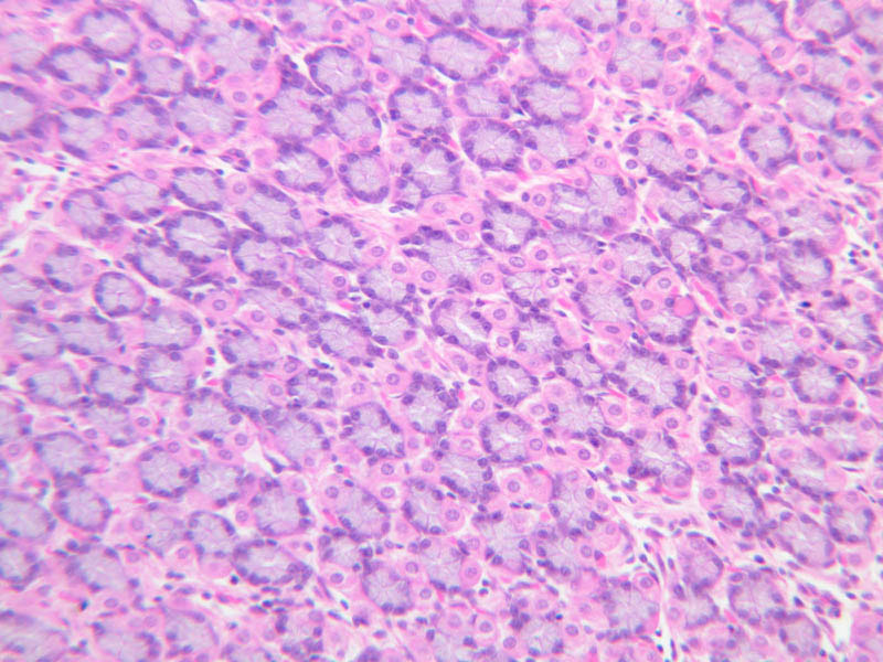
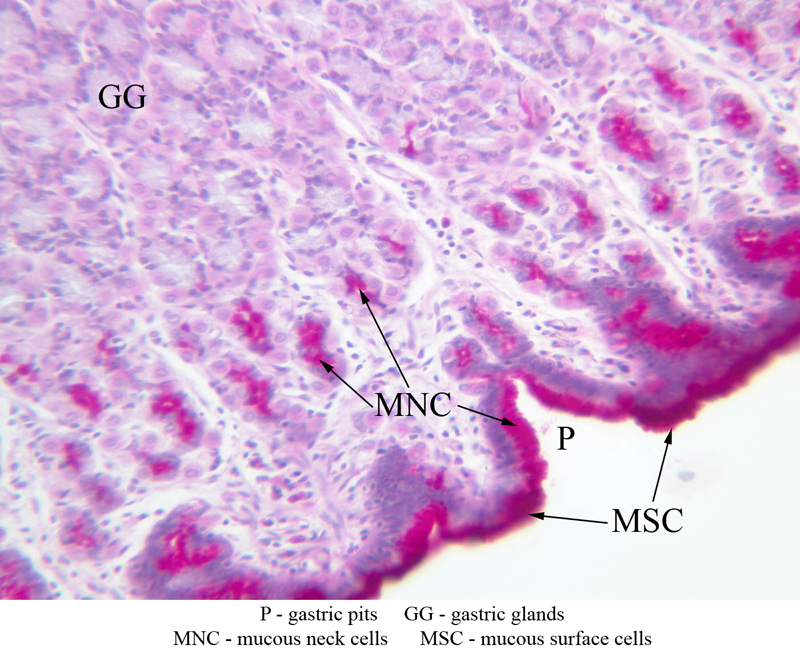
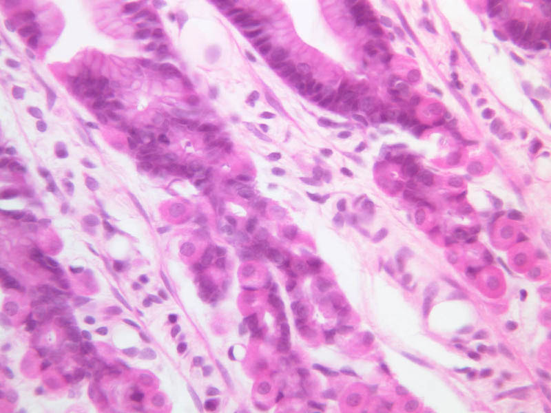
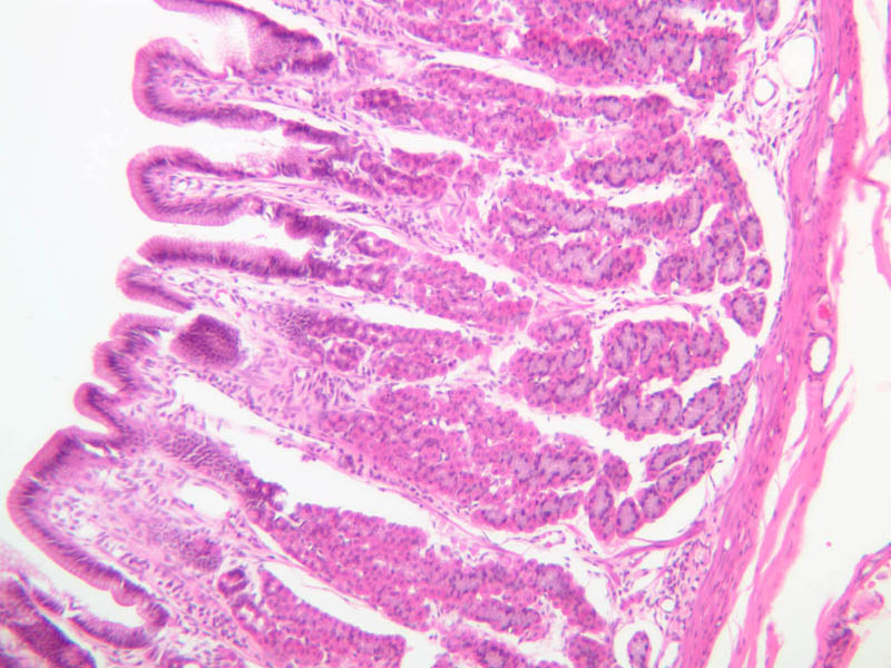
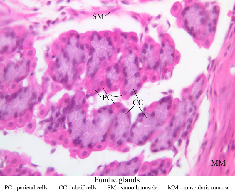
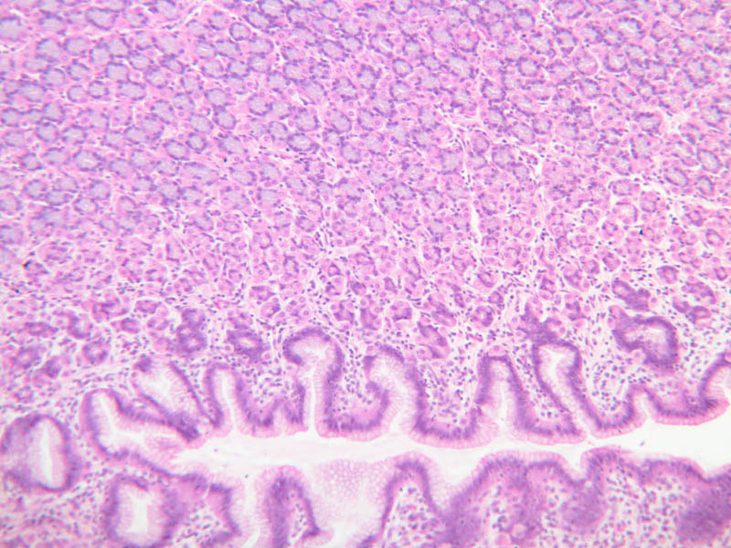
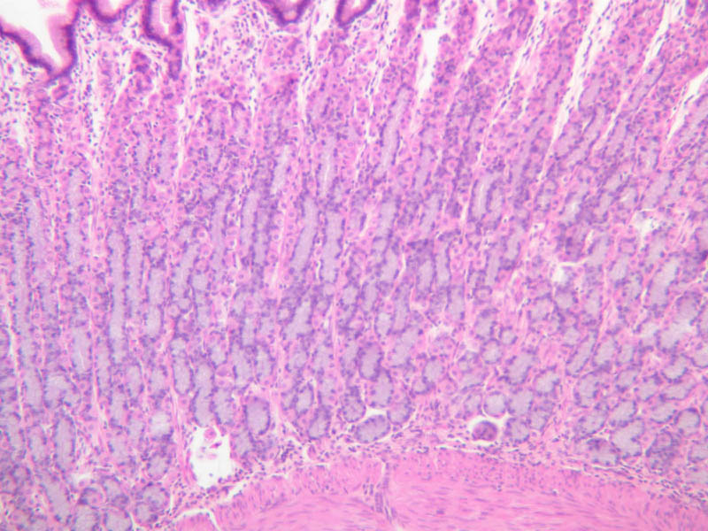
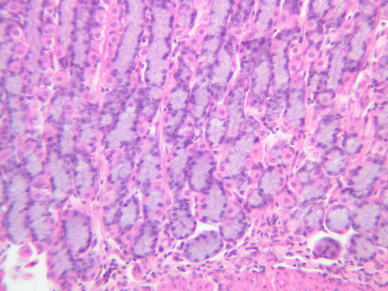
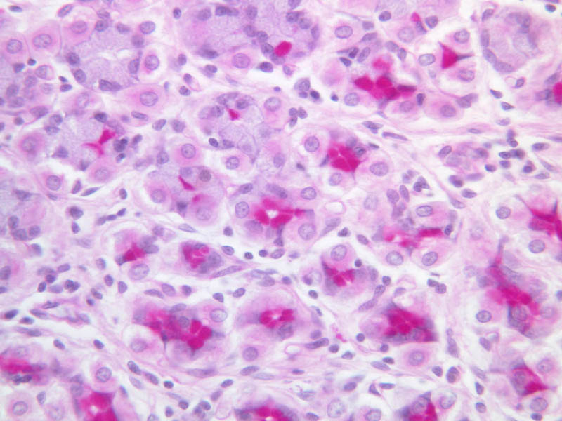
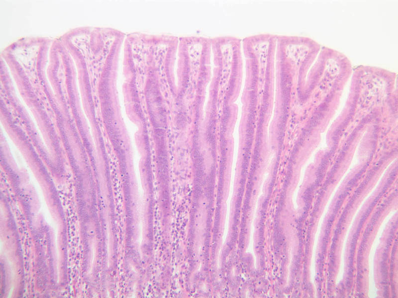
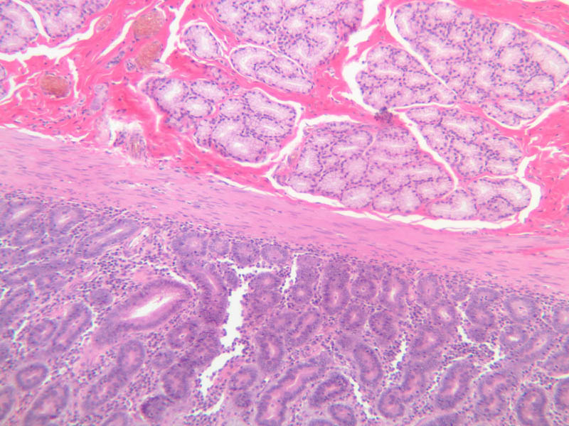
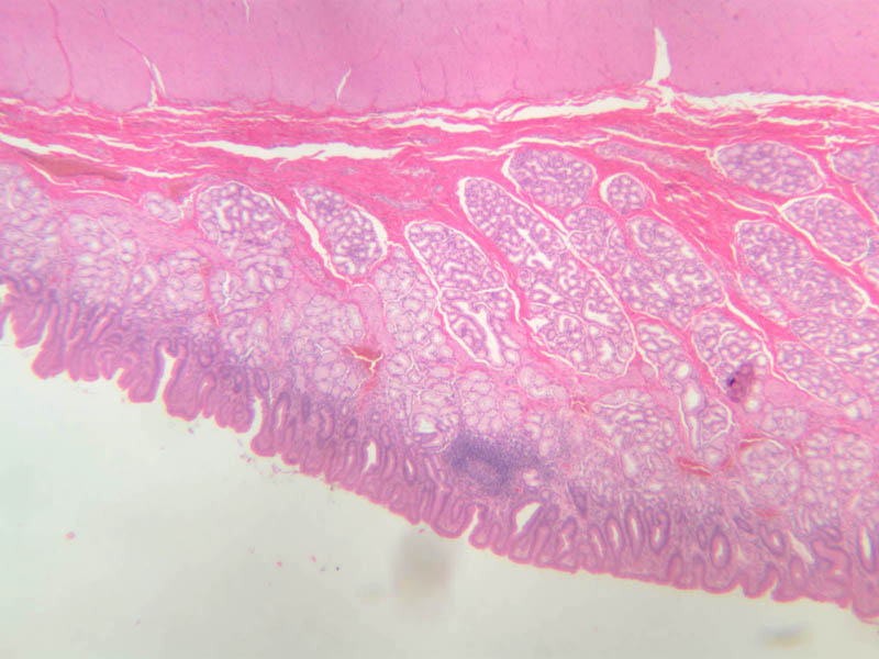
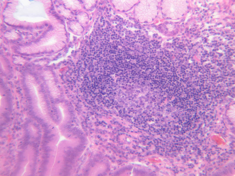
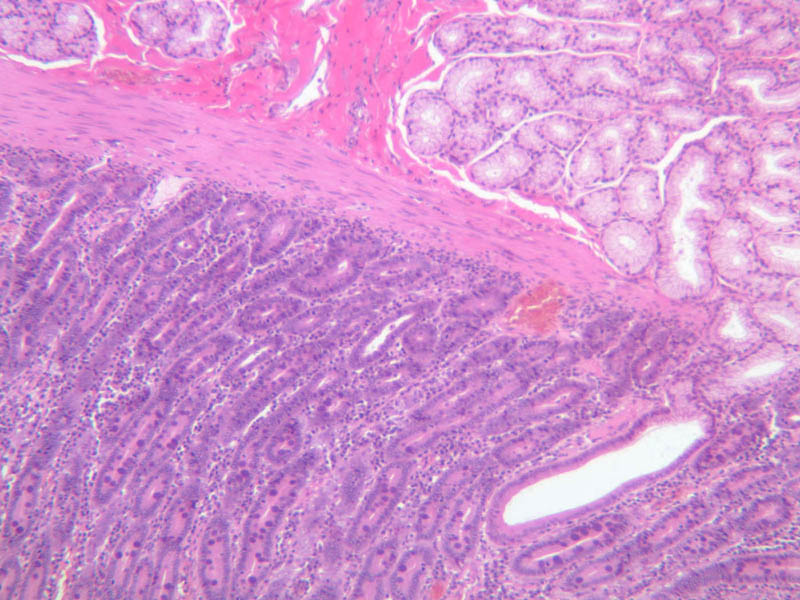
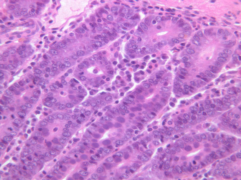
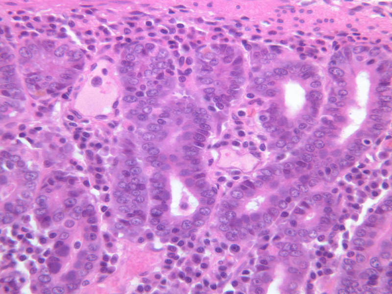
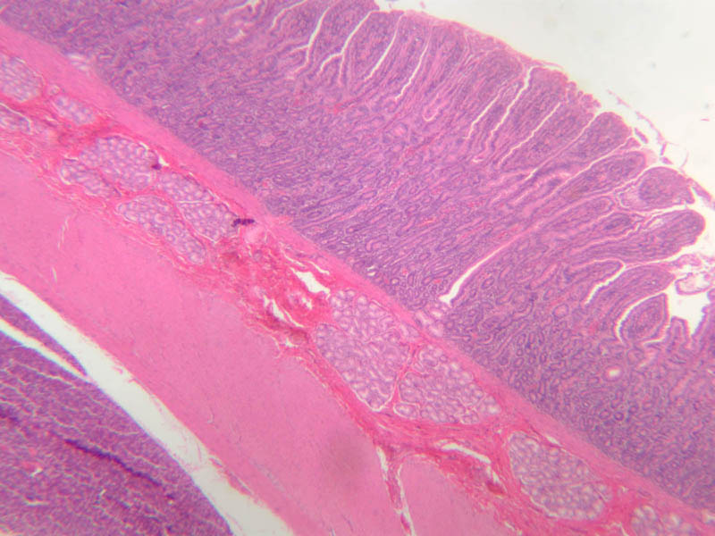
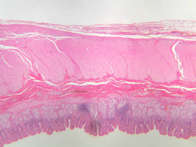
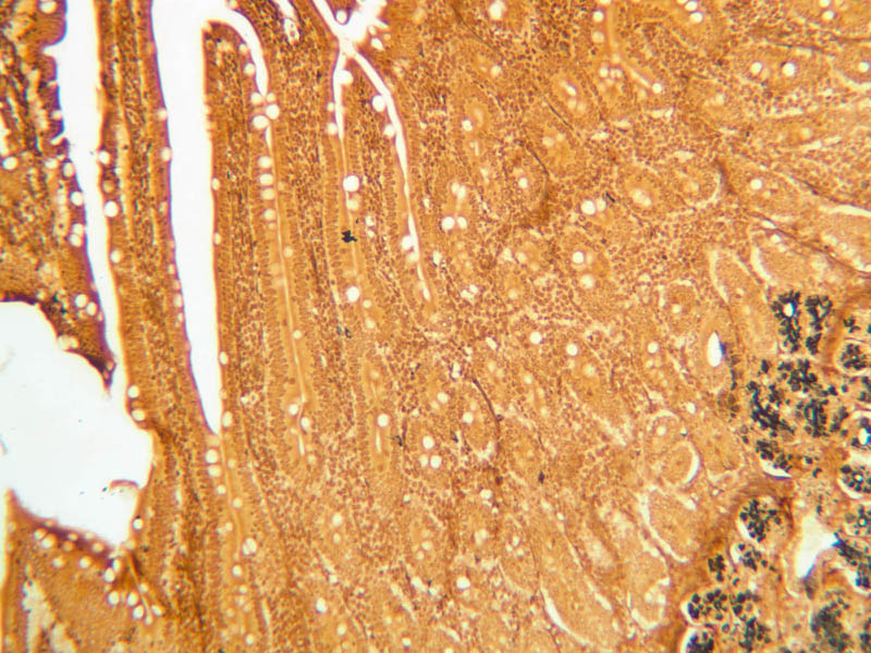
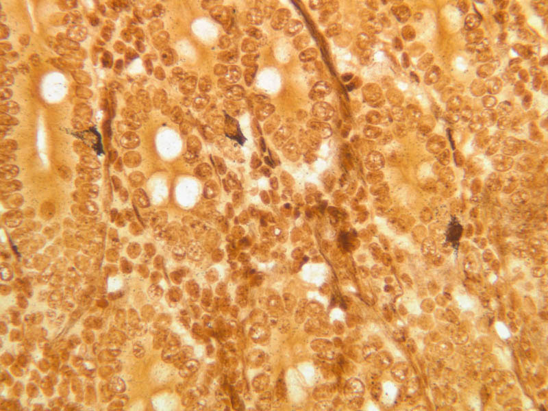
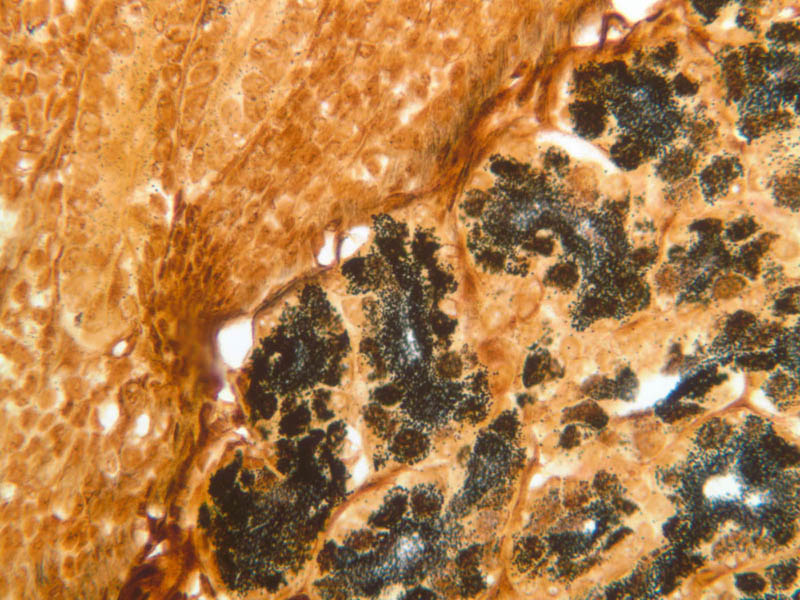
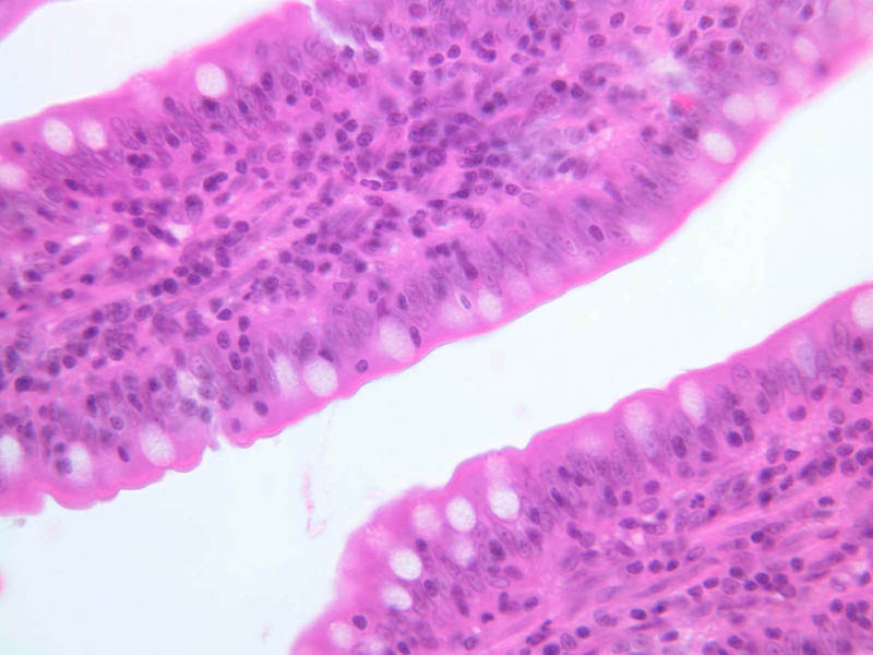
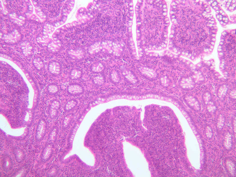
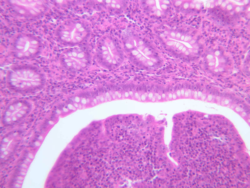
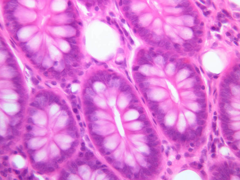
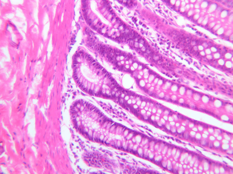
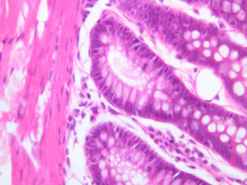
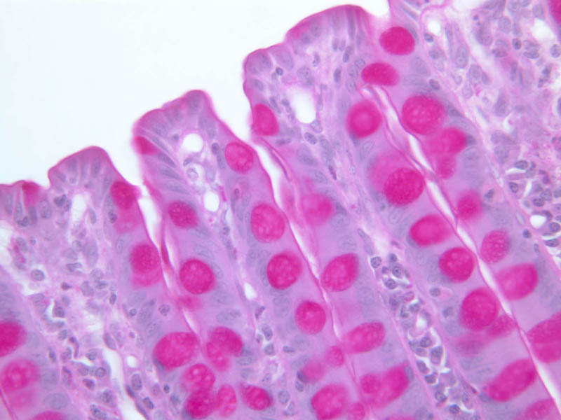
By holding slide B-4 or B-5 up to the light, you should be able to identify the mucosa, submucosa and muscularis externa (B-4, H&E [2.5x-labeled, 10x-labeled, 20x, 40x]; B-5, H&E [2.5x-labeled, 10x, 20x] [10x-labeled, 20x, 40x] [10x, 40x] [2.5x, 10x]). The mucosa appears as a basophilic band that is about 1.5 mm thick. The external muscular layer appears as a 2-3 mm thick band that runs a fairly straight course along the length of the section. The submucosa, which is sandwiched between the mucosa and muscularis externa, is usually thinner and more lightly stained than either of the other two layers. Since these sections were taken from a relaxed stomach, the luminal surface is thrown into large folds (rugae), which appear to consist of a fingers of submucosal connective tissue and their overlying mucosa. Rugae are transient adaptations to the cyclical filling and emptying of the stomach: as the stomach fills with ingested food the rugae are stretched out of existence, only to reappear as the stomach flushes its contents into the duodenum. Examine slide B-4 (H&E [2.5x-labeled, 10x, 20x, 40x-labeled]) or B-5 (H&E [10x, 20x, 40x] [40x]) under the intermediate and high-dry objectives of your microscope. Mucous-secreting simple columnar cells, known as mucous surface cells, line the gastric lumen and the periodic deep depressions, known as gastric pits. The mucous surface cells differ from goblet cells both in their appearance and in the type of mucous they secrete. In H&E preparations they have a rather clear cytoplasm, but do not exhibit the sort of well-defined, unstained vacuoles that are typical of goblet cells. By contrast, in the PAS-stained section of slide B-6 (PAS [2.5x, 10x, 20x-labeled, 40x]) they stand out dramatically by virtue of their magenta-colored secretory product. As a rule, several glands extend from each gastric pit to the muscularis mucosae, largely obscuring the lamina propria. Small slips of smooth muscle that extend from the muscularis mucosae towards the luminal surface are presumed to be involved in expressing the glandular secretions. The glands of the stomach exhibit regional variations in size, form, and cellular composition. The glands of the cardia (slide B-1) and pylorus (slide B-9) are coiled, often branched tubules that that are associated with very deep gastric pits and consist almost entirely of mucous-secreting cells. In the gastric fundus (slide B-4, H&E [2.5x, 10x, 20x, 40x] [10x, 20x, 40x-labeled] [40x]) and corpus (slides B-5, H&E [2.5x, 10x, 20x, 40x] [10x, 20x, 40x] [40x] & B-6, PAS [2.5x, 10x, 20x, 40x]), the glands are relatively straight and are known as fundic glands or gastric glands; they are made up of mucous neck cells, chief cells, parietal cells, and APUD cells. The lumina of fundic glands are much less evident than are those of cardiac and pyloric glands. Study slides B-4, 5 & 6 to familiarize yourself with the cell types found in fundic glands. As their name suggests, mucous neck cells are most numerous just proximal to the junction of gastric gland and gastric pit. In H&E-stained sections, these cells, like others which secrete mucous (e.g., goblet cells), tend to stain poorly on account of the loss of their secretory product during histological processing. However, in the section on slide B-6, which has been stained by the PAS technique, they are very easy to identify (bright pink). You should study the distribution of mucous neck cells in this slide and then attempt to identify them in H&E preparations by looking at corresponding regions in slide B-4 or B-5. What functions are served by the mucous-secreting cells of the gastric mucosa? The chief or zymogenic cells have a rather granular, basophilic cytoplasm and produce pepsinogen, which, upon encountering the acidic gastric juice, is converted into the proteolytic enzyme pepsin. As the name suggests, the chief cells are usually the most numerous cell type in the glands of the fundic region. The parietal cells are large and have conspicuously acidophilic cytoplasm. They are the source of both gastric HCl. and intrinsic factor, which is necessary for uptake of vitamin B12 in the ileum. The fine structure of parietal cells shows that their cytoplasm is filled with a very large number of surface invaginations or secretory canaliculi, which, in turn, are covered with microvilli. It is apparently this elaboration of surface area that allows parietal cells to secrete large amounts of HCl. Why are parietal cells eosinophilic? Many organs contain cells that have particular affinity for chromium and silver salts; thus, classical histologists were able to identify chromaffin and argentaffin cells. These cells are now generally known as amine precursor uptake and decarboxylation (APUD) cells. They tend to be rather small cells that show little cytplasmic detail and, thus, appear as clear cells in H&E-stained sections. Most of the APUD cells of gastric mucosa produce gastrin, which is important in the regulation of HCl secretion. Argentaffin cells are identifiable in the sections of pylorus on slide B-11 (Ag [2.5x, 10x, 20x, 40x-labeled]). Examine the submucosal, external muscular and serosal layers in one of the sections of stomach. Note the numerous blood vessels in the submucosa and try to find elements of Meissner's submucosal plexus. Study the muscularis externa, noting that it is not always possible to distinguish an inner oblique layer, since this layer occurs only in the corpus and is never as prominent as either the circular or longitudinal layers. Look between the outer two external muscular layers for profiles of the neural elements of Auerbach's myenteric plexus. It is through the contractions of the muscularis externa that gastric secretions are mixed with ingested food to yield the semifluid chyme, which is passed on to the duodenum. Although the connective tissue and mesothelium of the gastric serosa are disrupted in some of your sections, you should be able to find intact regions of serosa. What is the function of this layer? In the pylorus (slide B-9, H&E [2.5x, 10x, 20x, 40x] [2.5x, 10x, 20x, 40x] [40x, 40x]), a funnel-shaped part of the stomach that continues into the duodenum, the glands are similar in appearance to the cardiac glands (B-1, H&E [2.5x-labeled, 10x, 20x, 40x]) in that they are coiled tubules made up of mucous-secreting cells. However, the pyloric glands are longer and tend to be more highly branched than the cardiac glands. Gastric pits are deeper in the pylorus than in the cardia. There is also a prominent Auerbach's (Myenteric) Plexus (B-10, H&E [2.5x, 10x, 20x, 40x]) Examine a section through the pyloroduodenal junction (slide B-10, H&E [2.5x-labeled, 10x, 20x, 40x]). Compare the mucosal glands in the pylorus with those in the duodenum and note the relationship of the muscularis mucosae to the submucosal glands (Brunner's glands) of the duodenum (B-10, H&E [2.5x-labeled] [2.5x, 10x] [2.5x, 10x, 20x, 40x]). The thickened musculature of the pyloric sphincter should be apparent in this section. This region of transition may also show exceptionally large amounts of lymphoid tissue in the mucosa (B-10, H&E [2.5x, 10x, 20x, 40x]. In some of your sections goblet cells appear blue due to overstaining.Stomach Image Gallery


Stomach Table of Identifications
| Row | Structure | Abbreviation | Optimal Stain | Representative Section | Note |
|---|---|---|---|---|---|
| 1 | Ruga | (none) | H&E | |
|
| 2 | Gastric Pit | P, GP | H&E, PAS | |
|
| 3 | Fundic Gastric Gland | FG | H&E | |
|
| 4 | Submucosa | SM | H&E | |
|
| 5 | Blood Vessel | BV | H&E | |
|
| 6 | Meissner's (Submucosal) Plexus | MP | H&E | |
|
| 7 | Circular Layer, Muscularis Externa | CM | H&E | |
|
| 8 | Auerbach's (Myenteric) Plexus | AP | H&E | |
|
| 9 | Longitudinal Layer, Muscularis Externa | LM | H&E | |
|
| 10 | Lymphatic Nodule | LN | H&E | |
|
| 11 | Parietal Cell | PC | H&E | |
|
| 12 | Chief Cell | CC | H&E | |
|
| 13 | Lymphocyte | L | H&E | |
|
| 14 | Mucous Neck Cell | MNC | H&E, PAS | |
|
| 15 | Mucous Surface Cell | MSC | H&E, PAS | |
|
| 16 | Gastric Glands (Corpus) | GG | PAS | |
|
| 17 | Smooth Muscle | SM | H&E | |
|
| 18 | Muscularis Mucosa | MM | H&E | |
|
| 19 | APUD (Enteroendocrine) Cell | (none) | Ag | |
|
| 20 | Cardiac Stomach | (none) | H&E | |
|
| 21 | Fundus | (none) | H&E | |
|
| 22 | Mucosa | M | H&E | |
|
| 23 | Muscularis Externa | ME | H&E | |
|
| 24 | Glands (Pylorus) | G | H&E | |
|
| 25 | Pyloric Sphincter | (none) | H&E | |
|
| 26 | Pyloric Stomach | (none) | H&E | |
|
| 27 | Duodenum | (none) | H&E | |
|
| 28 | Brunner's Glands | BG | H&E | |
Back to Top of Page
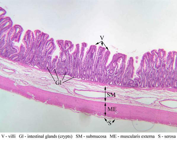
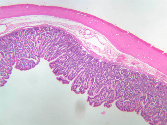
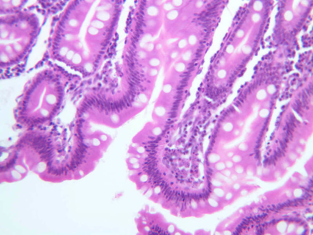
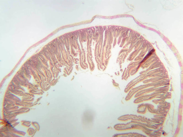
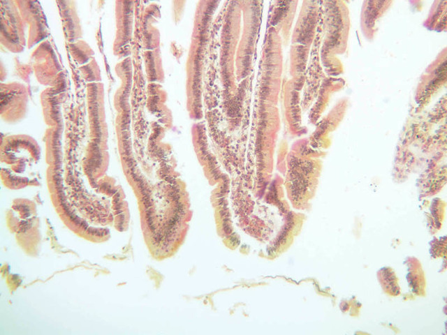
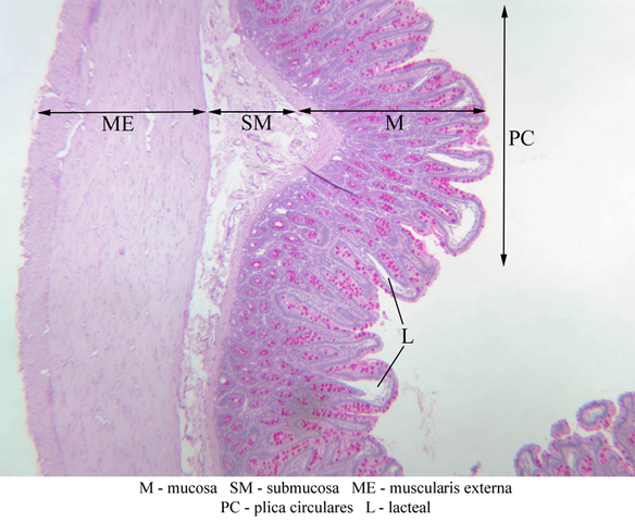
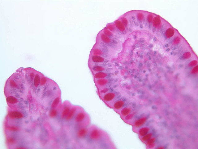
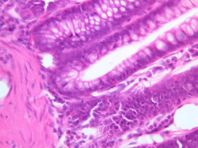
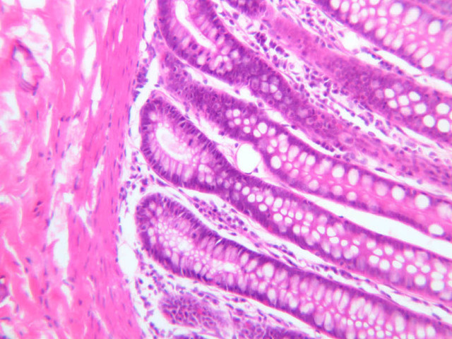
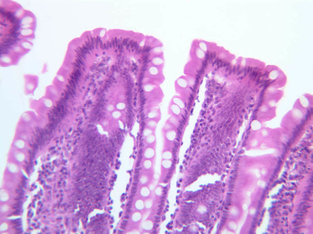
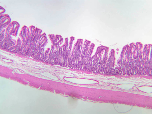
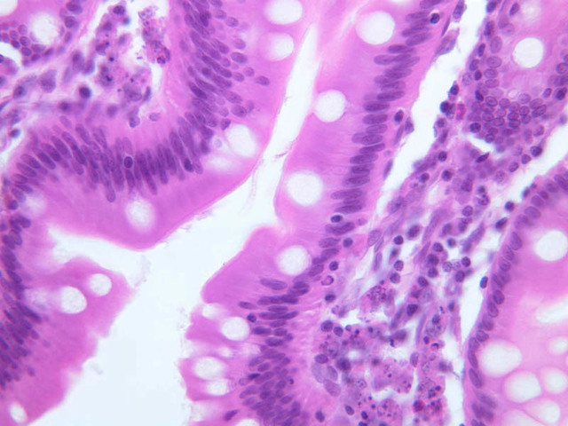
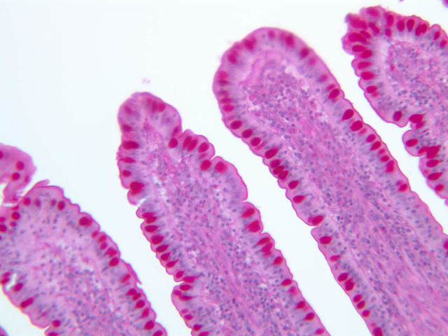
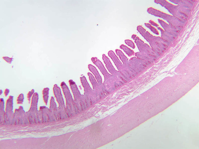
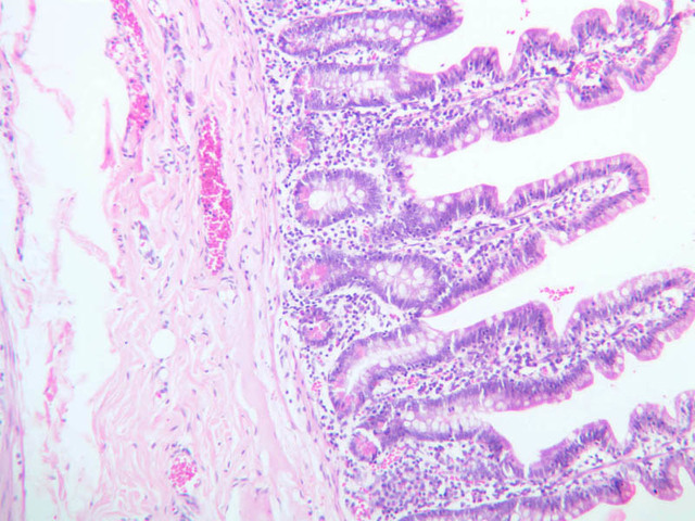
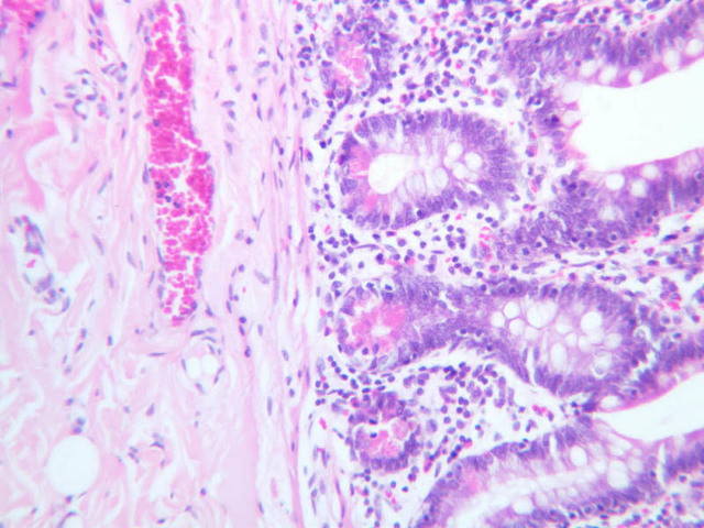
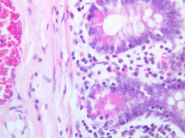
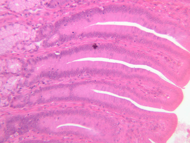
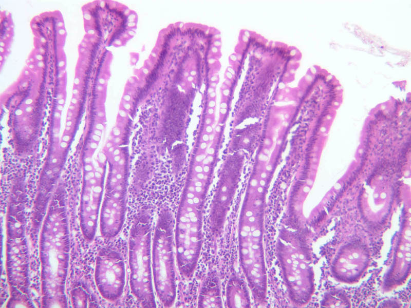
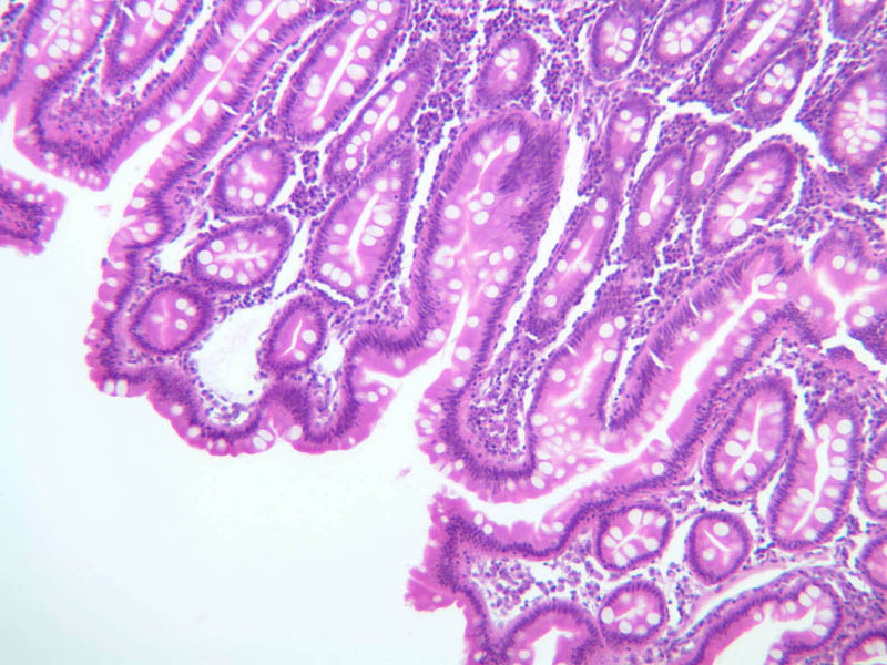
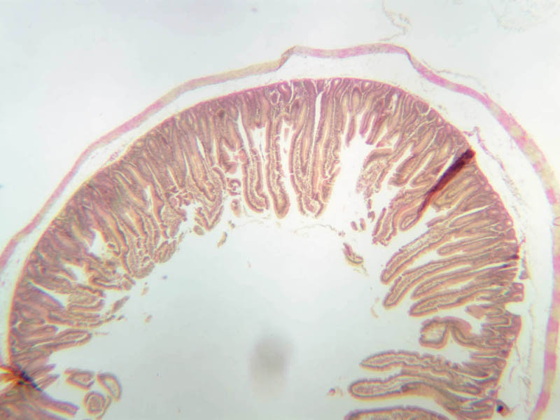
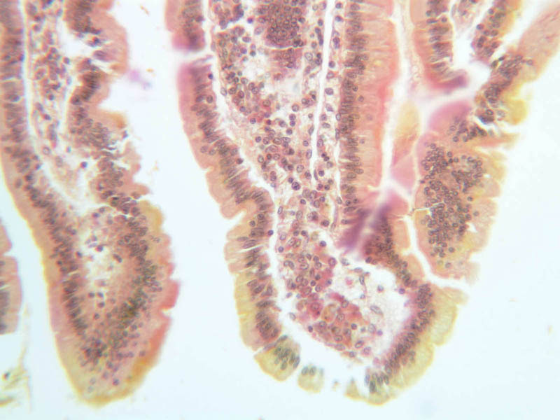
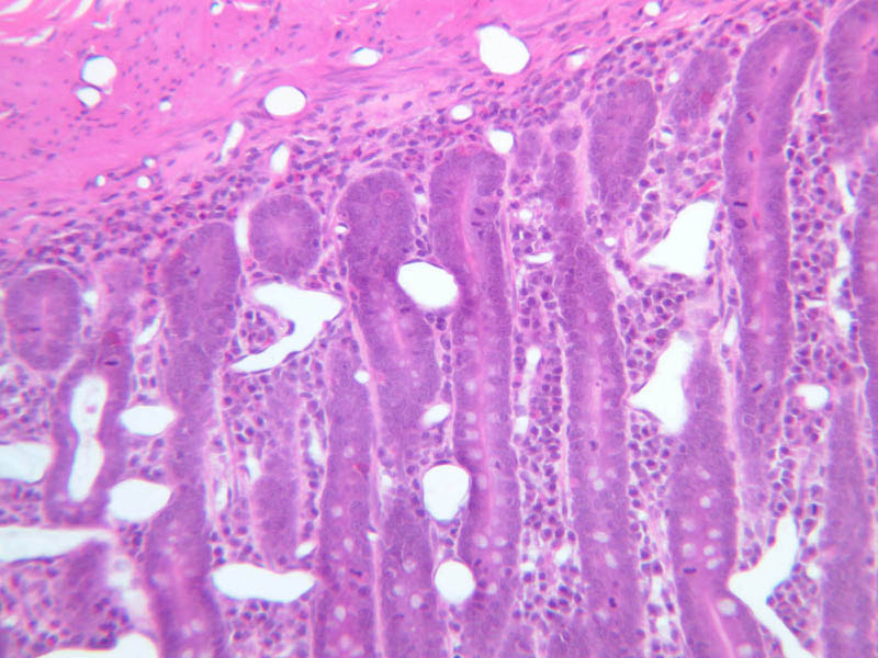
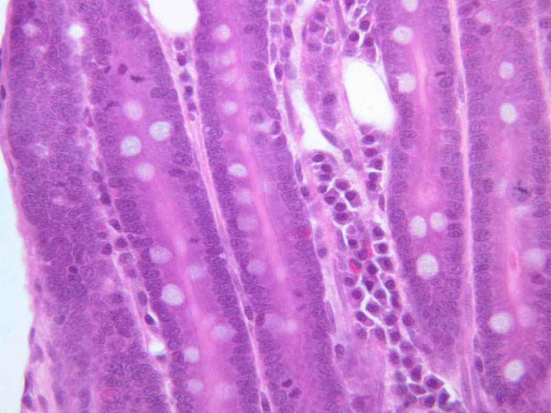
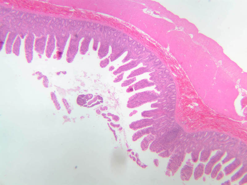
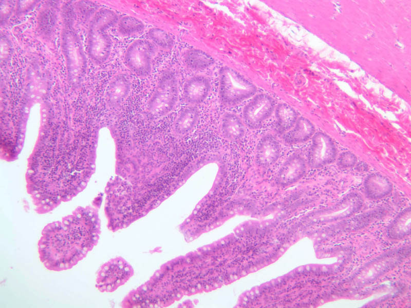
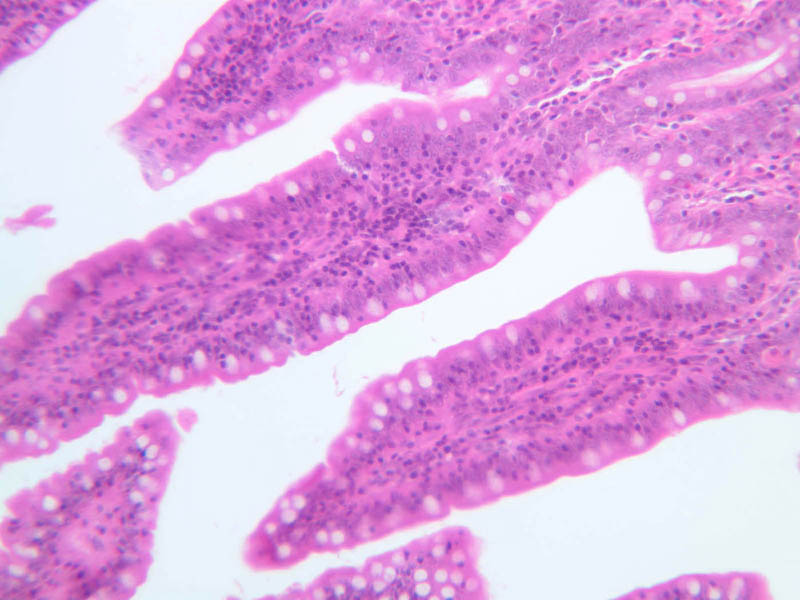
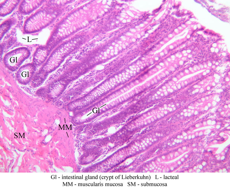
Small Intestine
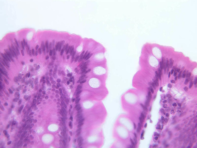
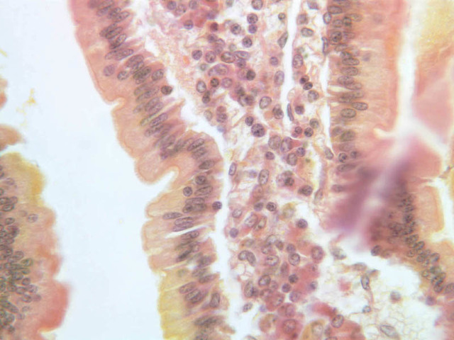
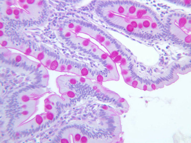
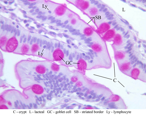
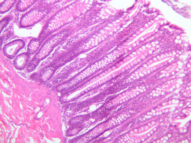
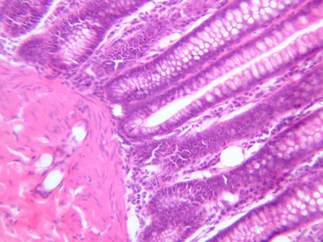
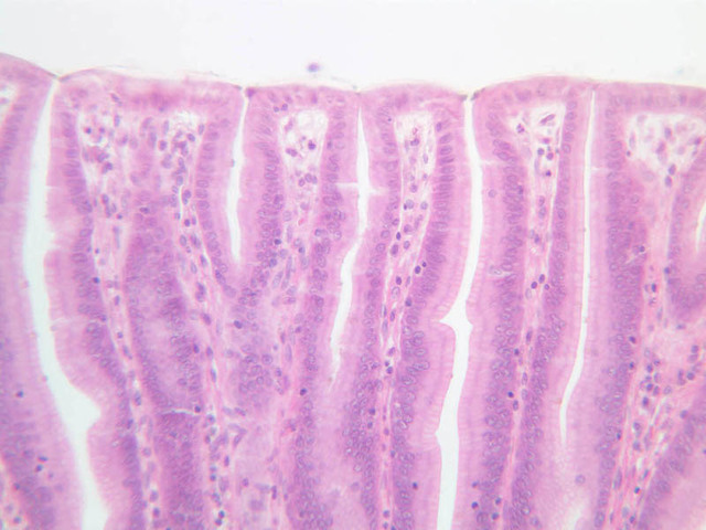
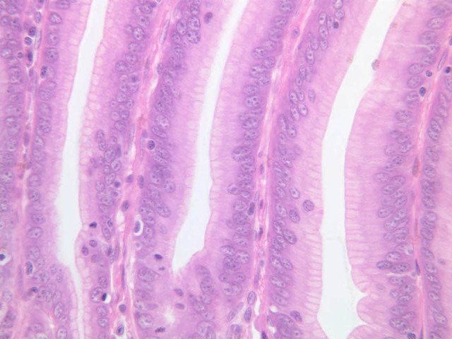
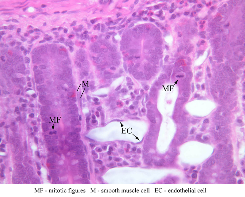
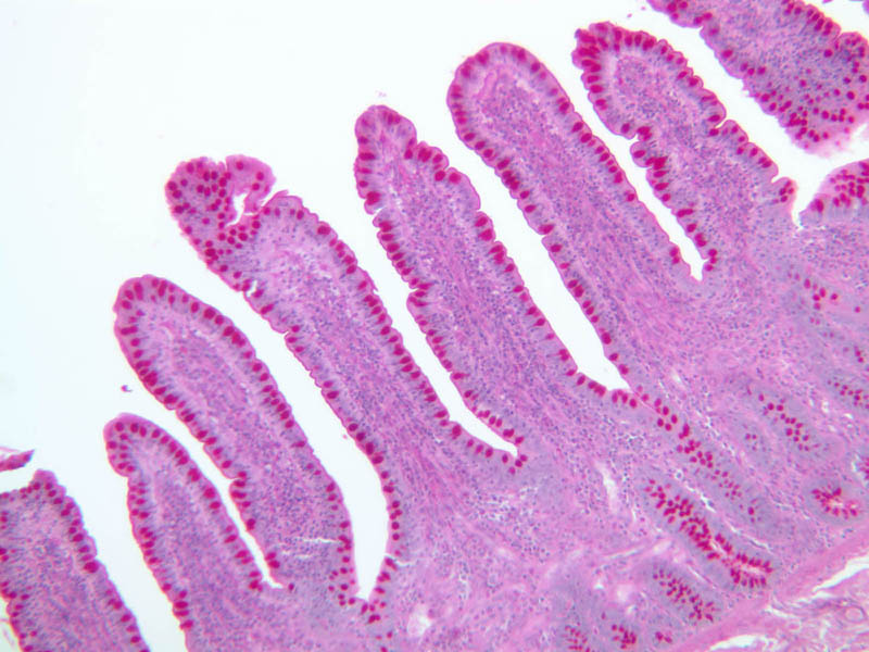
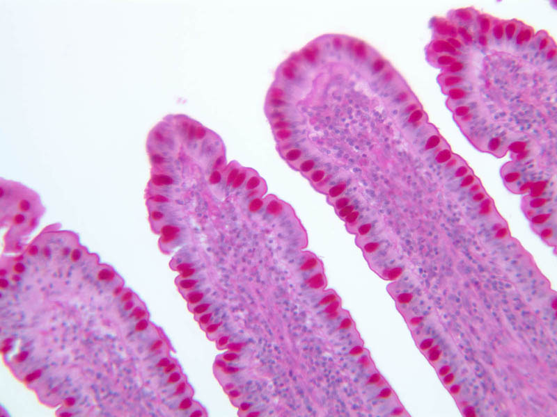
The small bowel is characterized by the presence of long finger-like villi that project into the lumen and small, simple tubular glands between them referred to as the crypts of Lieberk\xFChn. The small intestine also features periodic transverse ridges of submucosa and overlying mucosa, known as plicae circulares or valves of Kerckring. By careful examination of slides B-10, 12 & 16, you should be able to find clear examples of intestinal glands (i.e., crypts of Lieberk\xFChn) opening between adjacent villi. In this segment of the digestive tract, the process of digestion is completed and the end products of digestion are selectively absorbed into the blood and lymph vessels of the mucosa. The epithelial cells of the small intestine are thus specialized for absorption as well as secretion. Absorptive cells are located on the villi, which are projections of the mucosa that serve to increase the absorptive surface. Absorptive cells (enterocytes) have a prominent striated border composed of microvilli, another device to increase surface area. Electron micrographs of absorptive cells show that the microvilli are of surprisingly regular height. Secretory cells of three types are located in the intestinal glands. They are goblet cells, argentaffin cells (APUD cells), and Paneth cells. The APUD cells of the small intestine secrete serotonin, cholcystokinin-pancreozymin (CCK), secretin, and gastric inhibitory peptide (GIP). Paneth cells are pyramidal-shaped zymogen-producing cells located in the bottoms of the glands. Their secretory product is lysozyme, an enzyme that degrades bacterial cell walls. The high turnover rate of the intestinal epithelium is evidenced by numerous mitotic figures, especially in the more basal regions of the intestinal glands. Progeny of mitotically active, undifferentiated "stem" cells move up the crypts and onto the villi, assuming the definitive characteristics of either goblet cells or enterocytes along the way. Mature cells of both types degenerate and are shed as they reach the apices of the villi. Complete renewal of the intestinal epithelium takes less than a week.Back to Top of Page
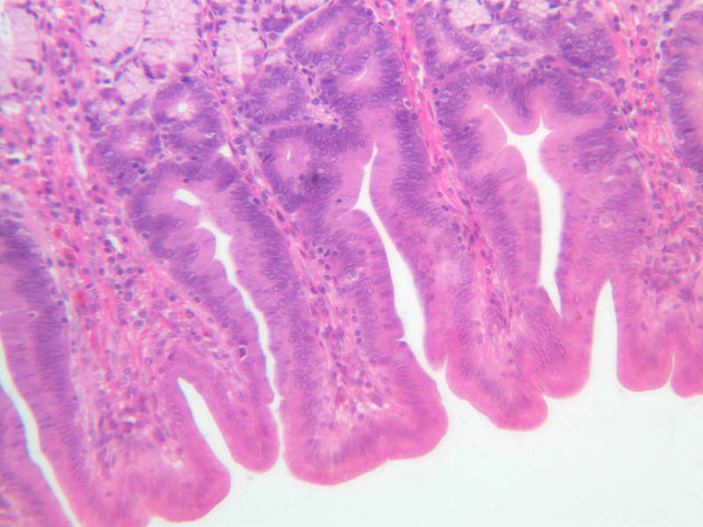
Duodenum
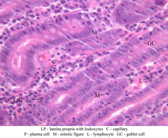
Examine a section of monkey duodenum (slide B-10, H&E [2.5x-labeled, 10x, 20x, 40x] [40x]) and note the characteristics just described for all segments of the small intestine. Note the so-called plicae circulares, which consist of folds of submucosa and its overlying mucosa. Note the shape of the villi, which are often described as leaf-shaped. Examine the epithelial cells lining the villi, distinguishing absorptive cells and goblet cells. Note the striated border which appears as a line along the free surface. Look at the glands and identify goblet cells. The other cell types present are not distinguishable in an H&E section. Look briefly for mitotic figures in the glands (B-10, H&E [2.5x, 10x, 20x, 40x-labeled]). Examine the lamina propria, especially that forming the core of the villus. It is an unusually cellular tissue, including numerous leukocytes and plasma cells, among other types. Note once again the lacteal which forms the axis of the villus and recall its function in absorption. Locate the capillaries of the villus just beneath the absorptive epithelium. Smooth muscle fibers extend vertically from the muscularis mucosae into the core of each villus and help to empty capillaries and lacteals. Look for Brunner's glands and elements of Meissner's plexus in the submucosa. Examine the muscularis externa and look for examples of the myenteric plexus (B-10, H&E [2.5x, 10x-labeled, 20x, 40x]). Also, try to identify the thin tunica serosa on the outer surface of the section. Finally examine the silver-stained section of duodenum on slide B-11 (Ag [10x, 20x, 40x, 40x, 40x]) for examples of argentaffin (APUD) cells. Which gastrointestinal peptides are produced by APUD cells of the duodenum?Duodenum Image Gallery


Duodenum Table of Identifications
| Row | Structure | Abbreviation | Optimal Stain | Representative Section | Note |
|---|---|---|---|---|---|
| 1 | Brunner's Glands | (none) | H&E | |
|
| 2 | Crypts of Lieberkuhn | (none) | H&E | |
|
| 3 | Villi | (none) | H&E | |
|
| 4 | Lamina Propria (with Leukocytes) | LP | H&E | |
|
| 5 | Capillary | C | H&E | |
|
| 6 | Plasma Cell | P | H&E | |
|
| 7 | Mitotic Figure | M | H&E | |
|
| 8 | Lymphocyte | L | H&E | |
|
| 9 | Goblet Cell | GC | H&E | |
|
| 10 | Circular Layer, Muscularis Externa | CM | H&E | |
|
| 11 | Longitudinal Layer, Muscularis Externa | LM | H&E | |
|
| 12 | Tunica Adventitia | TA | H&E | |
|
| 13 | Artery | A | H&E | |
|
| 14 | Vein | V | H&E | |
|
| 15 | Auerbach's (Myenteric) Plexus | MP | H&E | |
Back to Top of Page
Jejunum
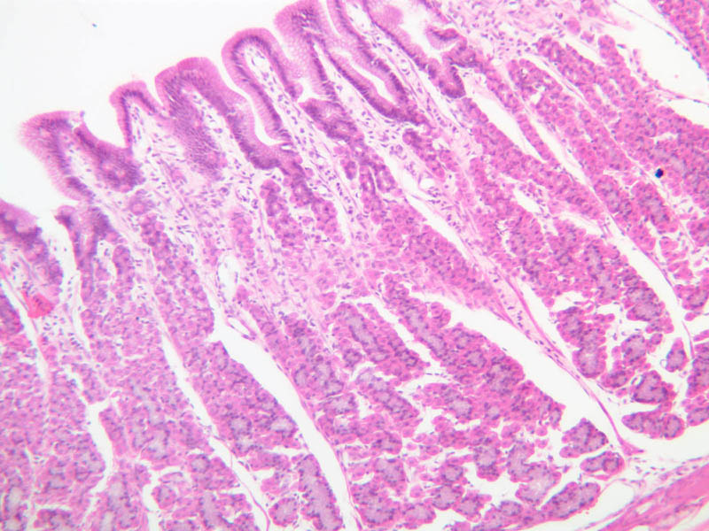
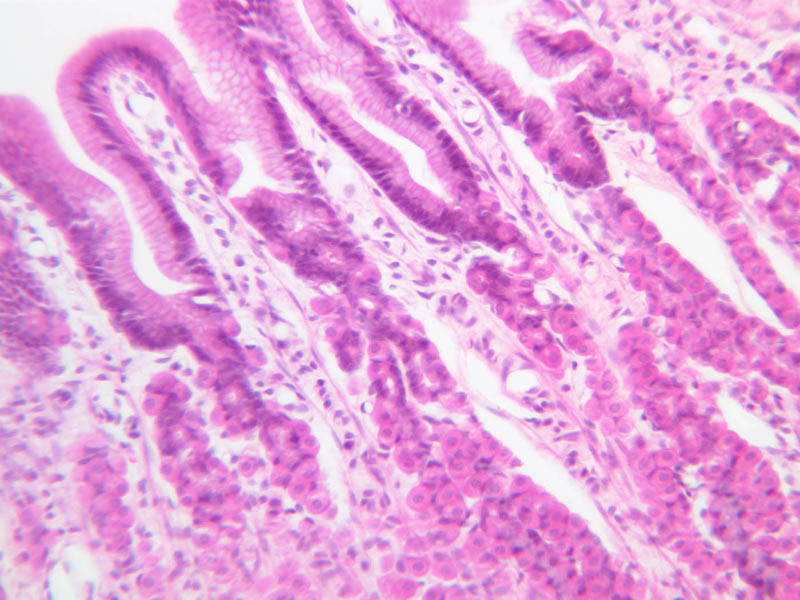
Examine a section of the jejunum (slide B-12, H&E [2.5x-labeled, 10x, 20x, 40x] [2.5x, 10x, 20x, 40x]; B-13, Fe [2.5x, 10x, 20x, 40x]; & B-15, H&E [2.5x, 10x, 20x, 40x, 40x-labeled]). Note that the villi are taller and narrower than those of the duodenum. Look at the distribution of goblet cells and note the striated border on the absorptive cells. The intestinal glands in the jejunum are limited to the mucosa. To fully appreciate the distribution of goblet cells and the extent of the striated border, examine a section of intestine which has been stained by the PAS technique for polysaccharides (slide B-13, PAS [2.5x-labeled, 10x, 20x, 40x-labeled]).Jejunum Image Gallery


Jejunum Table of Identifications
| Row | Structure | Abbreviation | Optimal Stain | Representative Section | Note |
|---|---|---|---|---|---|
| 1 | Villi | V | H&E | |
|
| 2 | Intestinal Glands (Crypts) | Gl | H&E, PAS | |
|
| 3 | Submucosa | SM | H&E, PAS | |
|
| 4 | Muscularis Externa | ME | H&E, PAS | |
|
| 5 | Serosa | S | H&E | |
|
| 6 | Mitotic Figure | MF | H&E | |
|
| 7 | Smooth Muscle Cell | M | H&E | |
|
| 8 | Endothelial Cell | EC | H&E | |
|
| 9 | Mucosa | M | PAS | |
|
| 10 | Plica Circulares | PC | PAS | |
|
| 11 | Lacteal | L | PAS | |
|
| 12 | Goblet Cell | GC | PAS | |
|
| 13 | Striated Border | SB | PAS | |
|
| 14 | Lymphocyte | Ly | PAS | |
Back to Top of Page
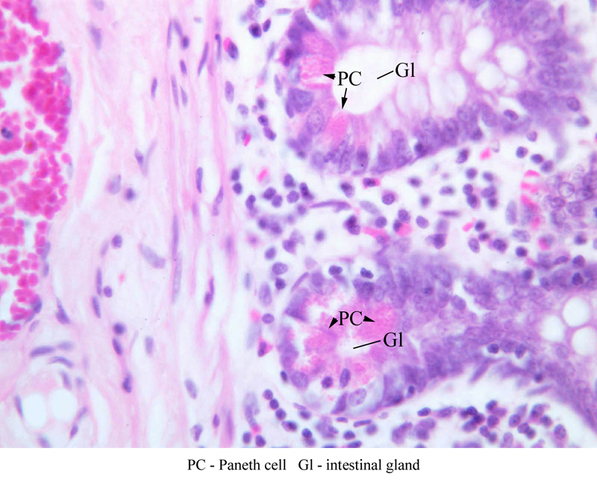
Ileum
Examine a section of the ileum (slides B-16, H&E [2.5x, 10x, 20x, 40x] [2.5x, 10x, 20x, 40x]; B-17, PAS [2.5x, 10x, 20x, 40x]; & B-18, H&E [2.5x, 10x, 20x, 40x]). Note that the villi here are short and club-like. Goblet cells are even more frequent than in the jejunum. The lamina propria is particularly filled with leukocytes; try to distinguish several types in addition to the ubiquitous lymphocytes. Do you see any glands in the submucosa? Compare the three segments of intestine and review the features described. Notice especially the accumulations of lymphoid nodules in the ileum. Collectively, they are known as Peyer's patches. Although Paneth cells occur throughout the small intestine the best examples in your slide sets are in the section of ileum on slide B-19 (H&E [10x, 20x, 40x-labeled]). To locate Paneth cells, which are distinguished by their content of strongly eosinophilic granules, examine the depths of several crypts of Lieberk\xFChn. What is the function of Paneth cells?Ileum Image Gallery


Ileum Table of Identifications
| Row | Structure | Abbreviation | Optimal Stain | Representative Section | Note |
|---|---|---|---|---|---|
| 1 | Paneth Cell | PC | H&E | |
|
| 2 | Intestinal Gland | Gl | H&E | |
Back to Top of Page
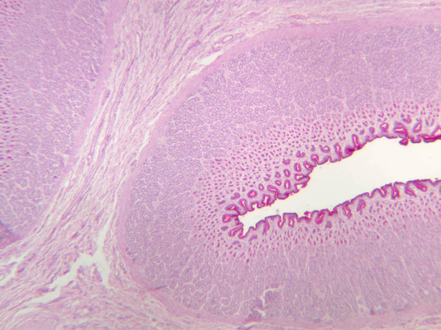
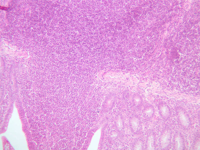
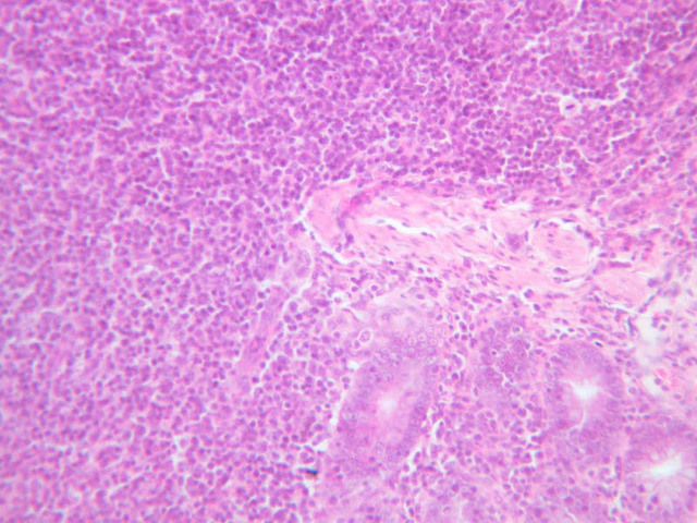
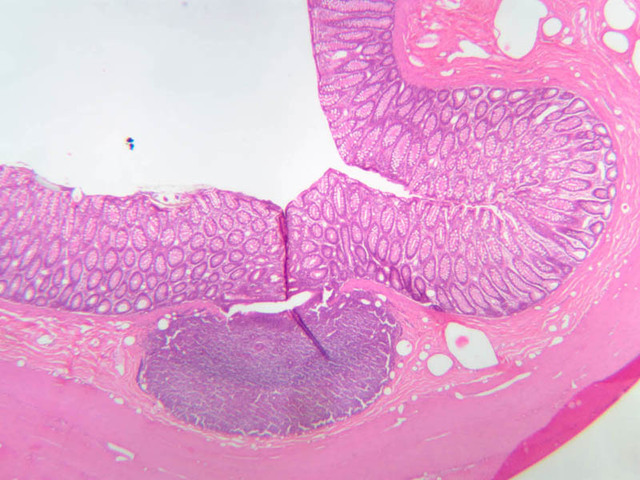
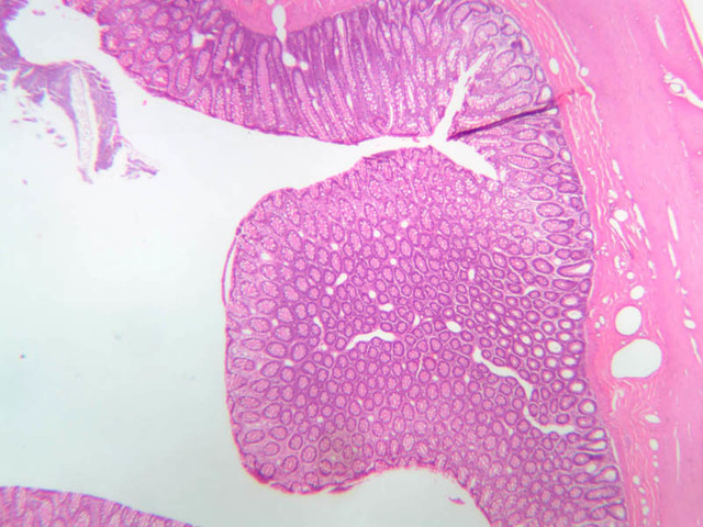
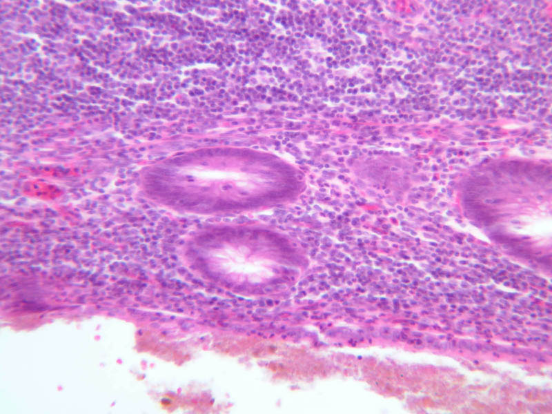
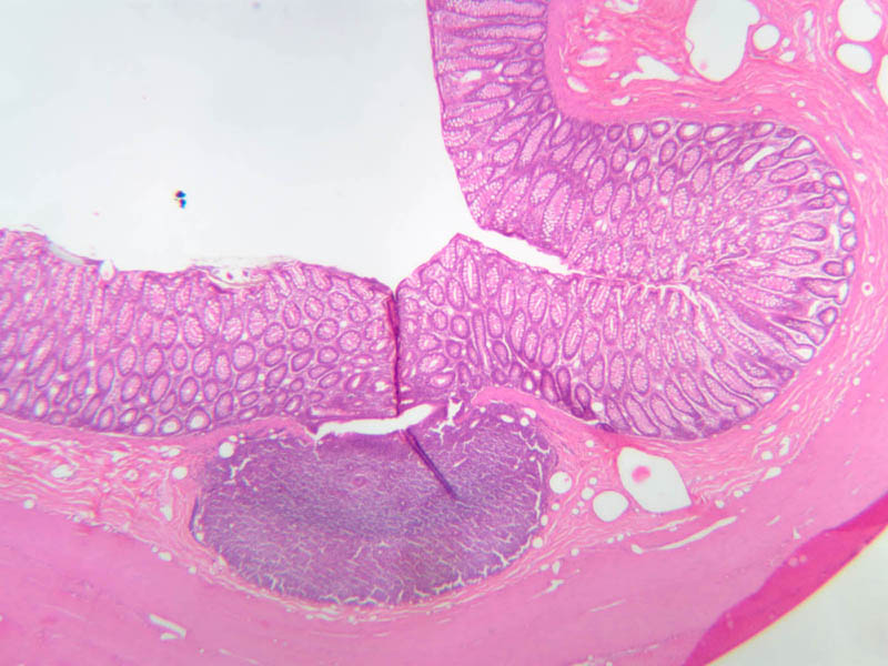
Vermiform appendix
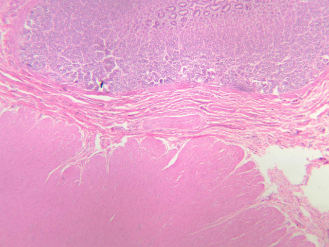
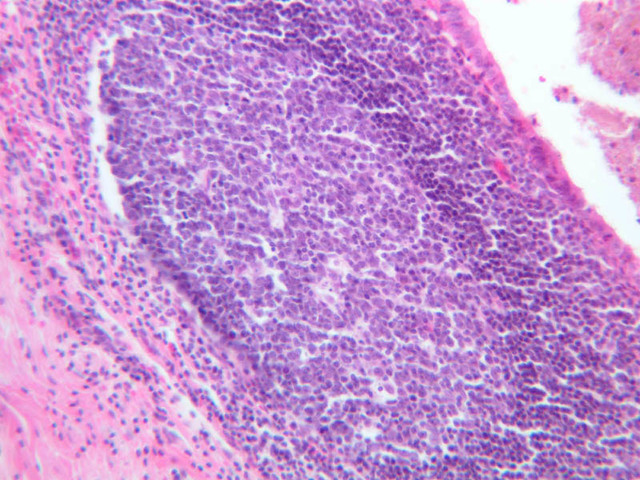
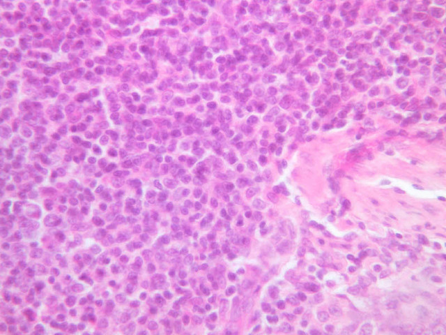
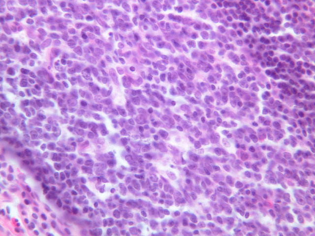
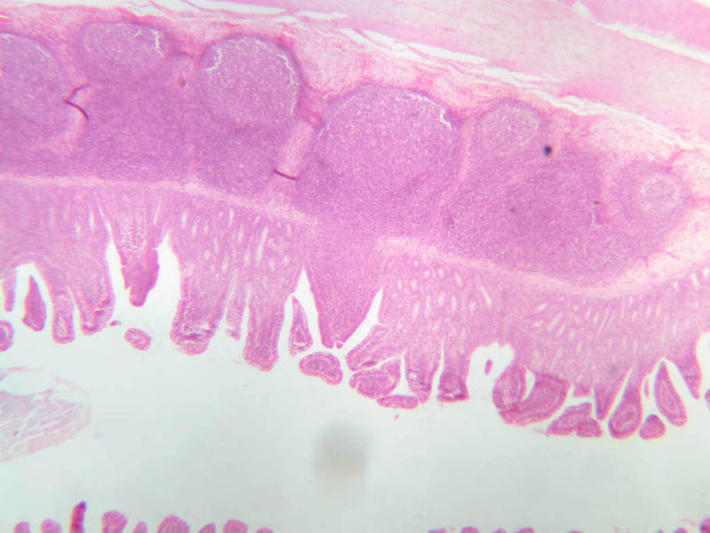
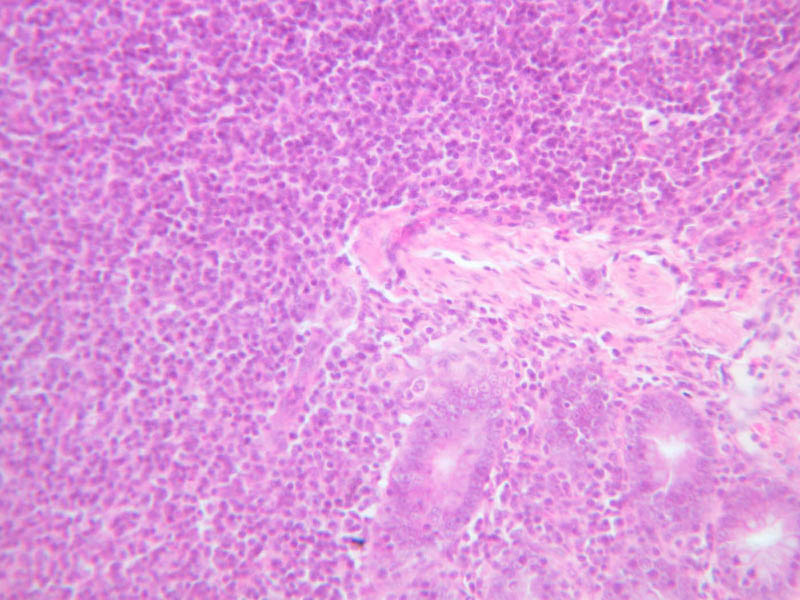
In the section of appendix (slide B-20, H&E [2.5x, 10x, 20x, 40x] [2.5x, 10x, 20x, 40x]), you will find that lymphatic tissue, which is distributed both diffusely and as nodules, distorts the crypts of Lieberk\xFChn and also blurs the usual features of the mucosa and submucosa. Nonetheless, you should be able to find recognizable profiles of intestinal glands as well as slips of the muscularis mucosae among the lymphoid elements. Note that in the appendix there are no absorptive cells and no villi.Vermiform Appendix Image Gallery


Back to Top of Page
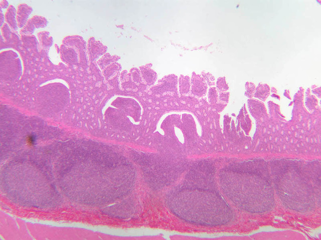
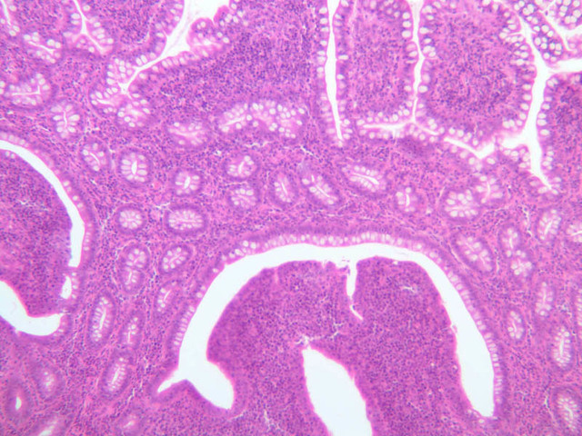
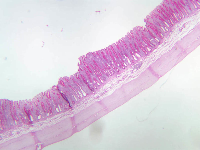
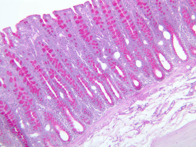
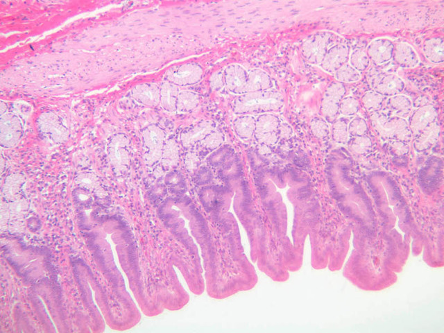
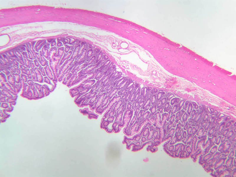
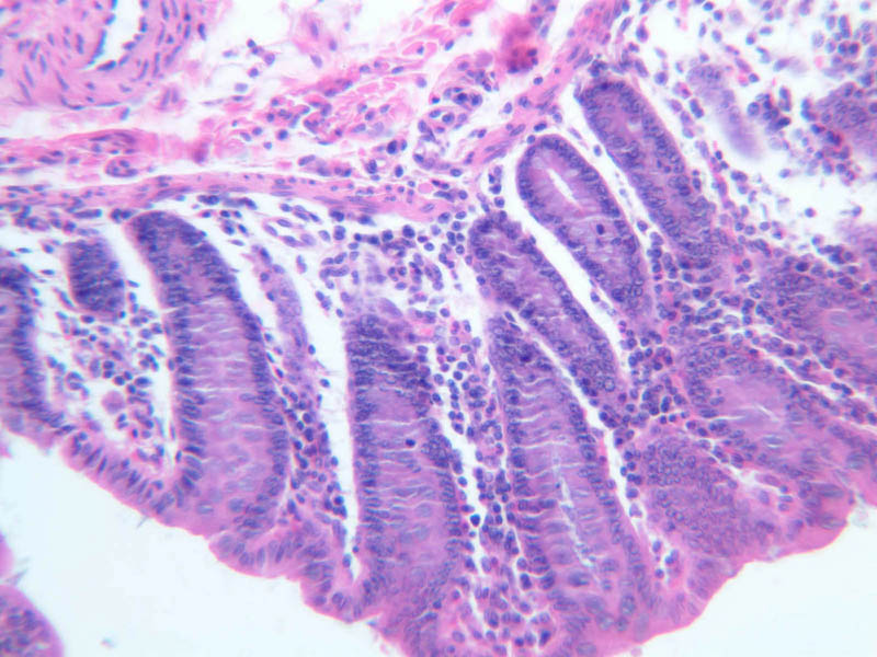
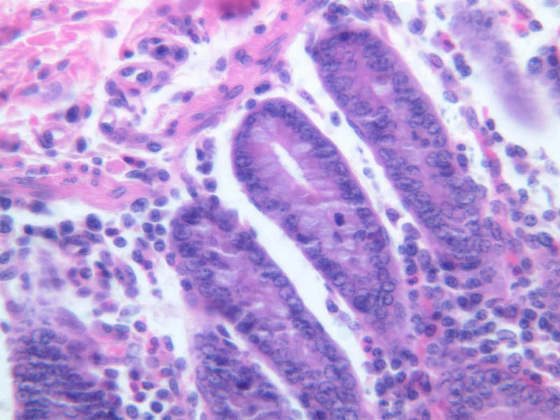
Colon
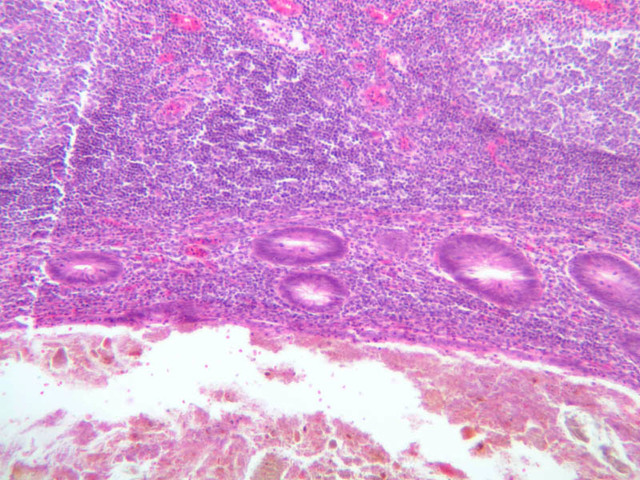
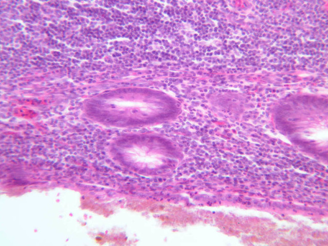
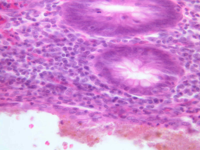
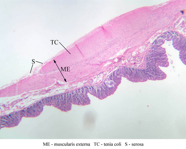
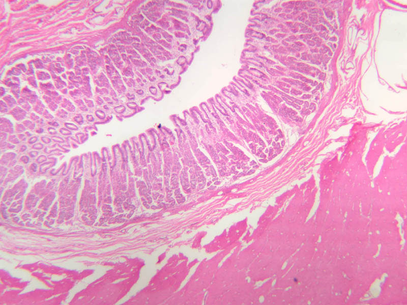
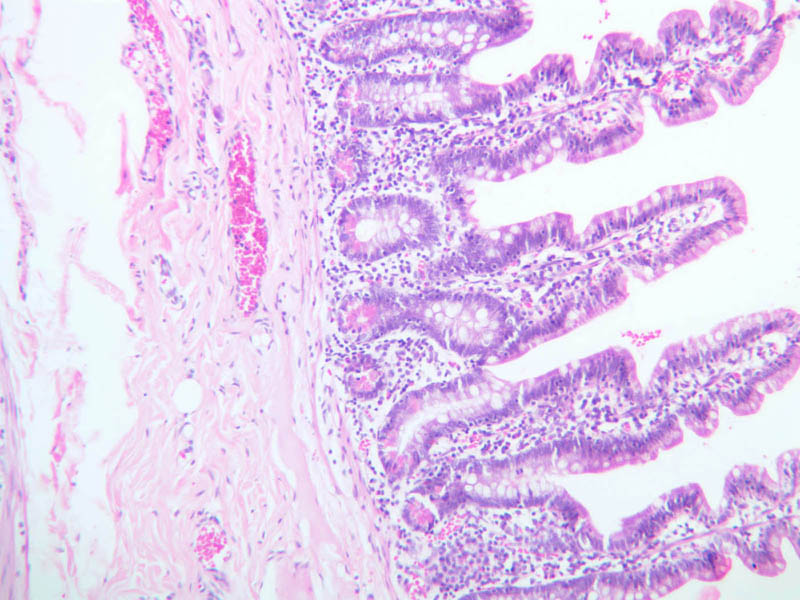
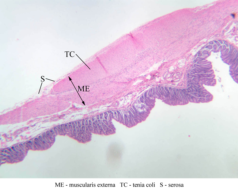
Examine slides B-23 (H&E [2.5x, 10x-labeled, 20x, 40x] [2.5x, 10x, 20x. 40x] [2.5x, 10x, 20x, 40x]), B-24 (H&E [2.5x, 10x, 20x, 40x]), and B-25 (PAS [2.5x, 10x, 20x, 40x] [2.5x-labeled, 10x, 20x, 40x]). Do you see any intestinal villi? Note the great number of goblet cells both in the surface epithelium and in the crypts of Lieberk\xFChn. To strengthen your understanding of regional differences in the frequency and distribution of goblet cells, you might wish to compare the PAS-stained sections of jejunum (B-13), ileum (B-17) and colon (B-25). As you examine the muscularis externa, you should note that over most of their extent both layers are thinner than in other regions of the gut; however, you should be able to find local thickenings of the outer longitudinal layer. These thickenings are known as tenia coli.Colon Image Gallery


Colon Table of Identifications
| Row | Structure | Abbreviation | Optimal Stain | Representative Section | Note |
|---|---|---|---|---|---|
| 1 | Intestinal Gland (Crypt of Lieberkuhn) | Gl | H&E | |
|
| 2 | Lacteal | L | H&E | |
|
| 3 | Muscularis Mucosa | MM | H&E | |
|
| 4 | Submucosa | SM | H&E, PAS | |
|
| 5 | Muscularis Externa | ME | H&E | |
|
| 6 | Tenia Coli | TC | H&E | |
|
| 7 | Serosa | S | H&E, PAS | |
|
| 8 | Mucosa | M | PAS | |
|
| 9 | Circular Layer, Muscularis Externa | CM | PAS | |
|
| 10 | Auerbach's (Myenteric) Plexus | MP | PAS | |
|
| 11 | Longitudinal Layer, Muscularis Externa | LM | PAS | |
Back to Top of Page
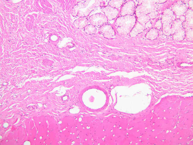
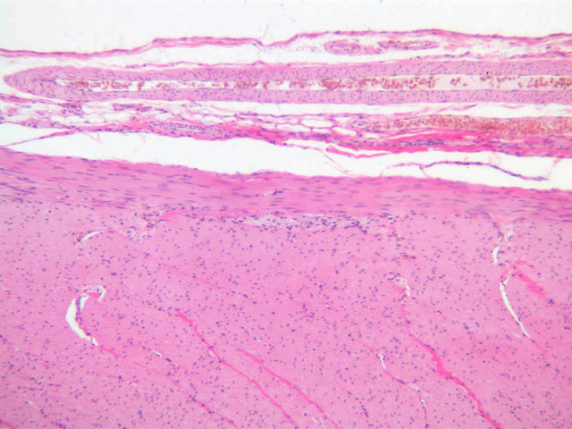
Anal canal
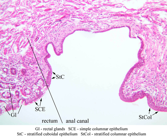
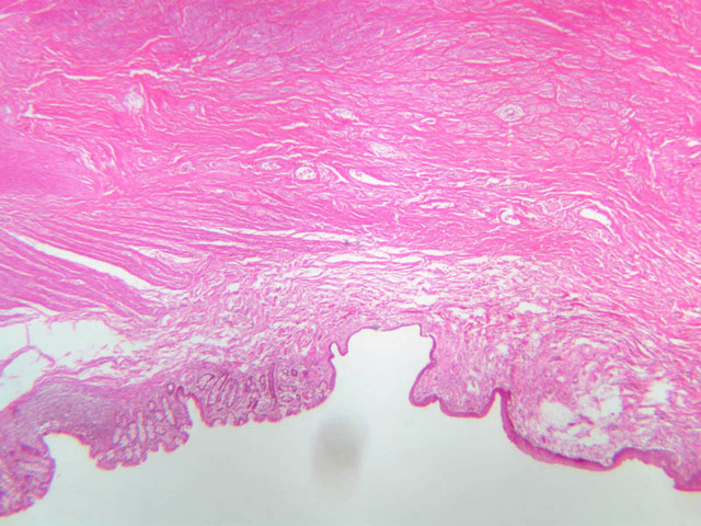
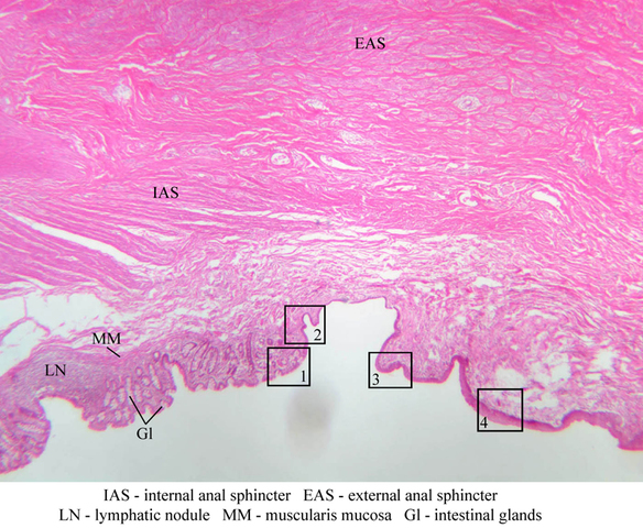
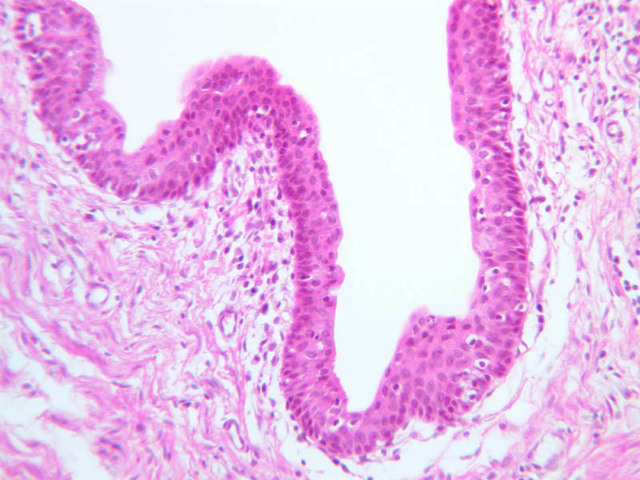
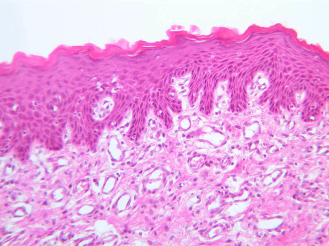
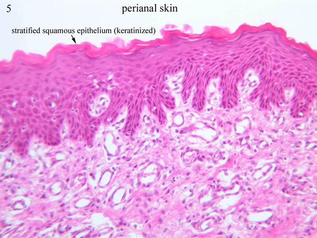
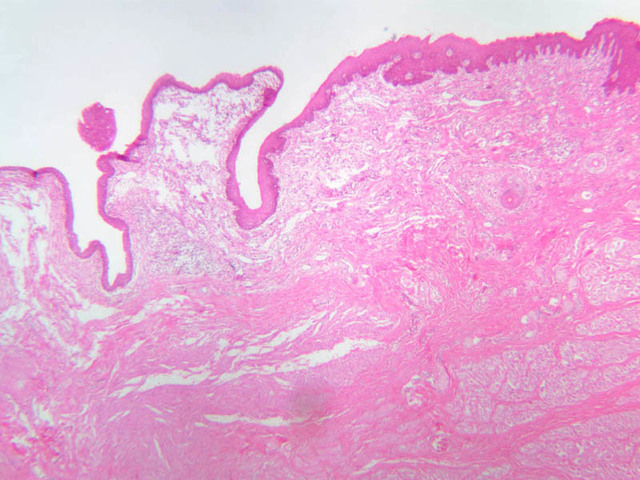
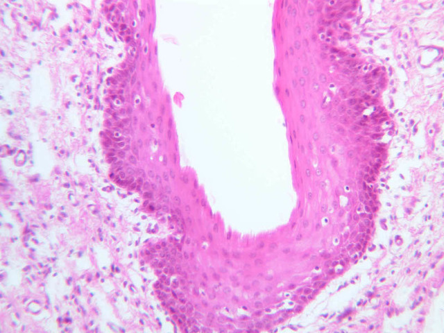
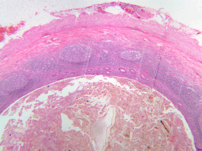
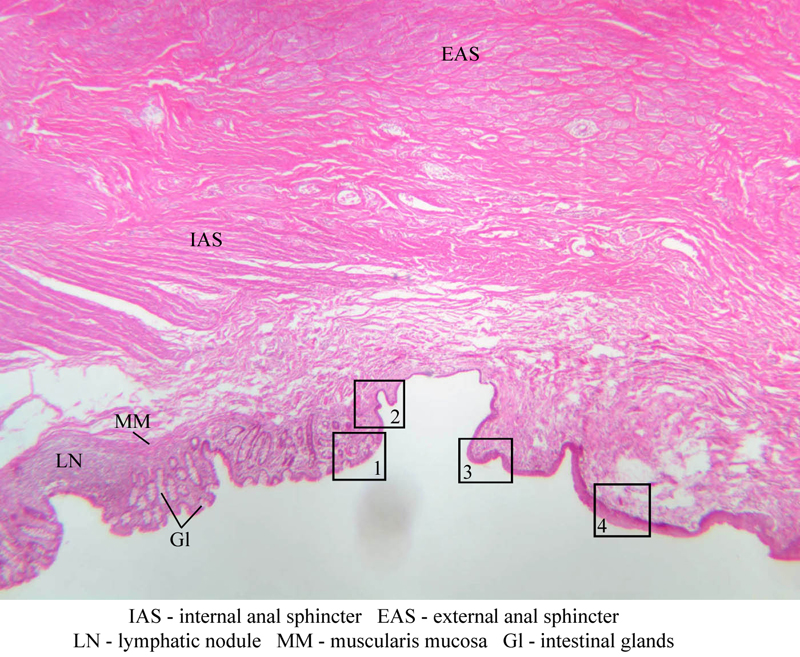
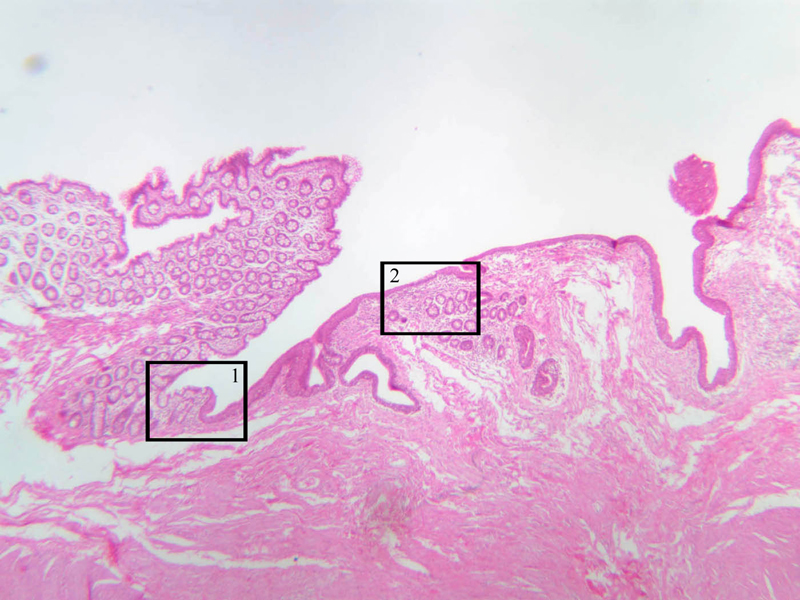
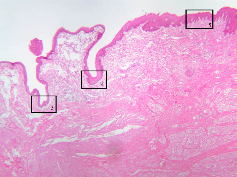
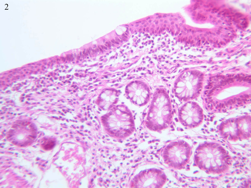
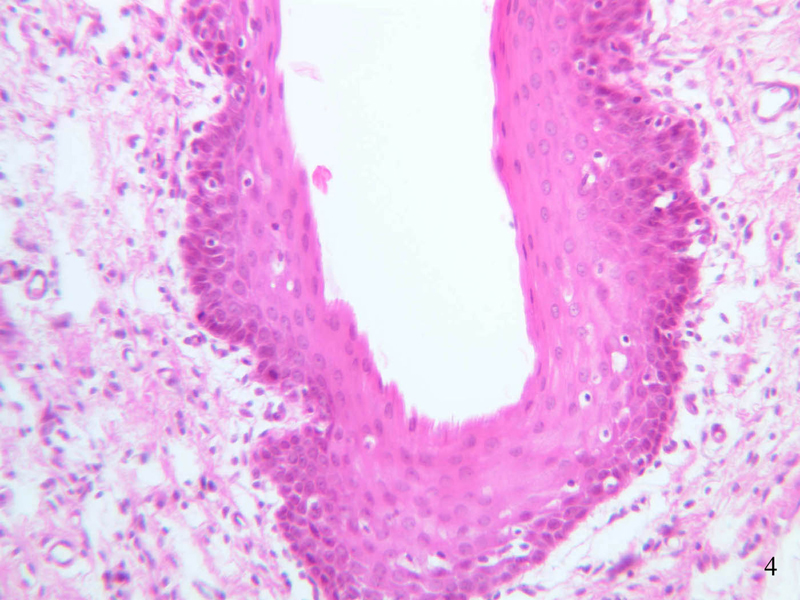
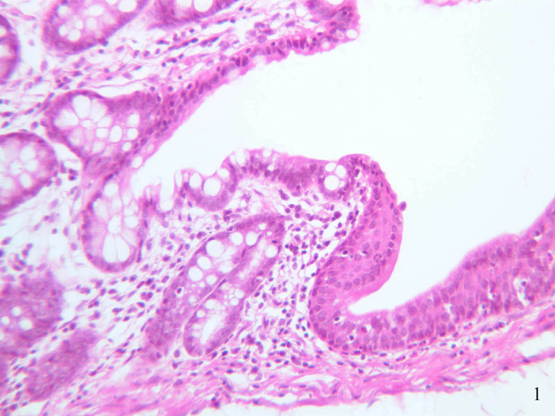
Using the low or medium power objective, scan the section of the rectoanal junction on slide B-26 (H&E [2.5x-labeled, 10x-labeled] [2.5x, 20x, 40x] [2.5x, 20x-labeled, 40x] [20x, 40x] [20x, 40x] [20x] [2.5x, 10x, 20x, 40x]) . Proximally, the rectal mucosa exhibits crypts of Lieberk\xFChn and the simple columnar epithelium is exceptionally rich in goblet cells. As you trace the epithelium towards the anus, you will find that the crypts disappear, then the simple columnar epithelium gives way to stratified squamous epithelium, often with a narrow intervening swath of stratified cuboidal or stratified columnar epithelium. Just proximal to the anal orifice keratin appears and just distal to the anal orifice you may be able to find sweat glands, sebaceous glands and hair follicles.Anal Canal Image Gallery


Anal Canal Table of Identifications
| Row | Structure | Abbreviation | Optimal Stain | Representative Section | Note |
|---|---|---|---|---|---|
| 1 | Internal Anal Sphincter | IAS | H&E | |
|
| 2 | External Anal Sphincter | EAS | H&E | |
|
| 3 | Lymphatic Nodule | LN | H&E | |
|
| 4 | Muscularis Mucosa | MM | H&E | |
|
| 5 | Intestinal Glands | Gl | H&E | |
|
| 6 | Rectum | (none) | H&E | |
|
| 7 | Anal Canal | (none) | H&E | |
|
| 8 | Rectal Glands | Gl | H&E | |
|
| 9 | Simple Columnar Epithelium | SCE | H&E | |
|
| 10 | Stratified Cuboidal Epithelium | StC | H&E | |
|
| 11 | Stratified Columnar Epithelium | StCol | H&E | |
|
| 12 | Perianal Skin | (none) | H&E | |
|
| 13 | Stratified Squamous Epithelium (Keratinized) | (none) | H&E | |
Back to Top of Page
Chapter Twelve Review
Review of Slides


Review of Identifications
| Row | Structure | Abbreviation | Optimal Stain | Representative Section | Note |
|---|---|---|---|---|---|
| 1 | Adventitia | (none), Ad | H&E | |
|
| 2 | Muscularis Externa | (none), ME | H&E, PAS | |
|
| 3 | Submucosa | (none), SM | H&E, PAS | |
|
| 4 | Mucosa | (none), M | H&E, PAS | |
|
| 5 | Stratified Squamous Epithelium | SSE | H&E | |
|
| 6 | Lamina Propria | LP | H&E | |
|
| 7 | Muscularis Mucosa | MM | H&E | |
|
| 8 | Circular Layer, Muscularis Externa | CM | H&E, PAS | |
|
| 9 | Auerbach's (Myenteric) Plexus | AP, MP | H&E, PAS | |
|
| 10 | Longitudinal Layer, Muscularis Externa | LM | H&E, PAS | |
|
| 11 | Mucous Glands | (none) | H&E | |
|
| 12 | Esophagus | (none) | H&E | |
|
| 13 | Cardiac Stomach | (none) | H&E | |
|
| 14 | Fundus | (none) | H&E | |
|
| 15 | Ruga | (none) | H&E | |
|
| 16 | Gastric Pit | P, GP | H&E, PAS | |
|
| 17 | Fundic Gastric Gland | FG | H&E | |
|
| 18 | Blood Vessel | BV | H&E | |
|
| 19 | Meissner's (Submucosal) Plexus | MP | H&E | |
|
| 20 | Lymphatic Nodule | LN | H&E | |
|
| 21 | Parietal Cell | PC | H&E | |
|
| 22 | Chief Cell | CC | H&E | |
|
| 23 | Lymphocyte | L, Ly | H&E, PAS | |
|
| 24 | Mucous Neck Cell | MNC | H&E, PAS | |
|
| 25 | Mucous Surface Cell | MSC | H&E, PAS | |
|
| 26 | Gastric Glands (Corpus) | GG | PAS | |
|
| 27 | Smooth Muscle | SM | H&E | |
|
| 28 | APUD (Enteroendocrine) Cell | (none) | Ag | |
|
| 29 | Fundus | (none) | H&E | |
|
| 30 | Glands (Pylorus) | G | H&E | |
|
| 31 | Pyloric Sphincter | (none) | H&E | |
|
| 32 | Pyloric Stomach | (none) | H&E | |
|
| 33 | Duodenum | (none) | H&E | |
|
| 34 | Brunner's Glands | BG, (none) | H&E | |
|
| 35 | Crypts of Lieberkuhn (Intestinal Glands) | (none), Gl | H&E | |
|
| 36 | Villi | (none) | H&E | |
|
| 37 | Capillary | C | H&E | |
|
| 38 | Plasma Cell | P | H&E | |
|
| 39 | Mitotic Figure | M, MF | H&E | |
|
| 40 | Goblet Cell | GC | H&E, PAS | |
|
| 41 | Artery | A | H&E | |
|
| 42 | Vein | V | H&E | |
|
| 43 | Serosa | S | H&E, PAS | |
|
| 44 | Smooth Muscle Cell | M | H&E | |
|
| 45 | Endothelial Cell | EC | H&E | |
|
| 46 | Plica Circulares | PC | PAS | |
|
| 47 | Lacteal | L | PAS, H&E | |
|
| 48 | Striated Border | SB | PAS | |
|
| 49 | Paneth Cell | PC | H&E | |
|
| 50 | Tenia Coli | TC | H&E | |
|
| 52 | Internal Anal Sphincter | IAS | H&E | |
|
| 53 | External Anal Sphincter | EAS | H&E | |
|
| 54 | Rectum | (none) | H&E | |
|
| 55 | Anal Canal | (none) | H&E | |
|
| 56 | Rectal Glands | Gl | H&E | |
|
| 57 | Simple Columnar Epithelium | SCE | H&E | |
|
| 58 | Stratified Cuboidal Epithelium | StC | H&E | |
|
| 59 | Stratified Columnar Epithelium | StCol | H&E | |
|
| 60 | Perianal Skin | (none) | H&E | |
|
| 61 | Stratified Squamous Epithelium (Keratinized) | (none) | H&E | |
Back to Top of Page
Comments
Top of page -- AshleyLPistorio - 27 May 2007Edit | Attach | Print version | History: r2 < r1 | Backlinks | View wiki text | More topic actions
Topic revision: r2 - 20 Jun 2015, LorenEvey

- Epithelium
- Connective Tissue
- Muscle
- Nervous Tissue
- Cardiovascular System
- Skin Appendages and Sensory Receptors
- Lymphatic System
- Cartilage and Bone
- Respiratory System
- Peripheral Blood and Bone Marrow
- Oral Cavity and Salivary Glands
- Esophagus and Gastrointestinal Tract
- Pancreas, Liver, and Gall Bladder
- Endocrine Organs
- Male Reproductive System
- Female Reproductive System
- Urinary System
Ideas, requests, problems regarding Medical Histology? Send feedback