 |
|
You are here: Medical Histology>Main Web>AtlasContents>UrinarySystemAtlas17 (20 Jun 2015, LorenEvey)Edit Attach
Chapter Seventeen: The Urinary System


Introduction
The series of organs in this system function to produce, store, and eliminate soluble waste products from the body. The filtrates from the kidney are transported by the ureter to the bladder for storage, and are eliminated by way of the urethra. A major function of the system is to salvage water, sugars, and ions from the filtrate and return them to the blood. In addition, specialized structures within the kidney secrete the vasopressor renin, which increases systemic blood pressure.Kidney
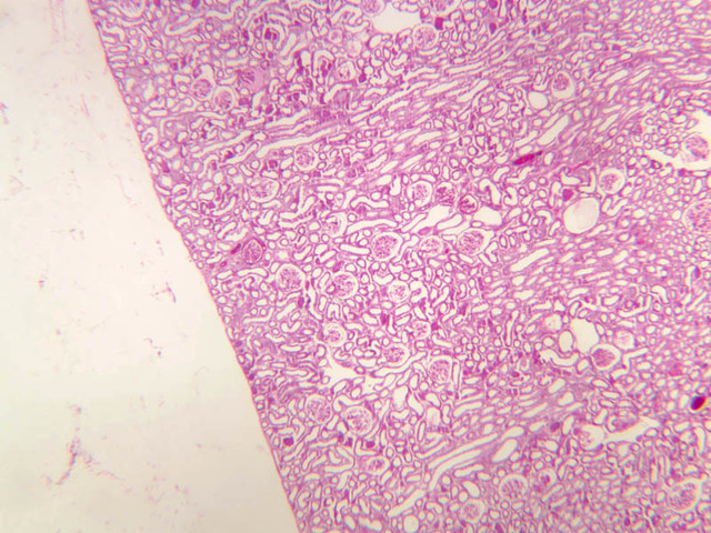
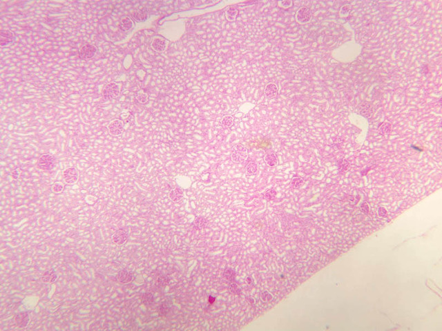
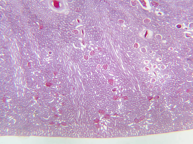
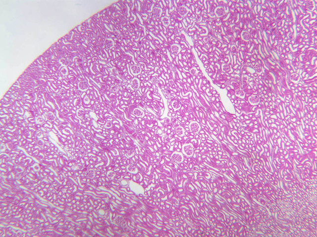
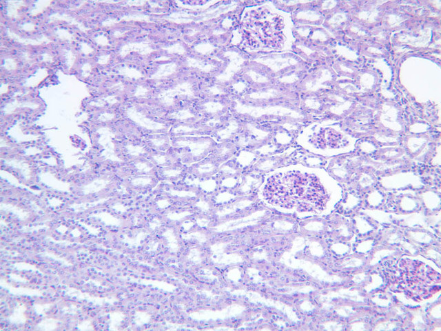
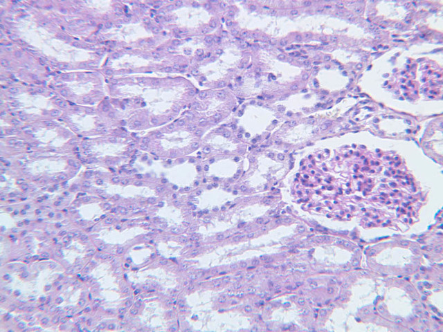
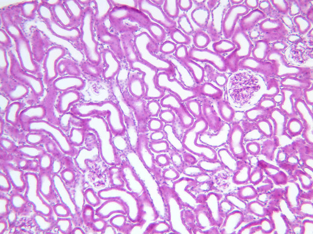
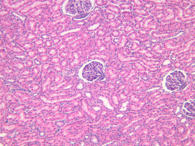
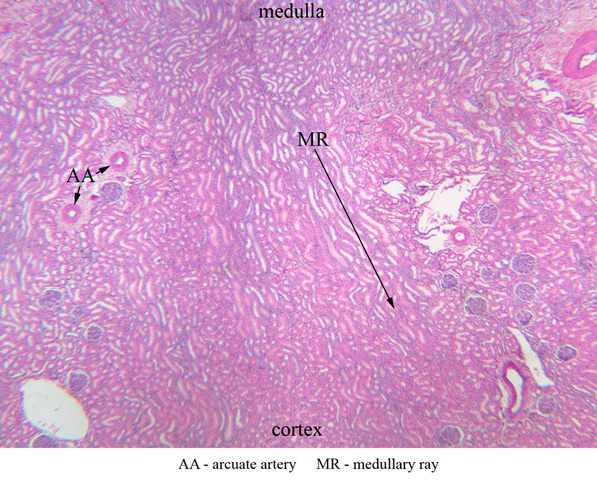
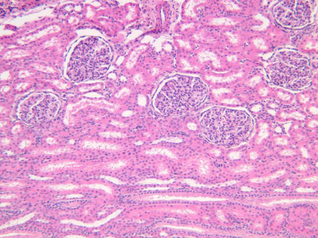
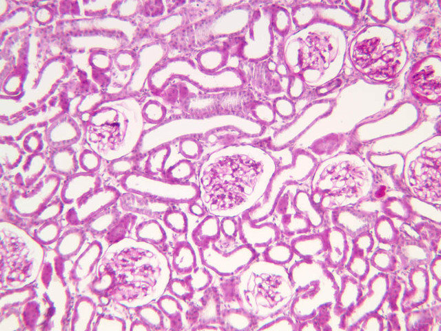
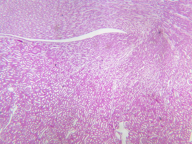
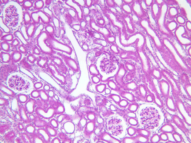
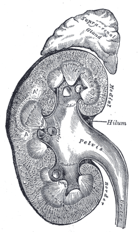
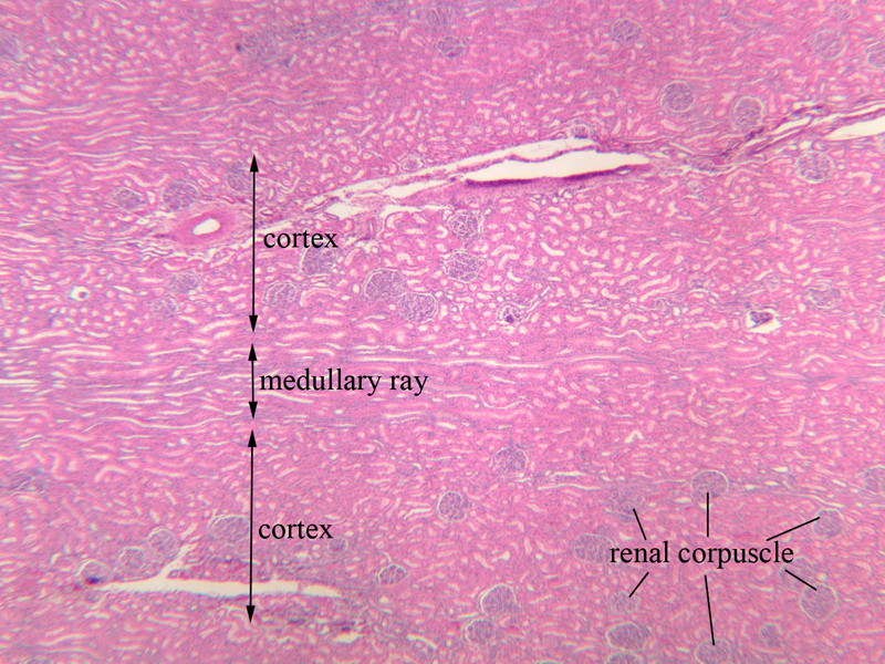
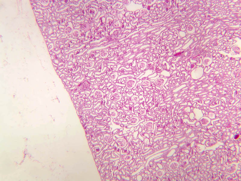
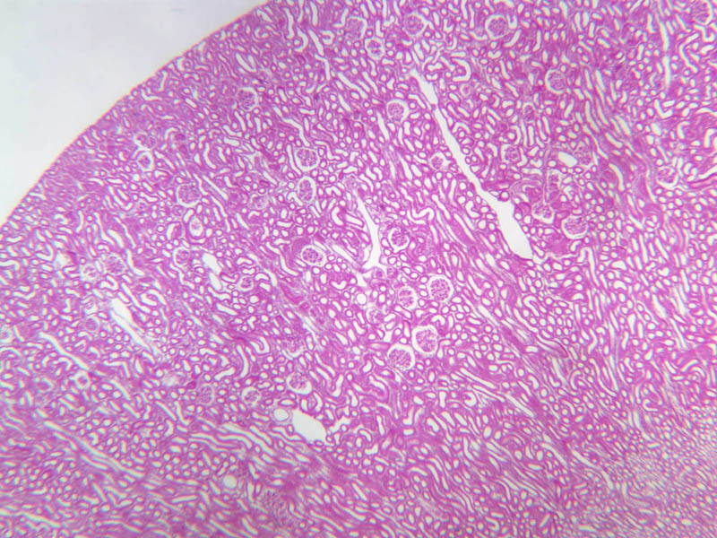
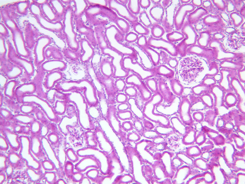
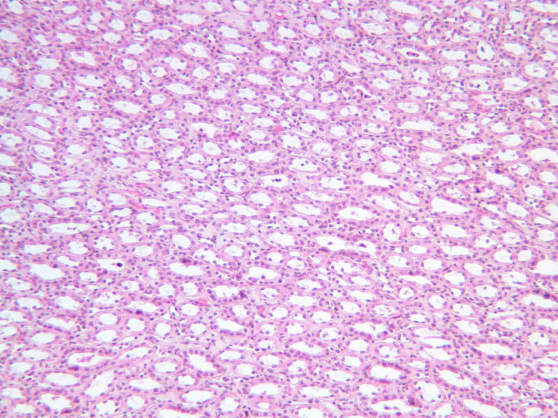
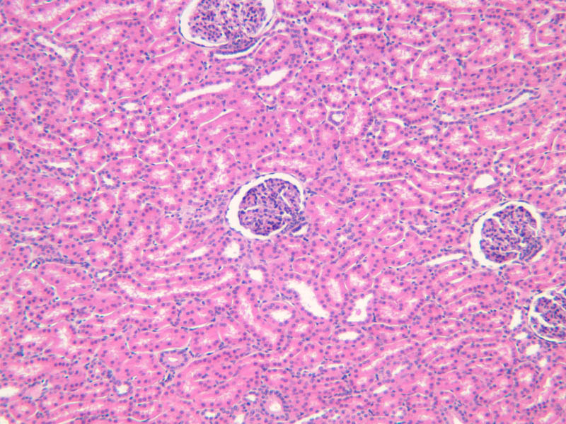
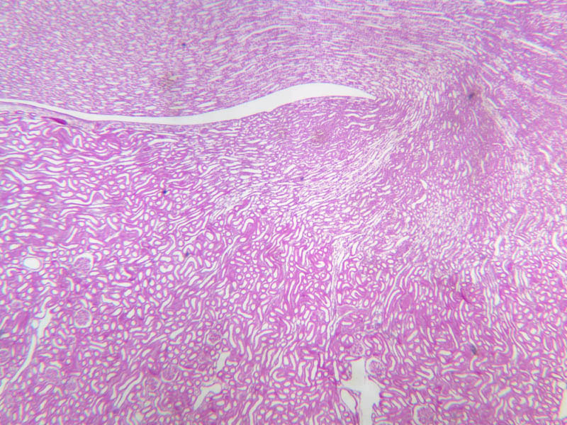
The kidney has a thin, tough connective tissue capsule beneath which lies the glandular tissue. The glandular tissue surrounds a large central cavity called the renal sinus. Adjacent to the renal sinus on the medial side of the organ is a notch called the hilus where the renal vessels and the ureter enter and leave the organ. The glandular portion of the kidney is composed of a number of conical pyramids, the renal lobes. Each renal lobe has its base on the capsule, and its apex (the renal papilla) projects into a minor calyx. The renal lobe consists of two regions, the cortex and the medulla. The cortex has a granular appearance due to the presence of renal corpuscles and renal tubules. These are components of the nephron, the functional unit of the kidney. The medulla has a striated appearance due to the presence of collecting tubules. Structures called medullary rays appear as vertical striations in the cortical substance. Renal columns are projections of cortical tissue between the bases of medullary pyramids.| Structure | Image |
|---|---|
| Gross Anatomical Location of Kidney | |
| Cross Section of Kidney | |
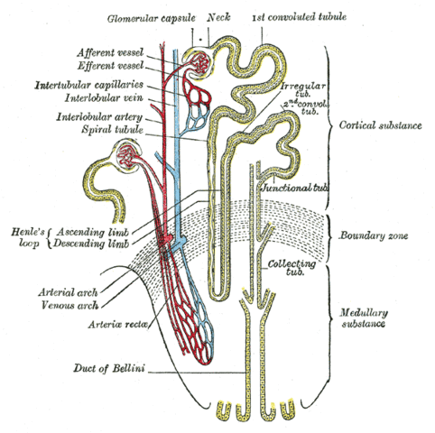
| The Nephron (kidney microanatomy) | |
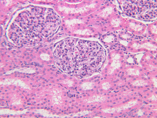
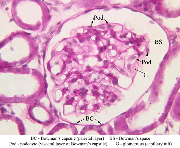
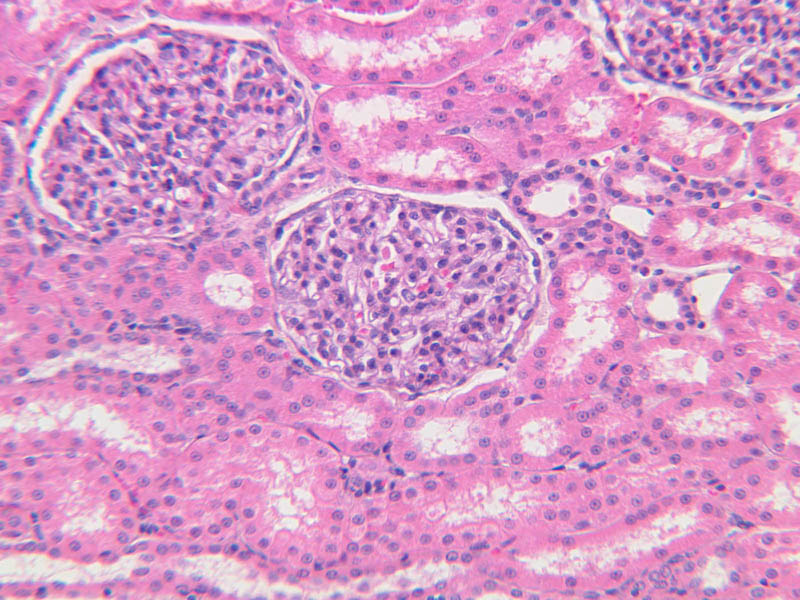
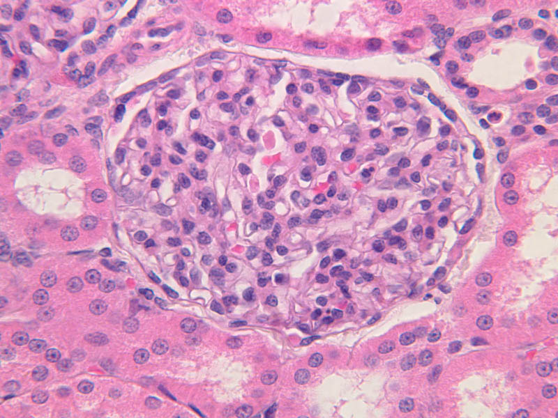
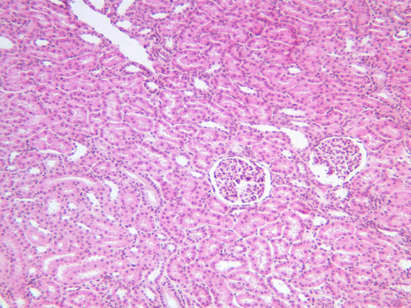
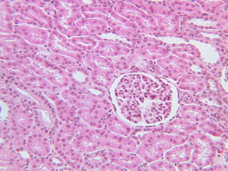
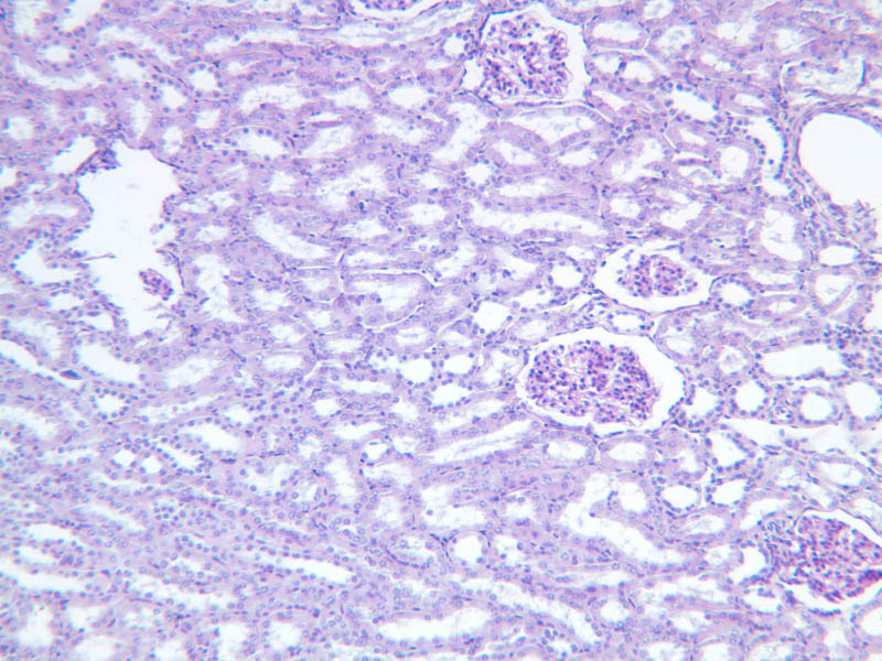
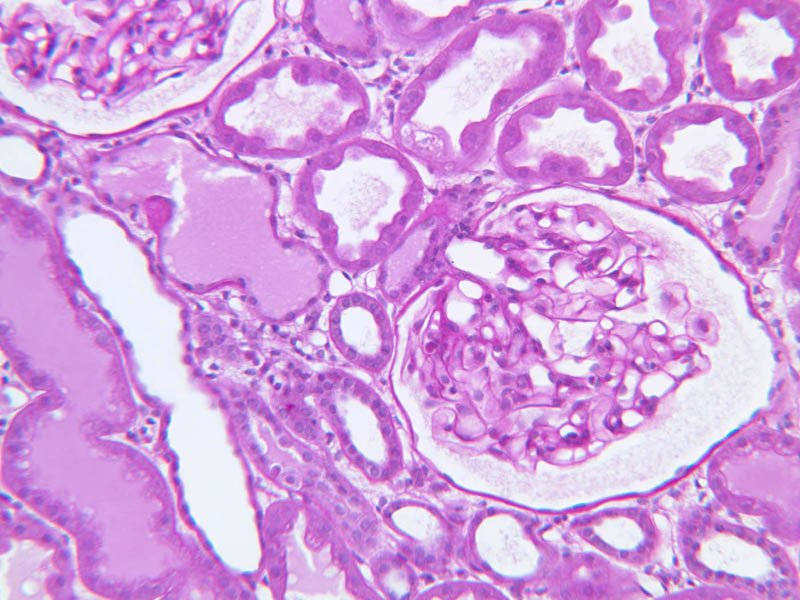
Renal Corpuscle
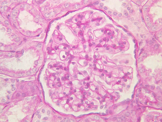
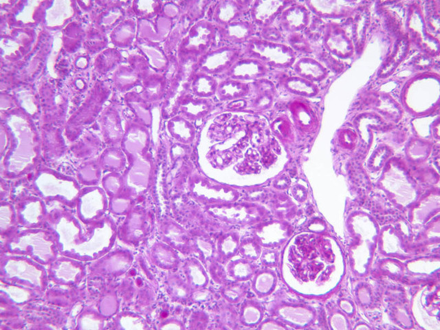
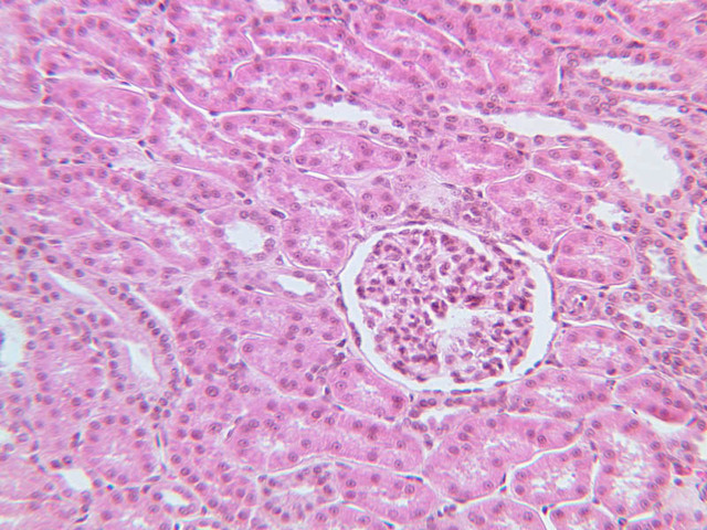
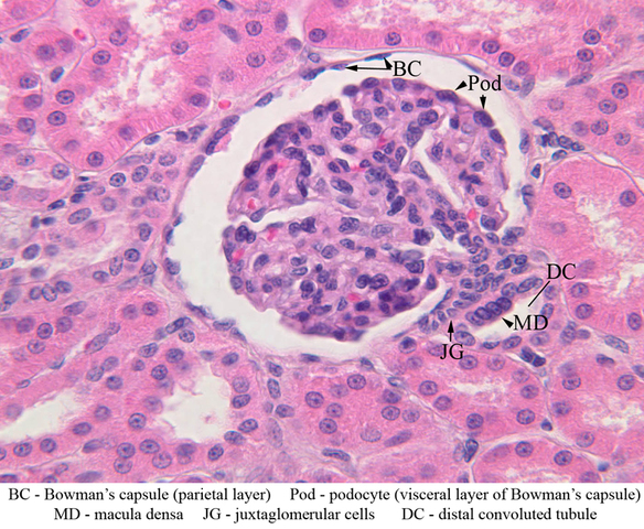
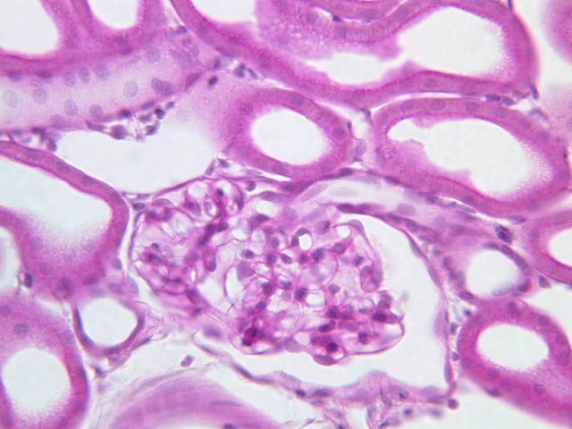
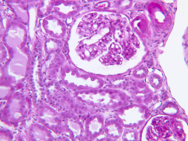
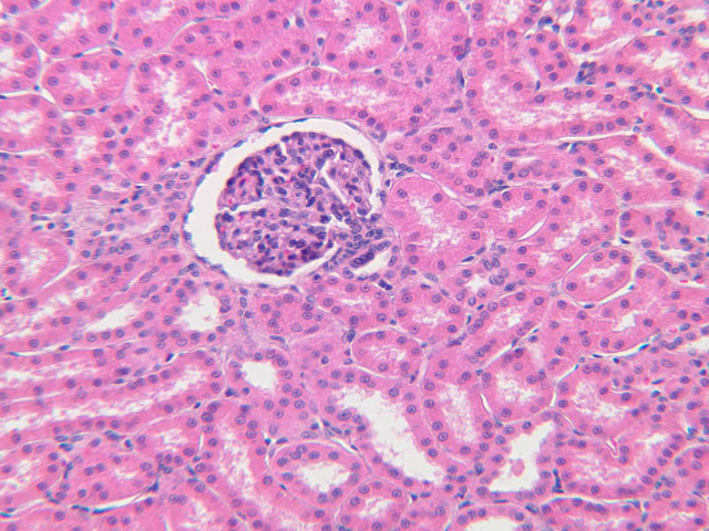
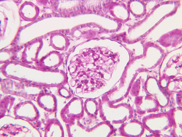
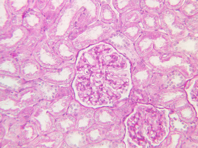
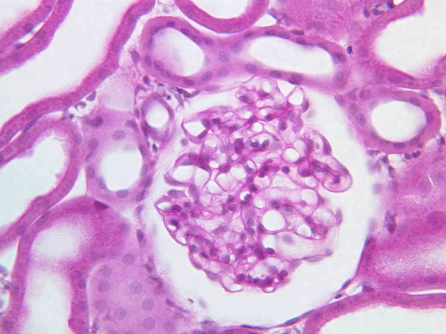
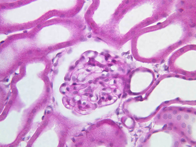
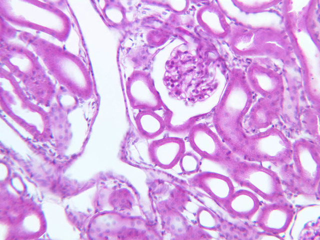
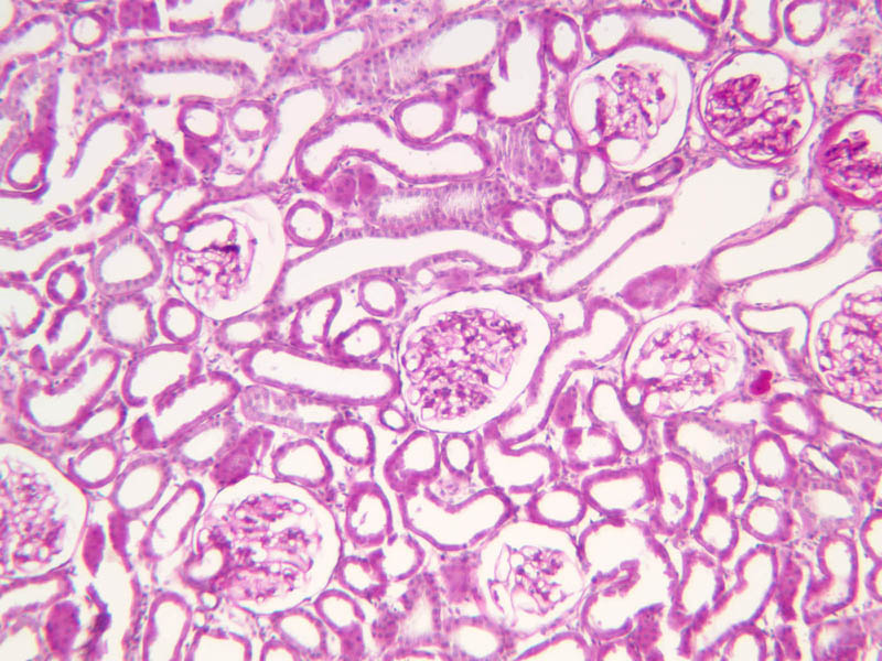
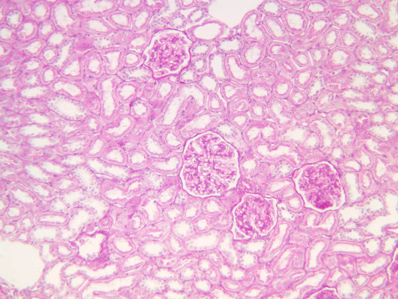
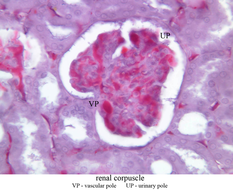
Study a section of the kidney, referring to R-P, Figs. 20.1, 20.3, 20.6 & Pl. 70. In the cortex, locate a renal (Malpighian) corpuscle (slide B-67, H&E [2.5x-labeled, 10x, 20x, 40x]; B-68, PAS [2.5x, 10x, 20x, [40x-labeled]; B-69, PAS [2.5x, 10x, 20x, 40x]; B-70, carmine [2.5x, 10x, 20x, 40x-labeled]; B-71, H&E [2.5x, 10x, 20x, 40x]). A renal corpuscle is the invaginated blind end of a renal tubule called Bowman's capsule. The capsule is composed of a simple squamous epithelium referred to as the parietal layer. Internally (the visceral layer) is a capillary tuft, the glomerulus, and specialized cells that are intimately associated with the capillary tuft. These cells are the podocytes. In H&E stained sections the podocytes are difficult to see but have pale cytoplasm, and lightly stained, slightly larger, oval nuclei. The podocytes extend finger-like processes (pedicels) that wrap around the capillaries. Between the pedicels are basal lamina-covered slits that act as a filter, allowing fluid from the blood to enter the corpuscle. In the electron microscope, podocytes are seen to consist of a central cell body and numerous foot processes applied to the basal lamina of the capillary. Note the pores between the foot processes and fenestrations in the capillary wall. The other major cell type within the corpuscle is the mesangial cell. These cells are difficult to distinguish from endothelial cells. Between the visceral and parietal layers of Bowman's capsule is Bowman's space, which fills up with the filtrate. The corpuscle is a polarized structure with a vascular pole and a urinary pole. At the vascular pole, the vessel bringing blood to the glomerulus is called the afferent arteriole, and the vessel taking blood away is called the efferent arteriole. These vessels anchor the glomerulus to the wall of Bowman's capsule.Renal Corpuscle Gallery


Renal Corpuscle Identifications
| Row | Structure | Abbreviation | Optimal Stain | Representative Section | Note |
|---|---|---|---|---|---|
| 1 | Renal Corpuscle | RC | H&E | |
|
| 2 | Cortex | C | H&E | |
|
| 3 | Medullary Ray | MR | H&E | |
|
| 4 | Bowman's Capsule (Parietal Layer) | BC | PAS | |
|
| 5 | Bowman's Space | BS | PAS | |
|
| 6 | Podocyte (visceral layer of Bowman's capsule) | Pod | PAS | |
|
| 7 | Glomerulus (capillary tuft) | G | PAS | |
|
| 8 | Vascular Pole | VP | Carmine | |
|
| 9 | Urinary Pole | UP | Carmine | |
Back to Top of Page
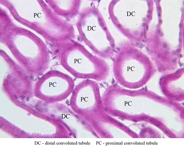
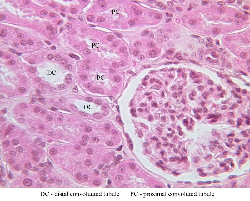
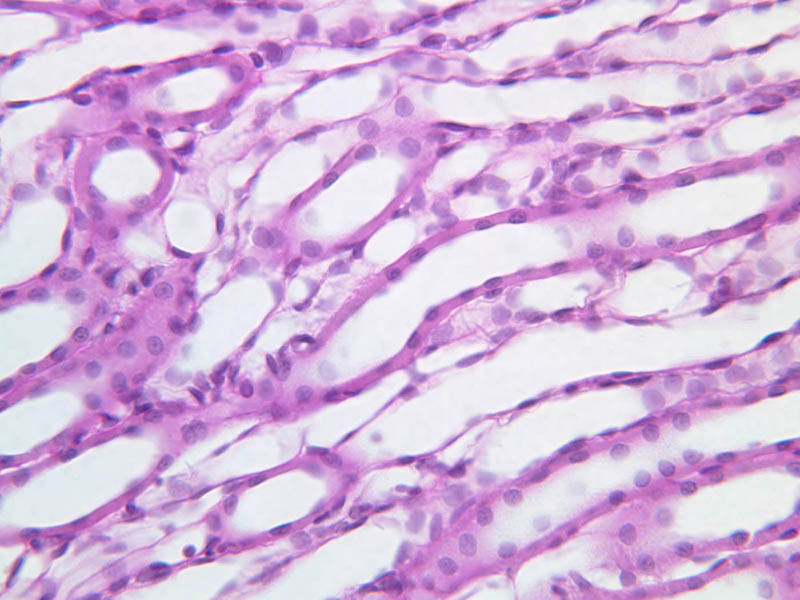
Proximal Convoluted Tubule (PCT)
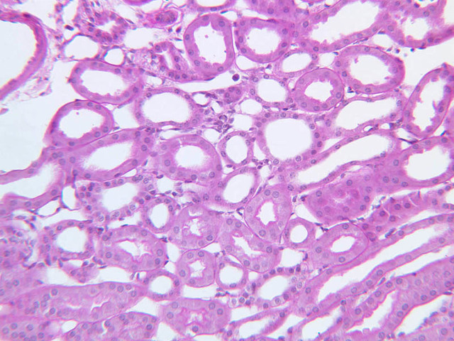
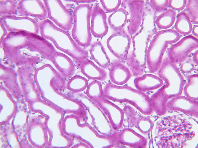
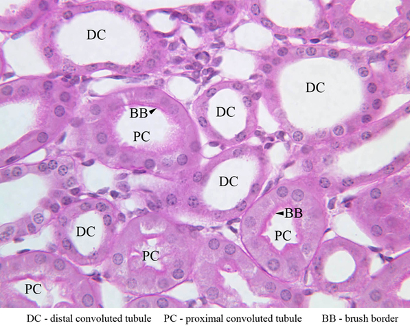
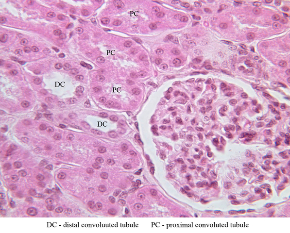
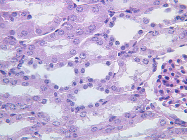
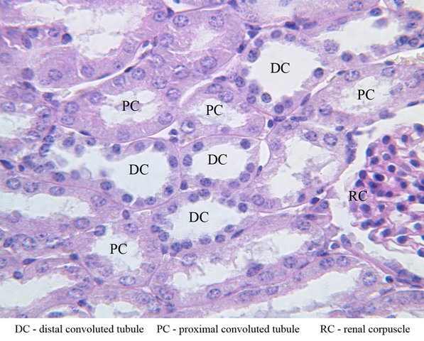
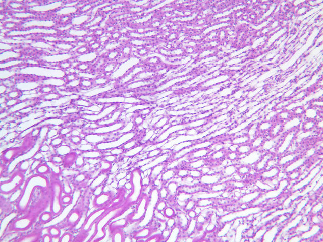
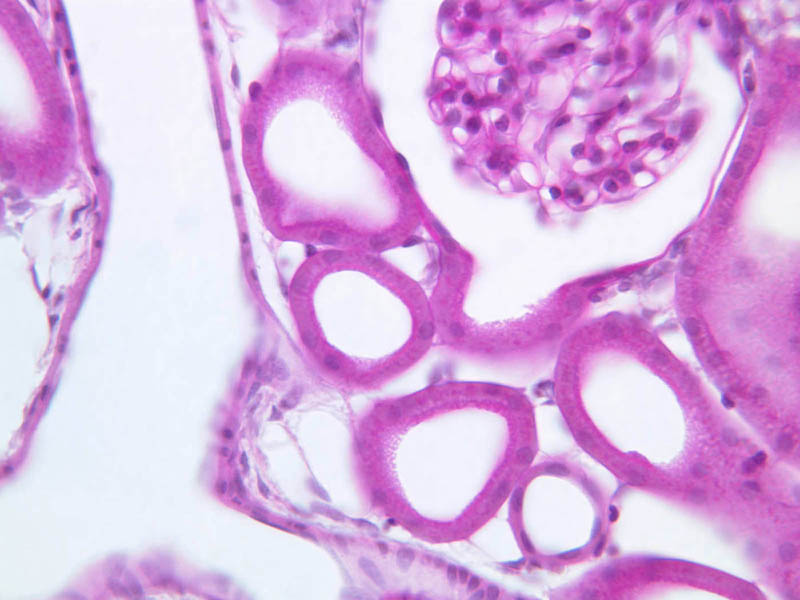
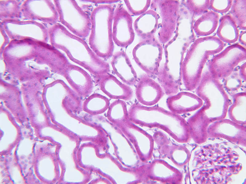
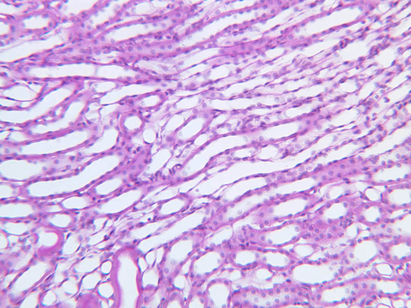
The filtrate formed in Bowman's space flows towards the urinary pole to enter the proximal tubule. The proximal tubule consists of an initial convoluted portion (the proximal convoluted tubule) and a distal straight portion (the pars recta). In cross section, the tubular epithelial cells are simple cuboidal cells, with a prominent brush border (microvilli) and basal striations (elongated mitochondria). The PCTs often have star-shaped lumens, which are generally larger in diameter than the distal tubules (see below). Cells of the PCT are generally more eosinophilic than cells of other tubules in the kidney, and stain particularly well with PAS (B-66, PAS [10x, 20x, 40x]; B-67, H&E [10x, 20x, 40x-labeled]; B-68, PAS [10x, 20x, 40x-labeled]; B-71, H&E [10x, 20x, 40x-labeled]). Try to identify PCTs. Also, examine regions near glomeruli in order to identify urinary poles in longitudinal section that are continuous with a PCT.Proximal Convoluted Tubule Gallery


Proximal Convoluted Tubule Identifications
| Row | Structure | Abbreviation | Optimal Stain | Representative Section | Note |
|---|---|---|---|---|---|
| 1 | Proximal Convoluted Tubule | PC | PAS | |
|
| 2 | Distal Convoluted Tubule | DC | PAS | |
|
| 3 | Brush Border | BB | PAS | |
Back to Top of Page
Thin Tubules
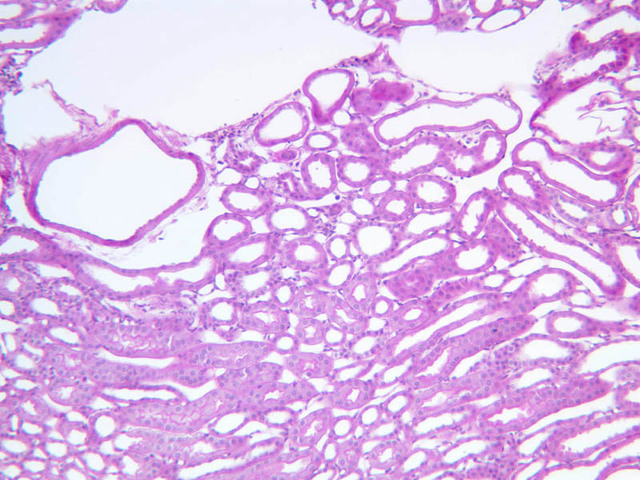
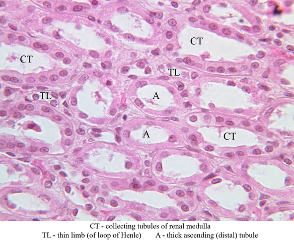
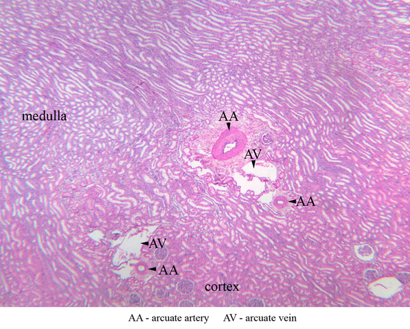
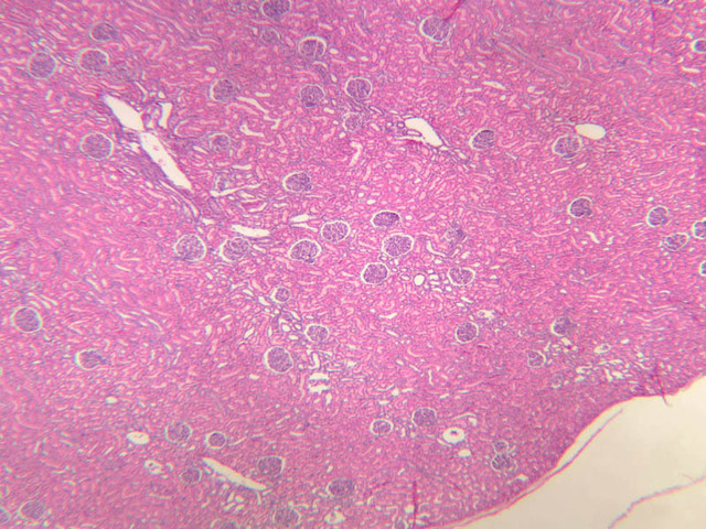
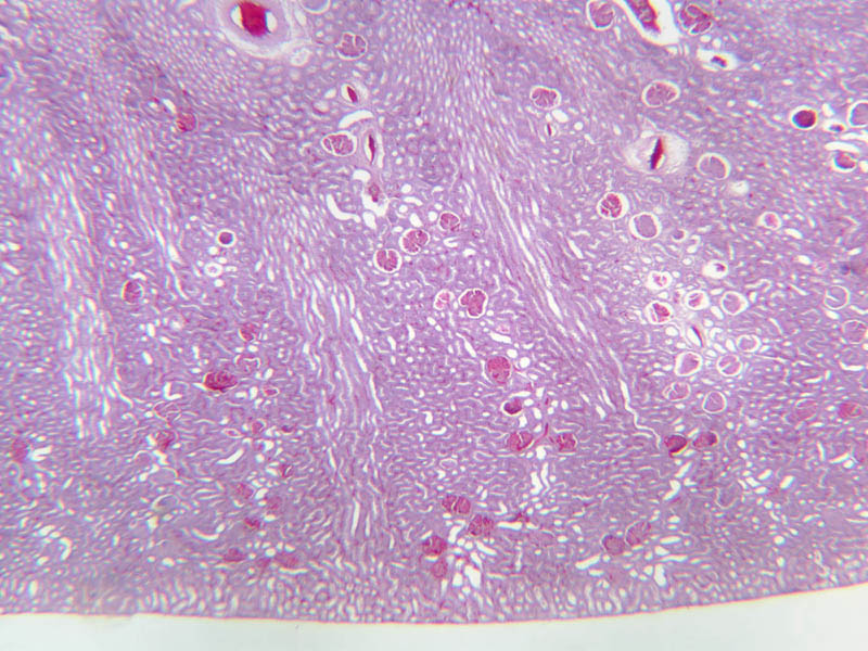
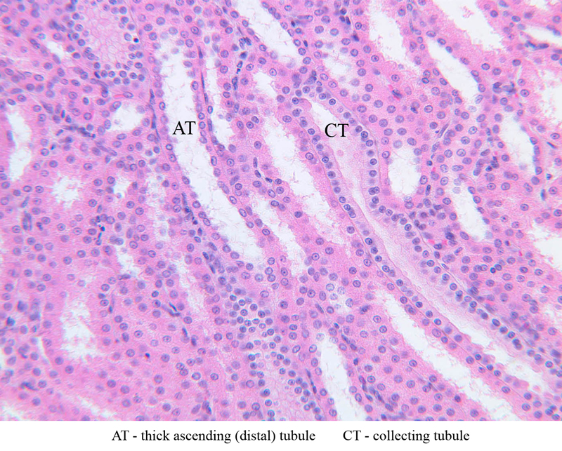
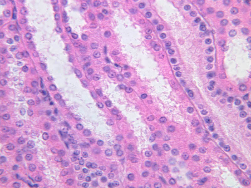
The straight portion of the proximal tubule (pars recta) enters a medullary ray and descends toward the medulla. Both parts of the proximal tubule are lined by cuboidal epithelial cells. The next tubular segment that the filtrate passes through is the thin tubule or thin loop of Henle which is mainly located in the medulla. Here the diameter of the tubule narrows markedly and the epithelium becomes simple squamous. The thin segment descends toward the apex of the pyramid. Before reaching the apex of the pyramid, however, it makes a hairpin turn and returns toward the cortex. Don’t confuse the thin segments of the tubules with the capillaries they associate with. Tubular epithelial cells are somewhat thicker, tubular diameter is somewhat greater, and the tubules do not contain RBCs. The next tubular segment is the distal thick tubule. The distal thick segment ascends through the medullary ray as the straight ascending segment. The straight ascending segment exits the medullary ray and returns to its corpuscle of origin. Try to identify each of these tubules by studying sections of the cortex and the medulla. Note the frequent appearance of arcuate artery and vein profiles along the boundary between the cortex and the medulla (B-67, H&E [2.5x-labeled, 10x, 20x, 40x-labeled]).Thin Tubules Gallery


Thin Tubules Identifications
| Row | Structure | Abbreviation | Optimal Stain | Representative Section | Note |
|---|---|---|---|---|---|
| 1 | Arcuate Artery | AA | H&E | |
|
| 2 | Arcuate Vein | AV | H&E | |
|
| 3 | Collecting Tubules of Renal Medulla | CT | H&E | |
|
| 4 | Thin Limb (of Loop of Henle) | TL | H&E | |
|
| 5 | Thick Ascending (distal) Tubule | TA | H&E | |
Back to Top of Page
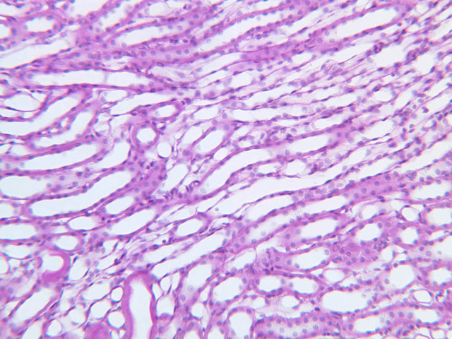
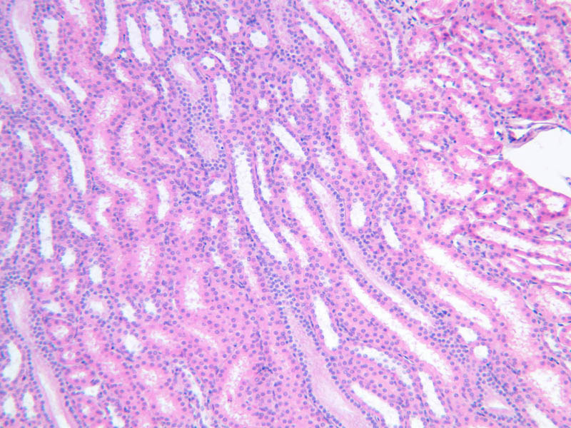
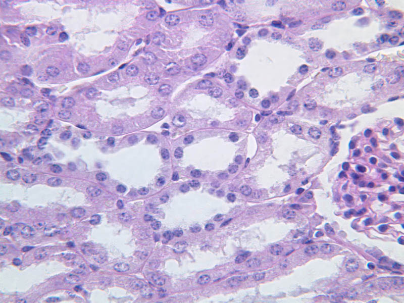
Distal Convoluted Tubule (DCT)
The distal tubule becomes convoluted and eventually empties into a collecting tubule. The cells of the distal convoluted tubule are cuboidal. In contrast to cells of the PCT, cells of the DCT are smaller, have less cytoplasm and hence appear more basophilic, and they have no brush border (B-67, H&E [10x, 20x, 40x-labeled]; B-68, PAS [10x, 20x, 40x-labeled]; B-69, PAS [10x, 20x, 40x-labeled]; B-71, H&E [10x, 20x, 40x-labeled]). Also, DCTs are smaller than proximal tubules. Look for DCTs in slides B-67 through B-71. Since the DCTs lack a brush border, they will be more readily distinguished from PCTs in sections stained with PAS.Distal Convoluted Tubule Gallery


Distal Convoluted Tubule Identifications
| Row | Structure | Abbreviation | Optimal Stain | Representative Section | Note |
|---|---|---|---|---|---|
| 1 | Proximal Convoluted Tubule | PC | PAS | |
|
| 2 | Distal Convoluted Tubule | DC | PAS | |
|
| 3 | Brush Border | BB | PAS | |
Back to Top of Page
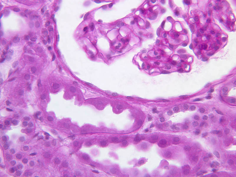
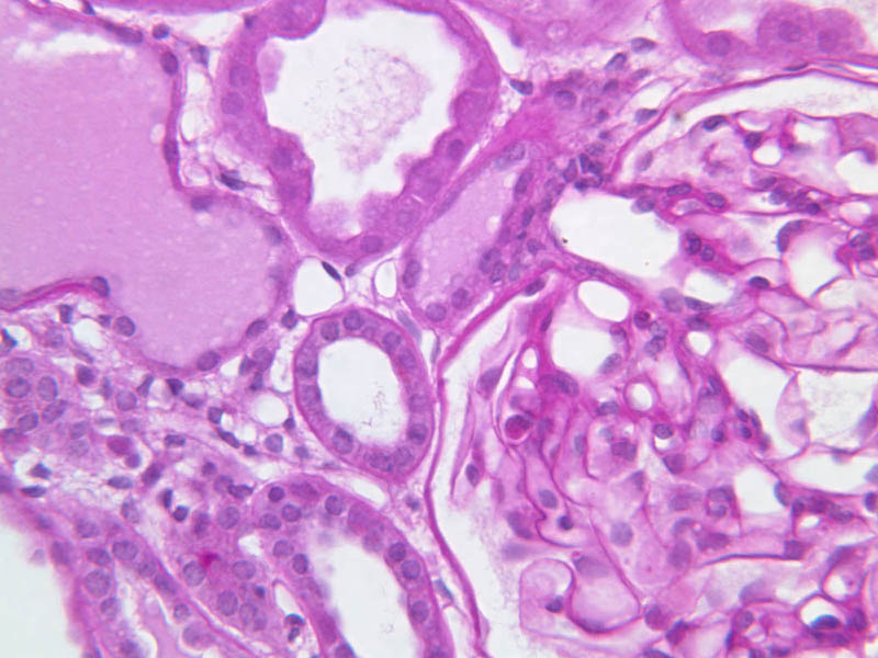
Juxtaglomerular Apparatus
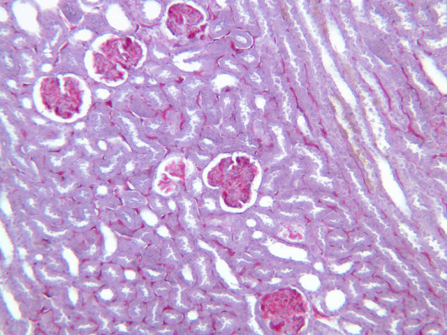
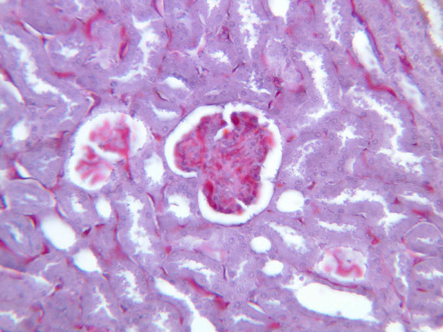
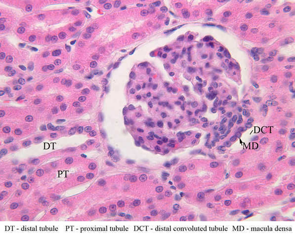
As the straight ascending segment of the distal tubule approaches its corpuscle of origin, it passes between the afferent and efferent arterioles and contributes to the juxtaglomerular apparatus (JGA). Locate a corpuscle showing an afferent arteriole entering and an efferent arteriole exiting at the vascular pole. Closely inspect the wall of the afferent arteriole. At this point, the vessel is closely applied to the wall of the distal convoluted tubule. The smooth muscle cells of the arteriole have spherical nuclei and are known as juxtaglomerular cells (JG cells). JG cells produce renin. Another structure to look for is the macula densa, which occurs where the cells of the DCT contact the afferent arteriole. Here they become taller and thinner forming the macula densa. Together, the macula densa and juxtaglomerular cells make up the juxtaglomerular apparatus (B-67, H&E [2.5x, 10x, 20x, 40x-labeled] [40x-labeled]; B-71, H&E [40x] [40x] [40x]).Juxtaglomerular Apparatus Gallery


Juxtaglomerular Apparatus Identifications
| Row | Structure | Abbreviation | Optimal Stain | Representative Section | Note |
|---|---|---|---|---|---|
| 1 | Macula Densa | MD | H&E | |
|
| 2 | Juxtaglomerular Cells | JG | H&E | |
Back to Top of Page
Collecting Tubules
The DCT ends in the collecting duct system which carries the urine through the medulla to the end of the papilla. Collecting tubules are regularly shaped in cross section as compared with the often irregularly shaped sections of the nephron. The lining epithelium of the collecting system gradually increases in height from a simple cuboidal epithelium in the arched tubules to a tall columnar epithelium in the papillary ducts. Look carefully at the epithelium lining the renal calyx at the apex of the medullary pyramid and identify collecting ducts. Collecting ducts have large, more regular, diameters, and are comprised of uniform, lightly stained columnar epithelial cells (B-67, H&E [2.5x-labeled, 10x, 20x-labeled, 40x]; B-71, H&E [2.5x, 10x, 20x, 40x]. Basement membranes are unusually important structures for the functioning of the kidney. Since a principal constituent of the basement membrane is polysaccharide, this structure stains well with the PAS stain. Study a PAS-stained section of kidney (B-66, B-68, B-69) and carefully note where the prominent basement membranes are found. The stain also shows the brush border of the proximal convoluted tubules and may selectively demonstrate granules in the juxtaglomerular cells of afferent arterioles (B-66 [20x, 40x]).Collecting Tubules Gallery
Error: no images found
Collecting Tubules Identifications
| Row | Structure | Abbreviation | Optimal Stain | Representative Section | Note |
|---|---|---|---|---|---|
| 1 | Collecting Tubules of Renal Medulla | CT | H&E | |
|
| 2 | Thin Limb (of Loop of Henle) | TL | H&E | |
|
| 3 | Thick Ascending (distal) Tubule | TA | H&E | |
Back to Top of Page
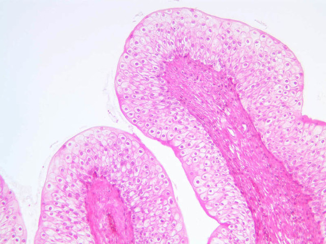
Ureter
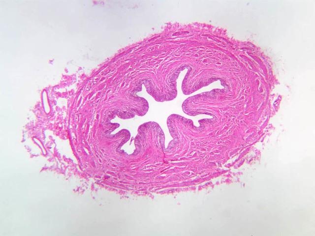
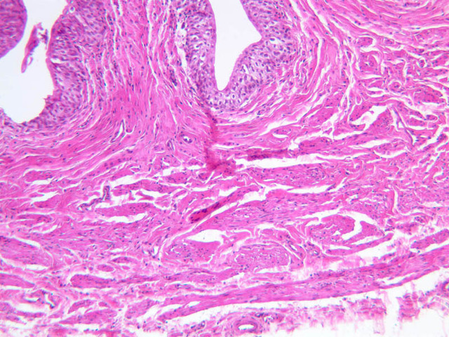
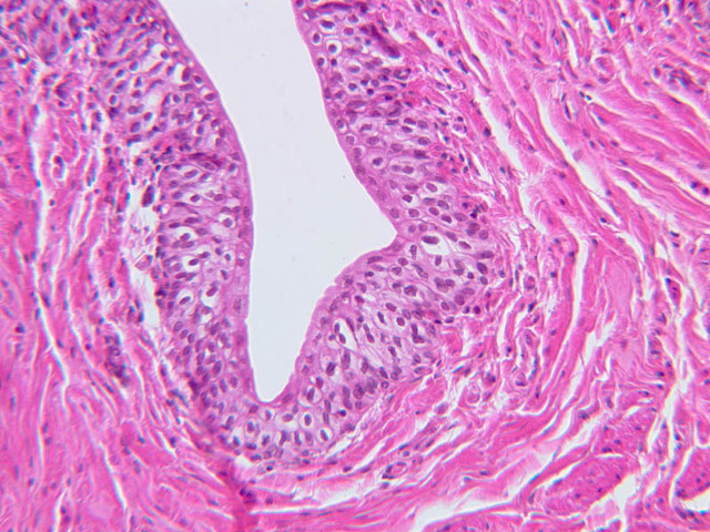
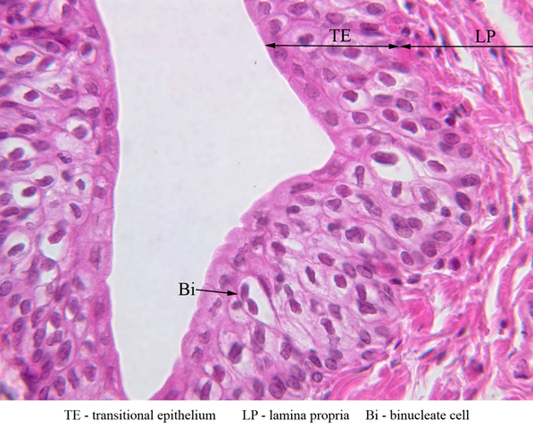
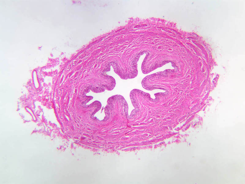
On slide B-72 (ureter, human, H&E [2.5x, 10x, 20x, 40x-labeled]), identify the mucosa (comprised of transitional epithelial cells), the lamina propria (which lacks a muscularis mucosa), and the muscularis. As these sections were obtained from the upper 2/3 of the ureter, there are only two layers to the muscularis. The inner smooth muscle layer is oriented longitudinally, and outer layer is circular smooth muscle. External to the muscularis is the tunica adventitia.Ureter Gallery
Ureter Identifications
| Row | Structure | Abbreviation | Optimal Stain | Representative Section | Note |
|---|---|---|---|---|---|
| 1 | Transitional Epithelium | TE | H&E | |
|
| 2 | Lamina Propria | LP | H&E | |
|
| 3 | Binucleate Cell | Bi | H&E | |
Back to Top of Page
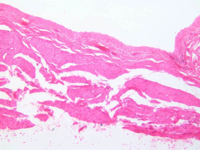
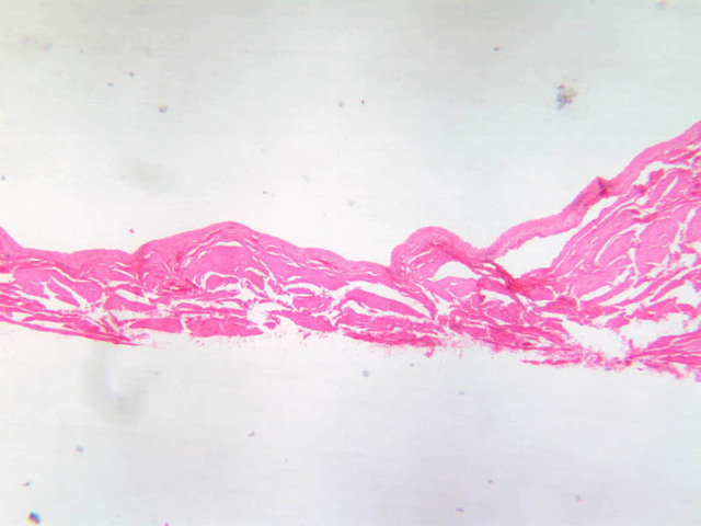
Urinary bladder
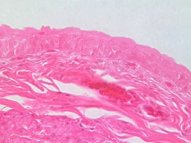
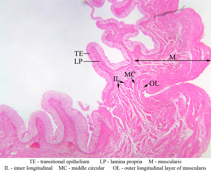
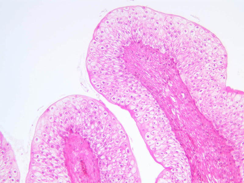
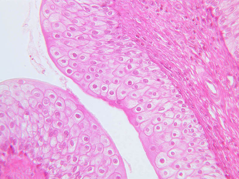
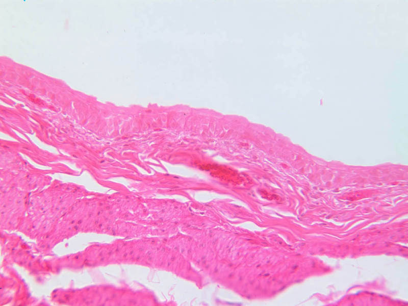
On slide B-75 (cat urinary bladder, contracted, H&E [2.5x-labeled, 10x, 20x, 40x]) identify the transitional epithelium. Notice that a transitional epithelium is usually thinner than a stratified squamous epithelium. The transitional epithelium of the urinary system is usually comprised of 6-8 cell layers in the contracted state with many of the cells on the luminal surface being dome-shaped. Identify the lamina propria and observe that like the ureter, there is very little (if any) muscularis mucosa. In the bladder, as in the lower 1/3 of the ureter, the muscularis externa consists of 3 layers of smooth muscle: an inner longitudinal layer, a middle circular layer, and an outer longitudinal layer. External to the muscularis, the tunica adventitia contains fat, blood vessels, and nerves of various sizes. Compare the thickness of the contracted bladder with a section of distended bladder (B-76, H&E [2.5x, 10x, 20x, 40x]). The transitional epithelium appears almost squamous due to distension. The epithelium is only 3-5 cell layers thick. The muscular layer also is thin. Transitional epithelial cells must be able to endure a highly concentrated urine to survive. To protect them they have membrane plaques that are thick and rigid. When the bladder is empty they fold into the cell, and return to the cell surface when the bladder is stretched. These structures render the epithelium impermeable to urine, and prevent water from diffusing in. This adaptive feature can be seen in the electron micrographs of transitional epithelium.Urinary Bladder Gallery


Urinary Bladder Identifications
| Row | Structure | Abbreviation | Optimal Stain | Representative Section | Note |
|---|---|---|---|---|---|
| 1 | Transitional Epithelium | TE | H&E | |
|
| 2 | Lamina Propria | LP | H&E | |
|
| 3 | Inner Longitudinal Muscularis | IL | H&E | |
|
| 4 | Middle Circular Muscularis | MC | H&E | |
|
| 5 | Outer Longitudinal Muscularis | OL | H&E | |
Back to Top of Page
Review of The Urinary System
Review of Slides


Summary of Identifications
| Row | Structure | Abbreviation | Optimal Stain | Representative Section | Note |
|---|---|---|---|---|---|
| 1 | Renal Corpuscle | RC | H&E | |
|
| 2 | Cortex | C | H&E | |
|
| 3 | Medullary Ray | MR | H&E | |
|
| 4 | Arcuate Artery | AA | H&E | |
|
| 5 | Arcuate Vein | AV | H&E | |
|
| 6 | Collecting Tubules of Renal Medulla | CT | H&E | |
|
| 7 | Thin Limb (of Loop of Henle) | TL | H&E | |
|
| 8 | Thick Ascending (distal) Tubule | TA | H&E | |
|
| 9 | Proximal Convoluted Tubule | PC | PAS | |
|
| 10 | Distal Convoluted Tubule | DC | PAS | |
|
| 11 | Brush Border | BB | PAS | |
|
| 12 | Bowman's Capsule (Parietal Layer) | BC | PAS | |
|
| 13 | Bowman's Space | BS | PAS | |
|
| 14 | Podocyte (visceral layer of Bowman's capsule) | Pod | PAS | |
|
| 15 | Glomerulus (capillary tuft) | G | PAS | |
|
| 16 | Vascular Pole | VP | Carmine | |
|
| 17 | Urinary Pole | UP | Carmine | |
|
| 18 | Macula Densa | MD | H&E | |
|
| 19 | Juxtaglomerular Cells | JG | H&E | |
|
| 20 | Transitional Epithelium | TE | H&E | |
|
| 21 | Lamina Propria | LP | H&E | |
|
| 22 | Inner Longitudinal Muscularis | IL | H&E | |
|
| 23 | Middle Circular Muscularis | MC | H&E | |
|
| 24 | Outer Longitudinal Muscularis | OL | H&E | |
|
| 25 | Binucleate Cell | Bi | H&E | |
Comments
Back to Top of Page -- AshleyLPistorio - 02 June 2007Edit | Attach | Print version | History: r2 < r1 | Backlinks | View wiki text | More topic actions
Topic revision: r2 - 20 Jun 2015, LorenEvey

- Epithelium
- Connective Tissue
- Muscle
- Nervous Tissue
- Cardiovascular System
- Skin Appendages and Sensory Receptors
- Lymphatic System
- Cartilage and Bone
- Respiratory System
- Peripheral Blood and Bone Marrow
- Oral Cavity and Salivary Glands
- Esophagus and Gastrointestinal Tract
- Pancreas, Liver, and Gall Bladder
- Endocrine Organs
- Male Reproductive System
- Female Reproductive System
- Urinary System
Ideas, requests, problems regarding Medical Histology? Send feedback